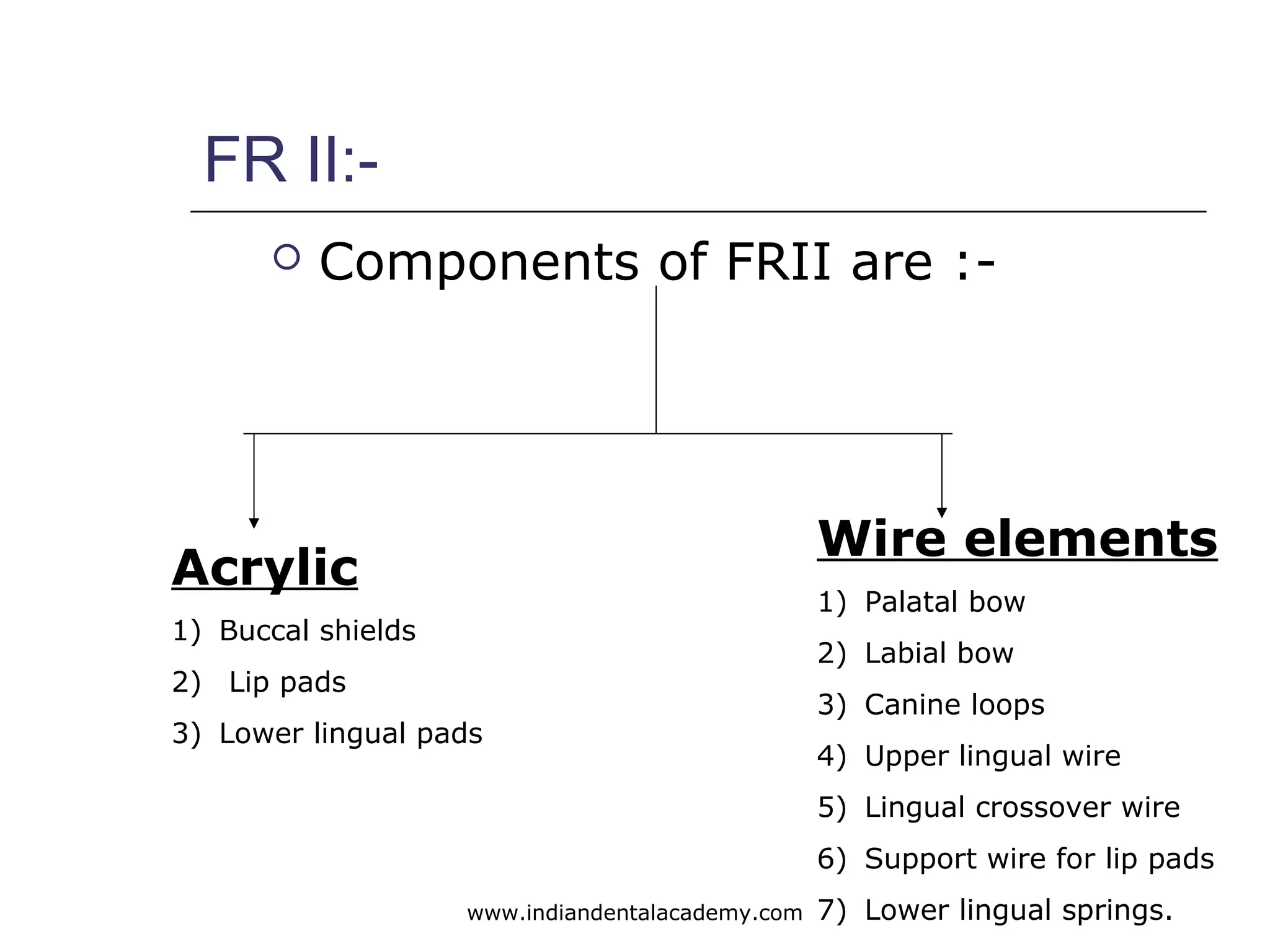
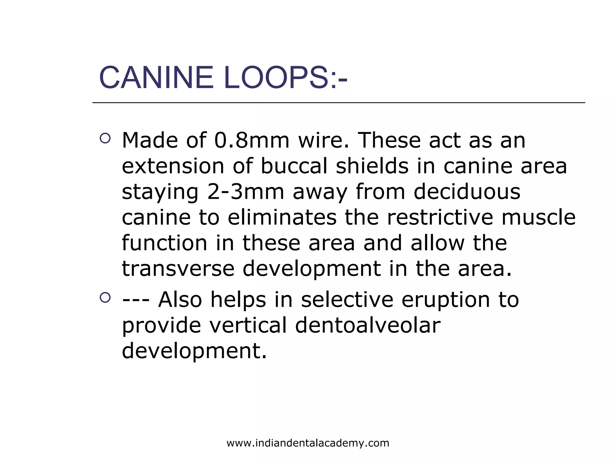
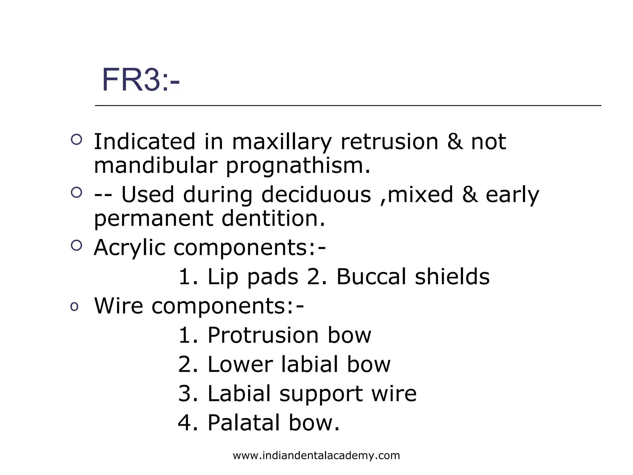
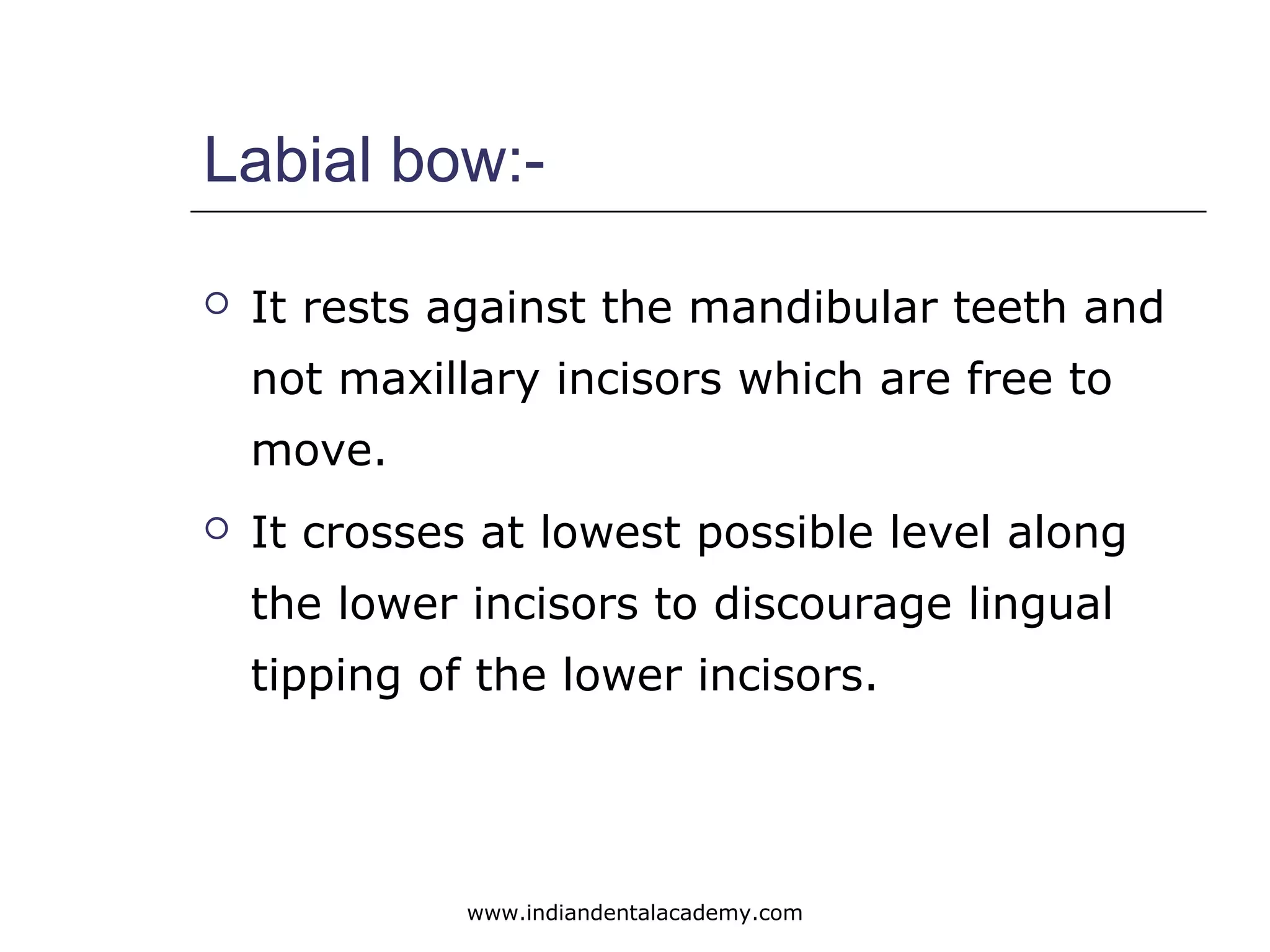
The Indian Dental Academy is the Leader in continuing dental education , training dentists in all aspects of dentistry and
offering a wide range of dental certified courses in different formats.for more details please visit
www.indiandentalacademy.com
2. Diagnostic aids for functional
appliances
Cephalometric analysis.
functional analysis.
www.indiandentalacademy.com
3. Cephalometric analysis
Four major areas of emphasis
which exist in Cephalometric
diagnosis for patients treated
during growth period e.g..
Functional appliances.
1. accomplishment of growth
increments and the direction or
vector of growth.
www.indiandentalacademy.com
4. 2. Assessment of the magnitude of
growth change–----
this helps in determining the
direction oh growth.
3. Inclination and position of the
upper and lower incisors----
to evaluate the probable
reciprocal growth increments of
the jaw bases.
www.indiandentalacademy.com
5. 4. Radiographic Cephalometric---
To allow the identification and
localization of anomalies and
abnormalities of size ,shape, and
spatial relations.
www.indiandentalacademy.com
6. FUNCTIONAL ANALYSIS
DIAGNOSTIC EXCERSISES:
a) Determination of postural rest
position of the mandible and
interocclusal clearance.
B) examination of the TMJ and condylar
movements.
C) assessment of the functional status
of the lips, cheeks, tongue with
[particular attention to the roles they
play in dentofacial abnormalities.
www.indiandentalacademy.com
8. Introduction:
Developed by balters in 1950’s
Is in common to Anderson –
activator.
Kantrowicz termed it as “ THE
SKELETON OF AN ACTIVATOR FROM
WHICH THERE IS NOTHING LEFT
BUT THE NAKED EMBODIMENT OF
ROBINS THOUGHTS”
www.indiandentalacademy.com
9. He assessed the appliance in 2 ways.
1.Bionator is less bulky, the anterior section of the
palate is free of acrylic thus children are free to
speak normally.
Thus, it can be worn day and night except at
meals.
2.For balters essential factor is tongue,
The equilibrium between tongue and cheeks
especially between the tongue and the lips in the
height, breadth and depth in oral space of
maximum size and optimal limits, providing
functional space for the tongue is essential for the
natural health of the dental arches. The tongue is
essential factor for the development of the
dentition. It is the center of reflex activity in the
oral cavity.
www.indiandentalacademy.com
10. Indications
For the treatment of sequelae of sucking habit
In correction of class II div 1 malocclusion with
well aligned arches, proclined incisors and
retruded mandible.
Treatment if deep overbite during mixed dentition
or even later.
Treating bruxism, periodontal disease and tmj
disorders.
In correction of class iii malocclusion.
Correcting open bite cases,.
www.indiandentalacademy.com
11. Treatment objective by Balters.
1. To accomplish lip closure and bring the back of the
tongue into contact with the soft palate.
2. To enlarge the oral space and to train its function.
3. To bring the incisors into edge to edge bite like what
beggs did.
4. Elongation of the mandible which will enlarge the oral
space and improve the tongue position
5. To achieve an improved relationship of the jaws,
tongue and the dentition as well as the surrounding
soft tissues.
www.indiandentalacademy.com
12. Case-selection:
Used basically for treatment of class II div 1
or class I with class II div 1 symptoms in
which lower lip cushions to the lingual of
the upper incisors constantly.
It intercepts the perverted perioral muscle
activity during the day when it is most
likely to deform the dentition.
www.indiandentalacademy.com
13. Types of the appliances:
1) Standard appliance
2) Class III appliance
3) Open bite appliance
www.indiandentalacademy.com
14. Standard appliance: it is used for the correction of
class II div 1 malocclusion in order to correct the
backward position of the tongue and its
consequences.
For the treatment of the narrow arches of class i
malocclusion.
It consist if acrylic parts and wire elements.
Acrylic parts: relatively slender acrylic body fitted to
the lingual aspects of the mandibular arch and part of
the maxillary dental arch.
Maxillary part: it covers only the molars and
premolars . anterior maxillary part from canine to
canine remains open. The relative position of the
joined upper and lower portions is determined by the
construction bite (usually edge to edge bite.)
www.indiandentalacademy.com
15. Acrylic extends about 2mm below the
mandibular gingival margin and about the
same distance above the maxillary gingival
margin.
Depending on the over jet: if the over jet is
increased then the acrylic extended to
cover the lower incisors.
www.indiandentalacademy.com
16. Wire elements:
1) Palatal arch.
2) Vestibular arch.
Palatal arch: Palatal arch is made from 1.2mm
diameter hard stainless steel wire. The egg
shaped palatal arch emerges from the upper
margin of the acrylic approx. Opposite to the
middle of the first premolar. Then it follows the
contour of the palate about 1mm distance from
the mucosa . the arch forms a wide curve that
reaches a line joining the distal surface of the
first premolar and follows mirror image on the
opp. Side.
www.indiandentalacademy.com
17. Mode of action:
It stimulate the distal aspect of the
dorsum of the tongue .Thus the
curve of the arch is directed
posteriorly effecting a forward
orientation of the tongue as well as
the mandible into class I relation. Of
the jaws.
www.indiandentalacademy.com
18. Vestibular wires:
Is made up 0.9mm dia. Wire. It emerges
from the acrylic below the contact point
between the upper canine and the 1st
premolar. The vestibular wire rises
vertically and is then bent down at right
angle to go distally along the middle of
the crown of the upper premolars. Just
anterior to the mesial contact point of the
1st
molar the wire is fashion in a round
bend toward the lower dental arch.
www.indiandentalacademy.com
19. The wire maintains a constant level at
height of the papillae , parallel to the
upper portion anterior to the
mandibular canine. At this point the
wire is bent to reach the upper canine
nearly touches the incisal third of the
incisors and from there in a mirror
image of the side already fabricated
proceeds posteriorly to the acrylic on
the opp. Side.
Vestibular wire: anterior part--labial wire.
lateral part–- buccinator bend.
www.indiandentalacademy.com
20. Mode of action:
Between the mucosa of the lips and
the incisors a slight negative
pressure is created. In the course of
treatment, this help to upright the
incisors , provide space for them
when the dental arch is widened lab
ally & sagitally & also inferior
development in the region of the
apical base.
www.indiandentalacademy.com
21. Buccinator bends:
1. They keep away the soft tissue of the cheeks
,which is normally drawn into the inter-occlusal
space in the malocclusion-thus bite is leveled &
eruption will proceed in the buccal segment.
2. They move the surface of the orofacial capsule
laterally increasing the oral space by virtue of
the forward positioning of the mandible which
relaxes the musculature while the vestibular
wire holds it away from the alveolar mucous
.,this favors the expansion or transverse
development of the maxillary dentition.
www.indiandentalacademy.com
22. Class III appliance :-
Meant for treatment of mandibular
prognathism to compensate for the
forward position of the tongue.
Acrylic part:- similar as that of the
standard appliance. a mandibular part
and 2 lateral maxillary parts extending
from the 1st
premolar to 1st
premolar are
joined together, opening the bite about
2mm to allow the upper incisors to
move labially past the lower incisors.
www.indiandentalacademy.com
23. This space is covered towards the tongue by an
extension of the mandibular portion of the
palate from canine to canine.
Upper incisal margin extend 2mm beyond the
upper margin of acrylic & 1mm of the
thickness of acrylic is removed from behind
the mandibular incisors.
Thus acrylic creates a barrier to prevent any
forward movement of the tongue toward the
vestibule. Its purpose is to teach the tongue
by proprioceptive stimuli to remain in its
retracted and proper functional space. Tongue
thus touches the anterior part of
palate(uncovered) and stiulates the forward
growth component.
www.indiandentalacademy.com
24. Wire elements:
Palatal arch
Vestibular wires.
Palatal arch: made up of 1.2mm dia.the
curve of the arch faces anteriorly
extending forward to a line connecting the
middle of 1st
premolars and then running
parallel along the palatal vault posteriorly
till the distal surface of the 1st
molar at
which point it enters the acrylic at a right
angle bend.
www.indiandentalacademy.com
25. Vestibular wire:
Made up of 0.9mm dia. Wire placed in
front of the lower incisors. It emerges
below the contact point of the upper
canines &1st
premolars. The buccinators
bends are fabricated and then wire runs
in a distal direction until it reaches a point
just behind the 2nd
premolar.then a round
bend is made and wire runs forward along
the lower incisors remaining the thickness
of a sheet of paper away from the labial
surface.
www.indiandentalacademy.com
26. OPEN BITE APPLIANCE:-
MEANT TO CORRET VERTICAL
DISCREPANCY---OPEN BITE.
Acrylic part: tongue is considered the main
cause which prevents the eruption of
maxillary and mandibular incisors allowing
over eruption of buccal segments causing---
open bite.
Thus acrylic covers anteriorly to prevent tongue
inserting into the aperture.this is free from
the teeth and alveolar bone so as not to
interfere with the expected growth changes.
www.indiandentalacademy.com
27. Mandibular part is joined with the
maxillary part by bite blocks in the
posterior region. These bite blocks
are made with indentation of the
teeth so as to disallow the eruption
and at the same time anterior are
free to erupt. This establishes the
inter-occlusal clearance and
postural vertical dimension. Block
must not be so thick as to prevent
lip seal.
www.indiandentalacademy.com
28. Wire elements:
Palatal arch
Vestibular arch
if the lips have the tendency to get
into open bite than lip shields may
be added to the appliance.
www.indiandentalacademy.com
29. Construction bite:-
Objective is to establish the class I relation.
Incisor teeth position is also important so
possibilities are:
• Preferably edge to edge relationship of all teeth
or at lest the lateral incisors. This provide
maxillary functional space for the tongue.
• If the overjet is too large- than step by step
protraction procedure is followed and the
mandibular incisors must be covered by a
grooved rim. After the reduction of overjet a
new appliance with edge to edge bite can be
fabricated.
www.indiandentalacademy.com
30. Construction bite:-
Class III: It is registered in the
most posterior position that is
possible for the mandible.
www.indiandentalacademy.com
31. Appliance modification:
I. Adding 3mm thin maxillary incisal capping from
distal surface of lateral incisor to lateral incisor
on the other side.
--- it secures the position of maxillary
incisors.
--- it also gives a intrusive force which
frees the occlusal surface of the buccal teeth
allowing a rapid leveling of bite.
---prevents any labial tipping of maxillary
incisors.( williamson &hamilton modification)
II. For class II div 1 with deep bite, lateral surfaces
remain free.
--- for deep bite with elongation of upper &
lower incisors. www.indiandentalacademy.com
32. Bionator and TMJ cases:-
Bionator is a effective means to treat tmj problems
in adult cases where tmj problems are coincident
with bruxism and clenching during rem period of
the sleep.
as it relaxes the muscle spasm- particularly lateral
pterygoid muscle when used in the night.
it also prevents the riding of condyle over the
posterior edge of the disk which causes the clicking.
Cause of correction:- it causes permanent
foreshortening of the pterygoid muscle of the
mandible.
www.indiandentalacademy.com
33. Role of the tongue:
According to balters class II malocclusion are due
to backward position of the tongue– disturbing
the cervical region– respiratory function in the
larynx is impeded– thus faulty deglutition– along
with mouth breathing.
Class III– due to forward posturing of the tongue
and cervical over development.
Class I– due to lack of transverse development of
the dentition as a consequence of weakness of
the tongue in comparison with the strength of
buccinators mechanism.
www.indiandentalacademy.com
34. Although the reasoning is teleological it does co-
relates with functional matrix concept of moss and
neurotrophic elements of functional matrix theory.
Thus, in treatment of class ii:- tongue is brought
forward by stimulation of the distal part of the
dorsum of the tongue.
-- Mandible is allowed to develop anteriorly &
brought in class i relation thus cervical viscera also
is brought forward.
-- This enlarges the respiratory space & enhances
the reflex of deglutition
In class III– tongue is brought backward & higher
thus idea is to reduce the anterior force vector &
bring the mandible in class I. This new position of
the tongue reduce cervical development.
Class i :- muscle exercise are used to strengthen the
tongue.
www.indiandentalacademy.com
35. Lip seal importance:-
For successful bionator therapy lip seal
closure is required for the treatment of all
kinds of malocclusion
According to balters this a precondition for
the free development of the growth
potential which is impeded by abnormal
function.
This expression of inhibited growth potential
is made possible by end to end incisal
biting position(ROLF FRANKEL)
www.indiandentalacademy.com
36. Frankel appliance:-
Introduction:-Developed by rolf
frankel a orthodontist from small
town zwickau,east Germany and he
called it as funktionsregler.
Also called as vestibular appliance,
oral gymnastic appliance.
www.indiandentalacademy.com
37. Concepts of frankel appliance:-
FR is largely confined to oral vestibule & hold
away the buccal 7 labial musculature from the
dentition in these areas in which pressure on
the dentoalveolar structures has restricted the
outward development of these structures
during the critical transitional phase of
development of dentition.
A major tenet of the frankel philosophy is that
the dentition is heavily influenced by the
functional matrix, the buccinator mechanism &
the orbicularis oris complex.Abnormal perioral
muscle function creates dynamic barriers to
optimal growth of the dent alveolar complex in
three-dimensional of space.
www.indiandentalacademy.com
38. I. Vestibular area of operation:-
These appliance provides a positive framework
and an optimized supportive structure on which
muscles that make up the stomatoganthic system
can work correctly.
Frankel stresses that appliance is an exercise device
for oral gymnastics. It gives the oral musculature
a proper skeletal matrix on which to function &
normal functional patterns can be established.
Adverse pressures are removed from the
dentoalveolar structures & result is seen in the
form of maxillary dental arch.
Thus when the appliance is finally removed the
dentition assumes the spatial relationship that is
similar to that established by the vestibular shields
& the lip pads.
www.indiandentalacademy.com
39. II. Sagital correction via tooth
borne maxillary anchorage:-
It is considered as second pillar of FR philosophy.
The forward posturing of the mandible is achieved
by an acrylic pad that contacts the alveolar bone
only behind the lower anterior segment.
The vestibular construction is fabricated so that the
forward posture is augmented by the acrylic wire
configuration making the lingual contact more of a
proprioceptive trigger for postural maintenance
than a pressure bearing area.
The appliance is anchored in the maxillary dentition
both in the molar & canine area
www.indiandentalacademy.com
40. III. Differential eruption
guidance:-
Mandibular posterior segment is
kept free to erupt for selective
differential eruption which corrects
the vertical dimension, but also
helps in sagittal correction of class
II malocclusion.
www.indiandentalacademy.com
41. IV. Minimal maxillary basal effect:-
There might be slight retrusive effect on maxilla
due to labial arch bow & due to musculature that
pulls the mandible but that is not our treatment
objective.
According to mcnamara maxilla in class ii
malocclusion usually normal & mandible
retrusion is our main concern
Maxillary dental correction like closure of
spaces can be done but i.E secondary and not
required & may cause adverse effects like–
tissue impingement, more tipping leading to
unseating of appliance.
www.indiandentalacademy.com
42. V. Buccal shields,lip pads, &
periosteal pull:-
Research by Enlow,Hoyt& Mofet has shown that
pull on periosteal tissues enhance growth beneath
it. FR causes outward periosteal pull by maximal
extension of shields and pads into the depths of
buccal & labial vestibule to the patient at which
the depth of the sulcus is under tension & the
bony shell beneath this area houses the erupting
permanent teeth, an outward growth of the
membranous bone, plus relief of any restrictive
tissue pressure, results in bodily transverse
changes in the posterior segment & bone
formation at the apical base contiguous to lip
pads.
This concept is still under research by ada
research institute and it is done on squirrel
monkey. www.indiandentalacademy.com
43. FR—VTO(visual treatment
objective)
It is a very simple yet very important
diagnostic maneuver.
Procedure:- the patient is asked to bite in
habitual occlusion & relax the lips & profile is
carefully studied & is photographed.
Than, the patient is asked to bring the mandible
forward into correct sagital relationship,
reducing the overjet and is photographed and
can be compared with original instant print
view with the teeth in occlusion.
www.indiandentalacademy.com
44. If this improves the profile ,then frankel
appliance is indicated. The photographs
can be shown to patient and used as a
very important motivational aid.
Patients with functional retrusion, deep
overbite& excessive interocclusal space
are good choice.
Patient with increased anterior face
height, shallow bite are poor choices.
www.indiandentalacademy.com
45. Types of FR
FR1:- used for treatment of class I
malocclusion & class II div1 Malocclusion
it is further divided into:-
FR1a:- used for treatment of class 1
Malocclusion with minor to moderate deep bite
and crowding.
FR1b:- used for treatment of class II div1
malocclusion where overjet exceeds 5mm
FR1c:- used for treatment of class II div 1
malocclusion where overjet is more than 7mm.
www.indiandentalacademy.com
46. FR2:- used for treatment of class II div 1 and
div 2 malocclusion.
FR3:- used for treatment of class III
malocclusion.
FR4:-used for treatment of open bite &
bimaxillary protrusion.
FR5:- they are the functional regulators that
incorporates head gear. Indicated in long face
patients having a high mandibular plane &
vertical maxillary excess
www.indiandentalacademy.com
47. Functional regulator 1:-
Components:- 1)shields. 2) pads. 3) bow
4)wires.
Buccaly,there is distinguishing vestibular shield,lip
pads, labial bow & canine loops.
Lingually, there is palatal wire or bow with
convexity facing distally & with lateral extension
crossing the occlusal surface in the embrasure
mesial to 1st
molar. These lateral extension arte
anchored in the shields.
www.indiandentalacademy.com
48. Appliance in the maxillary arch is
stabilized by firm insertion of wire
elements in the embrasure either
between 1st
molar & 2nd
deciduous molar.
The palatal bow ends are recurved in the
buccal region to rest in the central
groove of 1st
molar to act as molar
occlusal rest as stabilizing component.
Mandible has lingual bow with u loops
extending downward to the floor of the
mouth & below the incisors marginal
ridge.
www.indiandentalacademy.com
49. FR1a:--
It consists of:-1. Labial bow
2.canine loop
3. Palatal bow
4. Lingual bow
5. Lip pads.
6. Vestibular shields.
Mandible is brought forward & hold in this
position against maxillary teeth by cross-
sectional wires at the 1st
molar and at
canine- premolar embrasure.
www.indiandentalacademy.com
50. It prevent its dislodgement superiorly which
passes through 1st
molar- 2nd
deciduous
molar embrasure to end as occlusal rest
on 1st
permanent maxillary molar.It would
force the periphery of the shields into
sulcus.
It also prevent upper molar eruption where
as lower molar are free to erupt.
Palatal bow:-
www.indiandentalacademy.com
51. Canine loops:-
It locks the appliance into maxillary
dentition and is used for guiding
eruption of canines.
www.indiandentalacademy.com
52. Lingual wire bow:-
It guides or posture the mandible
forward. Its loops extend caudally
contacting the mucosa at the apical
base which acts as a main pressure
for the mandible.
Wire is free of lower incisors ,if
proclination of lower incisors
required then it contact them.
www.indiandentalacademy.com
53. Buccal shield:-
Approximate the buccal surface of the molars &
premolars.
Is carried deeply into the vestibular sulcus as
tissue attachments & comfort of patient will allow.
It stands away from the dentition & the basal
alveolar bone in the maxillary arch to relieve the
pressure from the contiguous musculature
allowing unrestricted alveodental development.
It provides constant exercise which will stimulate
periosteal pull with an intermittently outward
force.
www.indiandentalacademy.com
54. Lip pads:-
It eliminates the perverted perioral muscle
activity e.g. Hyperactive mentalis activity.
It prevents the lip trap, the deforming muscular
activity is controlled in both the arches.
Creates periosteal pull labially from the pad
pressue,exerts bone stimulus & reduces the
mentolabial sulcus.
It maintains the mandible in its mesial
construction bite.
www.indiandentalacademy.com
55. FR1b :--
It consists of lingual wire instead of
lingual acrylic pad in contrast to
FR1a.
www.indiandentalacademy.com
56. Lingual acrylic pad:-
It is anchored in the buccal shields & the
cross-over occlusal wires does not contact
the upper or lower deciduous molars.
[Premolars as it passes through inter-
occlusal space at the embrasures of
deciduous molars.
Advantage:- better tolerances for the
patient.
www.indiandentalacademy.com
57. Construction bite:-
A thin yellow or red wax wafer is prepared
in the form of horse –shoe shape &
softened by keeping in warm water for few
seconds.
For minor sagittal problems of 2-4mm,
construction is taken in an end to end
incisal relationships making sure there is no
obvious strain on facial muscles & the
balance between protractor & retractor
muscles is maintained.
While taking construction bite care is taken
that midline coincide.
www.indiandentalacademy.com
58. According to frankel– mandible should not
be moved further than 2.5 -3 mm &
vertical opening of about 2.5 -3mm in
buccal segments to allow the cross-over
wires to pass through.
Rule of thumb is the greater the horizontal
movement the less the vertical opening.
According to some clinicians like petrovic
who has also did a research on this that
correcting the sagittal discrepancies in two-
three stages is more effective as it causes
optimal prechondroblastic activity in the
region of the condyle.
www.indiandentalacademy.com
59. Model trimming:-
Another important step in frankel appliance
construction. The casts are carved or trimmed in the
buccal shield or lip pads area to increase the depth of
the sulcus.
It is done in order to produce the tissue tension
necessary to stimulate appositional bone development
in the basal area.
It is done with a pear shaped carbide bur & wax knife.
Application:- To prevent the soft tissue creeping under
the pads.
www.indiandentalacademy.com
60. Wire elements:-
Lower lingual support wire:- made
up of 1.25mm stainless steel wire.
The horizontal reinforcing wire
member follows the contours of the
apical base at approximate 1-2mm
from the mucosa & 3-4mm below
the lingual gingival margin of the
incisors.
www.indiandentalacademy.com
61. Lower lingual springs:-
Made up of 0.8mm dia. Wire. These
recurved springs are contoured to
the lingual surface of the lower
incisors, right above the cinguala
with free ends about 3mm below
the incisal margin.
-------- It prevents extrusion of the
lower incisors.
www.indiandentalacademy.com
62. Lower labial wires:-
Made of 0.9mm – it acts as a
support skeleton for the lip pads. It
is about 7mm below the incisal
gingival margin with the midline
wire “V” shaped to circumvent the
labial muscle attachment.
www.indiandentalacademy.com
63. Palatal bow:-
Made of 1mm wire- it has slight
posterior curve which increases wire
length to facilitate the lateral
expansion. It crosses in the
maxillary 1st
molar & 2nd
deciduous
molar embrasures and emerges
buccaly to lie between the maxillary
molar buccal cusps to act as a rest.
www.indiandentalacademy.com
64. Labial bow:-
Made up of 0.9mm wire.:- It lies in
the middle of the labial surface of
incisors to end in buccal shields.
www.indiandentalacademy.com
65. FR1c:--
The buccal shields are split horizontally &
vertically into parts .The antero- inferior
portion contain wires for the lingual
acrylic pressure pad or shield & for the
lower lip pads. This permits the forward
movement of the anterior section of the
appliance.
Thus it is used when multiple
advancement is needed.
www.indiandentalacademy.com
66. FR II:-
Components of FRII are :-
Acrylic
1) Buccal shields
2) Lip pads
3) Lower lingual pads
Wire elements
1) Palatal bow
2) Labial bow
3) Canine loops
4) Upper lingual wire
5) Lingual crossover wire
6) Support wire for lip pads
7) Lower lingual springs.www.indiandentalacademy.com
67. LINGUAL STABILIZING BOW:-
It is made up o9f 0.9mm wire which
provides added stability to the maxillary
arch. It originates in the vestibular shield
and passes through the canine -1st
deciduous molar embrasure. The wire
forms loops that contact the incisors at
the canine-lateral incisor embrasure.
--It prevents the lingual tipping of the
maxillary incisors which is not desired.
www.indiandentalacademy.com
68. CANINE LOOPS:-
Made of 0.8mm wire. These act as an
extension of buccal shields in canine area
staying 2-3mm away from deciduous
canine to eliminates the restrictive muscle
function in these area and allow the
transverse development in the area.
--- Also helps in selective eruption to
provide vertical dentoalveolar
development.
www.indiandentalacademy.com
69. FR3:-
Indicated in maxillary retrusion & not
mandibular prognathism.
-- Used during deciduous ,mixed & early
permanent dentition.
Acrylic components:-
1. Lip pads 2. Buccal shields
o Wire components:-
1. Protrusion bow
2. Lower labial bow
3. Labial support wire
4. Palatal bow.
www.indiandentalacademy.com
70. Lip pads:-
Situated in the maxillary dentition instead of mandibular.
They stand away from the mucosa in the labial vestibular
area .
Purpose:-1) they eliminate the restrictive pressure of the
upper lips on the under developing maxilla
2) to exert tension on the tissue & periosteal
attachment in the depth of the maxillary sulcus to stimulate
bone growth.
3) to transmit the upper lip force to mandible via the
lower labial arch for a retrusive stimulus.
www.indiandentalacademy.com
71. Labial bow:-
It rests against the mandibular teeth and
not maxillary incisors which are free to
move.
It crosses at lowest possible level along
the lower incisors to discourage lingual
tipping of the lower incisors.
www.indiandentalacademy.com
72. Buccal shields:-
They contact the mandibular teeth
& mandibular apical base so as to
eliminate the buccinators
mechanism and orbicularis oris
musculature effect acting on the
maxillary dentition.
www.indiandentalacademy.com
73. Construction bite:-
Is done by retruding the mandible as
much as possible with the condyle in its
most posterior position in the fossa.
Bite is opened enough to let the
maxillary incisors move labially past the
mandibular incisors but is kept minimum
to allow lip closure with minimal lip strain.
www.indiandentalacademy.com
74. FRIV:-
Used in construction of open bites & in
bimaxillary protrusion.
Appliance therapy is preferred in mixed
dentition
Same vestibular configuration as FRI and FRII
but canine loops 7 protrusion bow is absent.
Four Occlusal rest on the maxillary 1st
molar,1st
deciduous molars to prevent tipping of the
appliance.
Palatal bow is like FRIII & is placed behind the
last molar.
Occlusal rest prevent the shifting of the
appliance in the posterior or dorsal direction.www.indiandentalacademy.com
75. FRV:-
Appliance consists of posterior bite
blocks that prevent molar eruption
due to action of elevator muscles of
mandible. Head gear tubes are
incorporated for extra-oral
anchorage.
www.indiandentalacademy.com
76. Treatment objective of frankel
appliance:-
1) An increase in transverse & sagittal intra-oral space:-
• achieved primarily through buccal shields & lip pads
• They eliminate the harmful mechanical pressure on the
pressure sensitive membranous bone structure thus favoring
forces acting within the oral cavity (tongue).
• The constant outward periosteal pull that is exerted on the
connective tissue fibers & muscle attachments in the oral
vestibule is transmitted to the contiguous alveolar bone by
the fibers inserting into the periosteum & bone. This aids in
the lateral movement of the dentoalveolar shell
www.indiandentalacademy.com
77. 2) Increase in vertical intra-oral space:-
• This is possible because the construction bite is
taken so that bite is opened in the posterior segment
so as the mandible is held forward along with
differential eruption of teeth.
• Disturbance in vertical development is due to the
cheek & less due to the effect of the tongue.
www.indiandentalacademy.com
78. 3) Mandibular protraction:-
• The position of mandible is changed through the
gradual training of the protractor & retractor muscles
followed by condylar adaptation.
• The lingual pressure pad guides the mandible by
exteroceptive stimuli to more position. Whenever the
mandible is brought back the lingual pads apply
pressure on the lingual alveolar process .
• This causes protractor muscles to position the
mandible mesially.
www.indiandentalacademy.com
79. 4) Muscle function adaptation:-
a) Development of new patterns of
motor function.
b) Improvement of muscle tonus
c) Establishment of proper oral seal.
www.indiandentalacademy.com
80. It prevents abnormal perioral muscle
function from exerting a deforming
influence on the bony structures .
It provides a new & more normal shell for
the functional matrix to work against .
It massages the soft tissue to improve
blood circulation, loosening the tight
muscles & improve the tonicity of muscle.
www.indiandentalacademy.com
81. Thus frankel his appliance as an exercise
device & the exercise he recommended
are oral gymnastics.
The mandibular lip pads prevent the
hyperactivity of mentalis muscle,
eliminate lip trap & assist in establishing
proper oral lip seal.
Oral gymnastics are :- all normal function
like swallowing, speaking,& mimic
movement.
And keeping the lips closed all the time.
www.indiandentalacademy.com
82. Wear time:-
All time except meals.
During day for 2hours to 4 hours(1st
2 wk.)
4hours to 6 hours(after 3 wks)
full time wear (2 months later.)
www.indiandentalacademy.com
83. Twin block:-
They are simple bite blocks that
effectively modify the occlusal inclined
plane, these devices use upper & lower
bite blocks that engage on occlusal
planes.
They are designed for full time wear.
They achieve rapid functional correction
of malocclusion by transmitting favorable
occlusal forces to the occlusal inclined
planes covering the posterior teeth.
www.indiandentalacademy.com
84. The occlusal inclined plane:-
It is the fundamental mechanism of the
natural dentition.
In the normal development cuspal planes
play important role in determining the
relationship of the teeth as they erupt into
occlusion.
Occlusal forces transmitted through dentition
provide constant proprioceptive stimuli to
influence the growth rate & adaptation of the
trabecular structure of the supporting bone.
www.indiandentalacademy.com
85. The position & angulations of occlusal
inclined plane is very crucial foe efficiency
correcting arch relationships.
Upper & lower inclined plane interlock at 70
degrees and is most effective in guiding the
mandible into forward position.
Lower block covers the two premolar and
become thinner buccolingually in the canine
region.
It should be free of the mesial marginal
ridge of the lower 1st
molar which must be
free to erupt to reduce the overbite.
www.indiandentalacademy.com
86. Upper inclined block is angled from the mesial
surface of the upper 2nd
premolar to the medial
surface of upper 1st
molar.
Only the palatal cusp of the upper posterior are
covered as upper arch is wider than the lower
arch.
Other angulations:- 45°:-patient fails to
posture forward consistently and thus occlude
the blocks correctly.
90°:- earliest twin block, it forces the patient
to make a conscious effort to occlude in a
forward position which is not consistent and it
tends to drop out of occlusion which allows the
mandible to retrude to its original distal
occlusal position.
www.indiandentalacademy.com
87. It was developed by CLARK.
First developed in 1977 as a two
piece appliance resembling a
Schwarz double plate & a split
activator.
Development of twin block:-
www.indiandentalacademy.com
88. Bite registration:-
In classII div1 a protrusive bite is
registered to reduce the overjet and disto
occlusion by 5 to 10mm on initial activation
of twin block depending on the freedom of
movement in protrusive function.
Bite is activated edge to edge in the incisors
with 2mm interincisal clearance which is
around 5 or 6mm clearance in the 1st
premolar region. This usually leaves 2mm
of clearance distally in the molar region.
www.indiandentalacademy.com
89. Evolution of the appliance design:-
The earliest twin block were designed
with the following basic components:-
1)midline screws to expand the upper arch.
2)occlusal bite blocks
3)adams clasp on upper molars & premolars
4)adams clasp on the lower incisors
5)inderdental clasp on lower incisors.
6)labial bow to retract the upper incisor
7)spring to move individual teeth & improve the arch form
8)provision for extra-oral traction.
www.indiandentalacademy.com
90. Delta clasp:-
Designed by Clark in 1985 to enhance the fixation of
twin blocks.
- It has improved retention.
- It minimizes adjustment.
- Reduce metal fatigue, thus reducing
breakage.
• It has a interdentally tags, retentive loops & buccal
bridge.
• The essential difference is in the retentive loops
which are shaped in closed triangle,unlike the open
v-shaped loop in the Adams clasp.
• In the appliance delta clasp are there on upper 1st
molars and lower 1st
premolars delta clasp made
up of 0.7 0r 0.75 dia.Mm of stainless steel wire.
www.indiandentalacademy.com
91. Standard twin block:-
Suitable for treatment of uncrowded classII
div1 malocclusion with good arch alignment &
overjet large enough to allow unrestricted
forward translation of mandible for full
correction of distal occlusion.
During treatment a midline expansion screw is
routinely included in the upper arch to
accommodate the lower arch as mandible
translate forward.
www.indiandentalacademy.com
92. The occlusal inclined plane & delta
clasp are placed in position with ball
end clasp mesial to canine in lower
arch and distal to canine in upper
arch.
www.indiandentalacademy.com
93. Stages of treatment:-
1) Active phase:- During this time twin
block are worn full time.
• Normally overjet & overbite is
corrected in 6months.
• Lower molars they erupt in occlusion
in 9 months.
• Average wear time is around 6-
9months.
www.indiandentalacademy.com
94. 2) Support phase:-
• Anterior inclined plane is added.
• Objective– to retain the corrected incisor
relationship until buccal occlusion is
stabilized.
• To achieve this the anterior inclined plane
has to engage the lower incisors & canine.
www.indiandentalacademy.com
95. Orthopedic traction:-
It uses Concorde face bow
It combines the extra –oral traction
along with inter-maxillary elastics.
This face bow has a soldered labial
hook and this face bow is attached
to maxillary molar.
www.indiandentalacademy.com
96. Types:-
Sagittal twin block:- Used for treating classII
div 2 malocclusion and class II div1 with
retroclined incisors.
It is designed primarily for anteroposterior arch
development.
Two sagittal expansion screws are positioned in
the palate to advance the upper anterior teeth.
These screws are angled to move posterior
segments distally & buccaly along the line of
arch.
The position of anterior cut determine the
number of teeth included in anterior segment.
www.indiandentalacademy.com
97. In the lower part, curved expansion screws
are placed in the lower canine region to
advance the lower anterior segment.
These screws are activated twice a week in
growing children, one quarter turn of each
screw at midweek & at week end.
Less activation is required for older
patients.
www.indiandentalacademy.com
98. Reverse twin block:-
For correction of class III type of skeletal
base.
It is achieved by reversing the occlusal
inclined plane to apply a forward component
of force to maxilla & downward & backward
force to the mandible in the lower molar
region.
The bite:- It is propped open with contact
only on the inclined planes to increase the
activation. www.indiandentalacademy.com
99. Advantages of twin block:-
1) Designed for full time wear even during most sports(except
swimming).Full time wear allows the patient to adapt
completely to the appliance & provides continuous
application of light physiologic forces to stimulate the
maximal possible growth response to correct skeletal
relationship.
2) It causes less interference with normal function.
3) Appearance is not noticeably affected
4) No visible anterior wires
5) It achieves rapid correction of malocclusion as it a full time
wear appliance.
6) It allows independent control of upper & lower arch.
www.indiandentalacademy.com
100. Herbst appliance:-
Developed in the early 1900’s
By Emil herbst who introduced it at
the international dental congress in
Berlin at 1905.
This is a fixed functional appliance.
This appliance was later re-
introduced in 1979 by Hans
pancherz.
www.indiandentalacademy.com
101. Indications:-
1) The herbst appliance is given for
the correction of class II
malocclusions due to retrognathic
mandible.
2) They can be used as an anterior
repositioning splint in patients
having TMJ problems.
www.indiandentalacademy.com
102. Post adolescent patients:
Herbst appliance efficiently
completes the treatment in 6-8
months by using the residual
growth left in these patients.
Mouth breathers:-
It can be given to a mouth breather
unlike other functional appliance.
www.indiandentalacademy.com
103. Un- cooperative patients:- Being a
fixed appliance it is worn for full 24
hours by the patient.
www.indiandentalacademy.com
104. It is similar to a joint working between the
maxilla and the mandible.
The mandible is kept in the anterior position
by a bilateral telescopic mechanism.
It consists of a tube into which there is a
plunger.
The tube is fixed to distal end of the
maxillary molars while the rod is fixed to the
lower 1st
premolars.
www.indiandentalacademy.com
105. Types:-
1) Banded herbst
2) Bonded herbst
Banded herbst:-
Upper and lower 1st
premolars and
the 1st
molars are banded. The
tubes are fixed to pivots soldered
to the disto-buccal aspect of the
upper 1st
molar bands. The shafts
or rods are fixed to pivots soldered
to the lower 1st
premolar bands.
www.indiandentalacademy.com
106. Bonded herbst:-
• It is a wire reinforced acrylic splint that
covers the occlusal part and part of buccal
and lingual surfaces of all teeth except the
anteriors.
• The pivots are fixed to the wire framework at
the disto-buccal aspect of the upper 1st
molar
and the mesial aspect of the lower 1st
premolars .
• The tube is fitted onto the pivots in the
maxillary molar area while the shaft is fixed
to the pivots in the mandibular premolar
region. www.indiandentalacademy.com
107. Treatment effects:-
Class I molar relation or over-corrected class
I molar relation.
An increase in mandibular growth.
Distalization of maxillary molars
Reduction in overjet.
Growth inhibition of maxilla.
Anterior translation of glenoid fossa.
Increase in SNB and decrease in SNA angle.
www.indiandentalacademy.com
108. Advantages:-
1. Continuous nature of action as it is a fixed
appliance.
2. Duration of treatment is short .
3. Patient compliance is positive.
4. Can be used successfully in patients who
are at end of their growth.
5. Can be used in patients having mouth
breathing due to nasal airway obstruction.
www.indiandentalacademy.com
109. Dis-advantages:-
1. Increased risk for development of
dual bite.
2. Repeated breakage and loosening of
the appliance
3. Oral hygiene is compromised
4. Chances of posterior open bite at
the termination of the appliance.
www.indiandentalacademy.com
110. Jasper jumper:-
Developed by J.J JASPER in the
year 1980.
Similar to herbst appliance
But it is more flexible.
Fixed functional appliance
www.indiandentalacademy.com
111. Design :-
It uses a modular system – jasper jumper.
It is made up of stainless steel coil that is
attached at both the ends to stainless steel
end caps.
The coil is covered in a opaque
polyurethane for hygiene and comfort.
These modules are available in variable
sizes ranging from 26mm – 38mm in
length.
www.indiandentalacademy.com
112. End caps are attached to the fixed appliance
at the maxillary posterior and mandibular
anterior region.
The module is attached posteriorly to the
maxillary arch by a ball pin that passes
throu8gh the face bow tube of the maxillary
1st
molar.
Anteriorly it is anchored to the lower arch
wire distal to mandibular canine by a small
bayonet bend and a lexan bead.
www.indiandentalacademy.com
113. Indications:-
For correction of skeletal class II
malocclusion with maxillary excess
and mandibular retrusion.
www.indiandentalacademy.com
114. Mechanism of action:-
The force module is selected by measuring the
distance between the mesial aspect of the
upper face bow tube and the distal aspect of
the lexan bead distal to mandibular canine.
To this length 12mm is added to get the
required length of force module.
Thus when the teeth come in occlusion the
force module being longer tends to curve
producing a mesial force on the mandibular
arch & a distal force in the maxillary arch.
www.indiandentalacademy.com
115. Treatment effects:-
According to Rankin, Parker &
Blackwood it brings both the skeletal
and dental correction (40:60)
Skeletal changes:
1. Displaces the maxilla distally.
2. Rotation of the mandible in clockwise
direction.
3. Forward movement of the condyle.
www.indiandentalacademy.com
116. Dental changes:-
1. Intrusion of mandibular incisors.
2. Anterior translation and tipping of
all mandibular teeth.
3. Posterior tipping & intrusion of
upper molars.
4. Distal tipping of maxillary incisors.
www.indiandentalacademy.com
117. Advantages:
It produces continuous force.
Very good patient compliance.
Easy to maintain oral hygiene.
Mandibular movement is very good.
www.indiandentalacademy.com