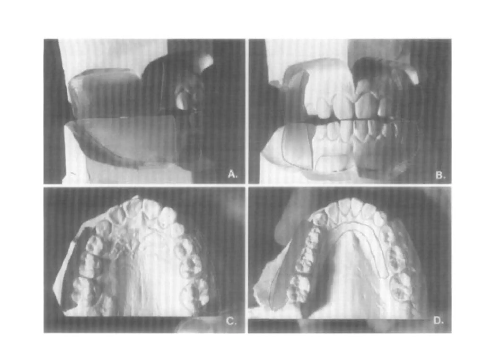
Frankel functional regulator
• Introduction.
• Epigenetic hypothesis
• Frankel philosophy.
• Visual treatment objective.
• Construction of function regulator.
• Mode of action.
• Modification of function regulator.
• Clinical management of the appliance.
• Review of literature.
• Conclusion.
C
O
N
T
E
N
T
S
Introduction
• We know that the cranio facial complex, forms itself as a bed for a
network of organs.
• During the same period of growth and development it presents itself
with a varied shadows to the clinician in the form of bizzare
craniofacial asymmetries and deformities that involve both cranial and
mandibular structures.
when?
problems
Where?
How
management
Prof Dr Rolf Frankel
• Outstanding contributor to functional appliance thought & the creator
of the Function regulator (Frankel) system of appliances.
Prof Dr Rolf Frankel
• Born -1908-march-29,leipzig.
• 1918-1927-completed secondary education.
• 1927-begin studying dentistry.
• Worked as assistant in oral surgery department in ESSEN
hospital.
EPIGENETIC HYPOTHESIS
• The theoretical concept of “Epigenetic control” was introduced by Van
Limborgh.
• The meaning of this term being the impact of “induction influences” on
embryonic development.
• These induction influences can be generally summed up as, all
biomechanical, bioelectrical, biophysical and biochemical parameters.
• The proponents of Epigenetic views the genome as providing
a set of formal, intrinsic and necessary causal factors that
when combined with the extrinsic and necessary epigenetic
causal factors, together are sufficient to account for the
regulation of development.
• Hence, this theoretical concept renews our understanding of
craniofacial morphogenesis and opens new perspectives.
• These experiments have
also revealed that certain
Osteogenic growth sites
are more sensitive to
biomechanical induction
than others and they
have different time
periods of sensitivity,
providing us a better
understanding of “the
Biologic targets in the
control process of facial
growth”.
Anatomy and histophysiology of the periosteum: quantification of the periosteal blood supply to the
adjacentbone with 85Sr and gamma spectrometry.
• The behavior of the entire bone remains closely influenced by periosteal activity.
• These principal functions are related to the cortical blood supply, osteogenesis,
and muscle and ligament attachments.
• Through its elastic and contractile nature, it participates in the maintenance
of bone shape, and plays an important role in metabolic ionic exchange and
physiologic distribution of electro-chemical potential differences across its
membranous structure
Buccinator mechanism
• Maintains integrity of dental arches and the relationship of teeth to each
other.
• Hence aberrations of muscle function can and do produce marked
malrelationships of the dental arches.
Guiding effect of Buccinator mechanism
Equilibrium theory
• From this perspective, we can say the dentition is in
equilibrium, when the muscle forces of the tongue and those
forces of the lips and cheeks are balanced except under certain
cases of muscular imbalances.
Frankels philosophy
THE FRANKEL PHILOSOPHY
• The concept of shielding the perioral musculature.
• The Function regulator of Frankel differs from other functional
appliances in the Sense:
The Frankel appliance is largely confined to the oral
vestibule.
Stresses the influence of the functional matrix, the
buccinator mechanism and the Orbicularis Oris complex.
The dynamic barriers to optimal growth of the cranio
facial complex in three dimensions of space.
The vestibular arena of operations
The classic concept of
“Pushing the dental arches
out from within”.
vestibular constructions act
as an artificial “Ought –to –
be” matrix.
Appliance as an exercise
device for oral gymnastics.
The primary role of the lip
and check musculature in
arch form development as
opposed to the tongue.
Sagittal correction
The major draw backs in the past.
The appliance is anchored to the maxillary dentition.
The role of acrylic pad.
Differential Eruption Guidance
• As the mandibular dentition is free .
• Rules out the demanding and precise grinding, so
often needed in other functional appliances.
Minimal Maxillary Basal effect
• According the Mc. Namara this effect is very little on
the maxilla.
Buccal shields & lip pads.
• Periosteal pull hypothesis.
• This working hypothesis, which was under research at the
American Dental Association Research institute by Graber et
al ( unpublished study, 1988), clearly shown that the buccal
shields stimulate bodily buccal movement of posterior teeth
and buccal plate.
Visual Treatment objective – As an Diagnostic
Test
• Gives a clue to the operator regarding the importance in
facial appearance and profile when subjected to any type
of functional appliance.
Frankel Treatment effects
• Template -against which the craniofacial muscles function.
• Influence on skeletal and dental development.
Types of functional regulators
• Classic classification.
• Current classification.
Treatment sequence
• An initial phase of fixed appliance therapy to idealize the
position of the incisors.
• An orthopedic phase in which the frankel appliance is used to
establish skeletal and muscular harmony.
• A final fixed appliance phase to position and detail the
dentition.
CONSTRUCTION OF FUNCTION REGULATOR
Parts of the appliance.
Separation of Teeth
• Recommendation for early separation of
teeth.
• Slicing of teeth.
IMPRESSION TAKING
IMPRESSION TRAYS
• Impression tray which does not cause overextension or lateral
distortion of the associated soft tissue should be used.
• Two types of trays used:
1. Thermal -sensitive arylic
2. custom trays.
Construction bite
• Procedures differ in both the degree of vertical opening and
amount of forward posturing of the mandible.
• One of the limitations of the Frankel appliance is the vertical
opening.
• In case of large overjet step by step advancement is
emphasized.
Construction bite
• According to Frankel:
-Mandible not more than 2.5-3.0mm.
• But according to mc namara et al upto 4-6mm Patient can
tolerate.
Preparation of working models
• Distance from the lateral extension of the base of the model to the
alveolar surface is at least 5 mm.
• One should not trim the posterolateral corners of the model bases too
severely.
• The first step in trimming the mandibular work model is to remove the
flash with either a laboratory knife or a rotary instrument until the
extensions of the vestibule have been reached
• The lower labial region should be carved with a pear-shaped
carbide bur and a laboratory knife.
• The extension of the lower labial relief is usually 12 mm. below
the lower border of the gingival margin, so that the lower
labial wire will lie approximately 7 mm. below the gingival
margin of the incisors.
• The lower labial extension in the anterior region of the model
shown is excellent and requires removal of only 2 to 3 mm of
plaster during the carving process.
Maxillary models
Wax relief
• 3.0 mm. in the maxillary vestibular area.
• 0.5 mm. in the mandibular vestibular area.
Wax relief for arch expansion
• Usually 3.0 mm of wax relief in the maxillary alveolar region
and 0.5 mm in the mandibular alveolar region is requested.
Fabrication of wire components
General rule
• All the wires should be bent with
smooth curve.
• The vestibular wires are placed 1.5
mm from the alveolar mucosa.
• Wires should follow the natural tissue
contours.
• The distance between the wires and
the wax padding should be
approximately 0.75mm.
Labial bow
Canine loop
Palatal bow
Cross over
wire
Wire components of FR 1
Lower lingual
wires
Cross over
wires
Labial bow
Palatal bow Canine loop
Buccal shields
• Extension
• Thickness 2.5mm
• Expansion of the
capsule
Lip pads
• extension and
• Tear drop shape
• Smoothen sulks
• Lip posture and seal .
5 mm
Lingual shields
• extension
• Over comes the poor
posture of mandibular
muscles
• Action -advancement
Labial bow
• Position and
extension
• Stabilizing
• Connecting
• ‘Function
activated’
Lower labial wires
• The 0.036 inch wire should pass from the buccal shield in a
slight antero inferior direction.
• The ends of the wires which will be embedded in the future
buccal shield are straight and positioned parallel to each other
so that the lip pads can be advanced or retracted as necessary.
Lower labial wires
Upper labial wire
• Incisors –midpoints.
• The loop portion must lie approximately 2 mm away from the
tissue above the canines.
• The arch of the labial bow should be bent in an ideal contour,
not to the contour of malpositioned teeth
Palatal bow
• Extension
• Occlusal rest on
maxillary molar
• Stabilizing action
• Intermaxillary
anchorage
Maxillary wires
Canine loop
• Extension
• Guide eruption of
canine
• Intermaxillary
anchorage
Canine extensions
• The canine extensions, made of 0.032 inch wire, act as an
extension of the vestibular shields in the canine region
• The canine extensions are placed 2 to 3 mm. away from the
deciduous canines.
• If permanent canines are present, the canine arms can be
located in a slightly closer position.
Lower lingual wires
• Extension
• Prevent lingual
movement of incisors
• Function activated
element in deep bite
and retruded anteriors
Cross over wires
• Run b/w 1st
and 2nd
premolars
• Not to be lodged
interdentally
• Cause movement
of buccal segments
FR1a and FR1 b
Lower lingual loops
Overjet 5mm
Lower lingual shield
Overjet 7mm
FR 1C
Step by step opening
in the anterior and
vertical direction
Overjet > 7mm
Lower lingual spring
Upper lingual wire
• The upper lingual wire can be used to stabilize the incisors
during treatment.
• The presence of the upper lingual wire, because it lies at the
level of the cingulum, was designed to prevent lingual tipping
of the incisors during treatment
Fabrication of vestibular segments
• The upper and lower models are locked
together with a fixator.
The FR – 2
• 80-90% use.
• Canine loop.(0.8mm)
• Improve mandible sagittal relationship.
• Selective eruption.
FR 2
Canine loop
and labial
bow
Upper lingual
wire
Lower lingual component
Upper lingual wire
• Runs b/w canine and lateral
• Stabilizing effect
• Prevents lingual tipping of
anteriors in div 2 cases
corrected in pre fr phase
The FR- 3
FR 3
Lower labial
wire(0.09-
mm)-18g
Upper
lingual
wire .06-.
07
Upper lip pads -3 fold
action.
Occlusal rests
Lowest possible
Level-prevent lingual tipping
18 g-Palatal bow-anterior stimulus,stabilizing component
More tear drop shape
Palatal bow and occlusal rests
• Palatal bow not
lodged
interdentally
• Additional occlusal
rest on lower
molar in deep bite
Construction bite
• Vertical limit-only allow maxillary incisors to
move past the mandibular incisors for
crossbite correction.
• Condyle –most superior position.
FR –IV
Stability of appliance&
prevention of eruption
FR 4
4 occlusal rests
Palatal bow( resembles FR 3)Last
molar.
Lower labial pads and buccal
shields
upper labial bow
Features
• No canine loops.
• No protrusion bows.
• Often used along with vertical extraoral chin
cup therapy.
MODE OF ACTION OF THE FUNCTION
REGULATOR
• Interception of aberrations of muscle function.
• Frankel appliance with holds muscle pressure
acting on the developing jaws and the dento
alveolar area.
• Passive expansion achieved through relief of
force from the neuromuscular capsule (the
buccinator mechanism).
FR in treatment of class II
• Mandible displaced anteriorly- retractor
muscle force
• Major dental changes – Proclination of
lower incisors
FR in the treatment of class 2
• Change in position brought by lingual shields
• Initial bite 2-3 mm
• Advancement in small steps for biologic
reasons.
FR in the treatment of class 2
• Step by step advancement by splitting the buccal
shields
• Suspending muscles are not overstrained
• FR advancement in steps stability in post retention
periods
FR in the treatment of class 3
Expansion of
upper oral
space
Tongue
space not
diminished
FR in the treatment of class
3
• Septo premaxillary ligament pull translates
upper incisors bodily
• FR3 promotes max basal bone
development and translates maxilla
forward
• Appliance should not be locked in the
maxilla by wires
FR in the treatment of skeletal open
bites
• Aimed at correcting the
poor lip valve
mechanism.
• Marked activity of
temporalis and masseter
when lips are closed
• Acc to Frankel tongue
thrust is compensatory
CHANGES BROUGHT BY FRANKEL
APPLIANCE IN THE OROFACIAL
COMPLEX.
Increase in Intra oral space (Sagittal and
Transverse)
• Achieved mainly through buccal
shields and lip pads.
• Favouring the forces acting from
within the oral cavity (the
tongue).
• Movement of the teeth in the
direction of least resistance.
• Effect of stretch on the
contiguous soft tissue.
• Supporting effect on the
lower lip.
• Establishment of a
competent lip seal.
• The physiological
relevance of hermetic
closure of the oral
functioning space.
Enhancing basal bone
development.
Vertical space increase
• According to Frankel, the
disturbance in vertical
development is more
often caused by the
cheeks, than the effect of
the tongue.
• screening effect of buccal
shields
Mandibular protraction
• “Sensory feed back mechanism” through
lingual acrylic shield.
Step wise advancement
Muscle function adaptation
• Development of new pattern of motor
functions.
• Creation of a new shell by the appliance.
• Massaging effect of pads and shields.
• The appliance loosens up the tight muscles
and improves tonicity.
MODIFICATIONS OF FRANKEL FUNCTIONAL
REGULATOR
FR with Buccal Tubes
• Incorporation of extra oral force at night.
• This was based on studies done by Mc
Namara.
FR with lingual crib spurs
• The presenting malocclusion.
• Eliminate all potent local factors.
• Treatment protocol.
• The activator head gear combination.
CLINICAL MANAGEMENT
• Relevance of strict clinical discipline.
• The appliance is checked for smoothness and
stability.
• Instruction to the patient/parent regarding the
appliance wear.
• Intra- oral check up.
• The patient / parent
should be shown the
dramatic profile change.
• In the treatment of class
III malocclusions.
Advocation of lip-seal exercises
• Establishment of a competent oral seal.
• Exercises can be initiated as early as the
schedule.
The schedule of appliance wear
• The first couple of weeks 2-4 hrs.
• The next period of 3 weeks 4-6 hrs.
• Speech improvement.
• The third appointment.-lip exercises
• Treatment progress checked regularly at 4
Week intervals.
• As treatment progresses there should be a
gradual improvement in the facial appearance.
Effect of FR on dento facial structures
Effect on arch width
• Greatest amount of expansion occurred in the premolar and
molar region. The average amount of posterior expansion was
4-5mm and 3-4mm in the maxillary and mandibular arches,
respectively.
Frankel and Frankel (1989),
• Report on a sample of 80 patients with severe tooth size /
arch size discrepancies and narrow arches all patients were
treated with either the FR-1 or R-2.
• Treatment began at an average of 8 years of age, and the
appliance was worn approximately 2 ½ years for at least 18hrs
per day.
Effects on the mandible
• Frankel and Reiss (1983), reported a forward displacement of
pogonion and point B at a rate of 4mm per year in 6-9 year
old children and 6mm per year in children 9 to 13 years of
age.
• Mc Namara (1982), found that the FR resulted in an average
increase of 1.2mm per year in mandibular growth.
• Reghellis (1983), also found that the increased mandibular
length by 1.8mm per year.
Mc Namara and co workers (1985)
• studied 100 patients treated with the Frankel appliance.
• Greater increase in mandibular growth increments were
noted in the older treated group than in the younger treated
group, and both treatment groups had larger mandibular
growth increment than their respective matched control
groups.
Effects on the maxilla
• Righellis (1983) found no significant horizontal
effect on the maxilla in patients treated with
the FR as compared with untreated class II
subjects.
Effects on the dentition
• Frankel 1984), indicates that the FR could not
cause flaring of the mandibular incisors since
the appliance does not contact them.
• Nielsen (1984),supported this assertation
who found that the FR did not induce
proclination of the mandibular incisors to the
cranial base.
Effect on the soft tissue profile
• Nielsen (1984) examined 10 class II div I
patients with the FR all patients showed an
improvement in their soft-tissue profile
because of improved lip position.
Comparison of FR with other
functional appliances
FR Vs twin blockMc namara AJO 1999
• 4O PTS WITH TWIN BLOCK AND FR COMPARED TO
CONTROLS
• Results
• Increase in mandibular length
Twin block – 3mm > controls
FR – 1.9MM
• Vertical dimension & dentoalveolar changes TB > FR
• TB -mandibular skeletal & dentoalveolar changes
• FR – more skeletal and less dentoalveolar
FR Vs herbst appliance
mc namara , ajo 1990
• 45 herbst and 41 FR pts compared with controls
• Results
• Both appliance – no effect on maxilla
• herbst – prevented vertical eruption
• Lower proclination L1 – herbst > FR
• mandibular length
Control - 2.1mm/yr
Herbst - 4.8mm
FR – 4.3mm
FR Vs fixed mechanotherapy
CREEKMORE,RADNEY AJO 1983
• FR compared to edgewise with headgear
• Edgewise had greater retractive force on maxilla
• Retraction of u1 > FR
• Pog forward 1mm< FR
Fr therapy in cleft palate patients
AJODO 1981
• 9 pts treated with Fr for 6-18 months
• To treat collapsed maxilla and cross bite
Results
• Not clinically useful in cleft patients
The Effect of Frankel II and Modified Twin Block Appliances
on the ‘C’-axis: The Growth Vector of the
Dentomaxillary Complex
• The growth axis (‘C’-axis), which describes the growth vector
of the dentomaxillary complex is not altered in any clinically
meaningful manner through the use of either the Frankel II or
the modified Twin Block appliances.
Stability evaluation of occlusal changes obtained with
Fränkel’s Function Regulator-2
• This study showed that the FR-2 appliance proved to
be effective for the occlusal correction of Class II
malocclusion, acting mainly on the improvement of
the overjet, overbite, and molar relationship.
• These occlusal effects were stable in the evaluated
period of 7.16 years after orthopedic treatment
Skeletal and Dental Changes Following the
Use of the Frankel Functional Regulator
• According to the results of this study, the major skeletal effect of the FR
was the apparent restraint of the normal forward growth of the maxilla.
• The major effect of the FR on the dentition was the retroclination of the
upper incisors and the proclination of the lower incisors which together
accounted for 54 percent of the total correction seen.
Skeletal and dental effects produced by
functional regulator-2 in pre-pubertal class II
patients: a controlled study
• The FR-2 treatment does not have relevant skeletal effects on
class II patients treated during the pre-pubertal growth phase,
while the overjet reduction is mainly due to dental effects.
Class II treatment effects of the Fränkel
appliance
• An increase in mandibular body length.
• A proportionally greater increase in mandibular growth as compared with maxillary
growth.
• An increase in LAFH without altering the facial growth pattern.
• Pronounced vertical development of the mandibular molars.
• A reduction in the overbite and overjet, and improvement in the molar relationship.
• Retrusion and palatal tipping of the maxillary incisors.
Meta-analysis of skeletal mandibular changes during Fränkel
appliance treatment
• Specifically, it appeared to have an effect on total mandibular
length with a low-to-moderate clinical impact.
CONCLUSION
• No universal or cook-book formula is available
within the orthodontic literature.
• A no of tools are available to the clinician to
attempt correction of maxillo-mandibular
malrelationships.
• Regardless of the appliance used there is a
large and similar amount of variation in
individual patient response to treatment.
THANK YOU

frankel functional regulator orthodontics.pptx

  • 1.
  • 2.
    • Introduction. • Epigenetichypothesis • Frankel philosophy. • Visual treatment objective. • Construction of function regulator. • Mode of action. • Modification of function regulator. • Clinical management of the appliance. • Review of literature. • Conclusion. C O N T E N T S
  • 3.
    Introduction • We knowthat the cranio facial complex, forms itself as a bed for a network of organs. • During the same period of growth and development it presents itself with a varied shadows to the clinician in the form of bizzare craniofacial asymmetries and deformities that involve both cranial and mandibular structures.
  • 4.
  • 5.
    Prof Dr RolfFrankel • Outstanding contributor to functional appliance thought & the creator of the Function regulator (Frankel) system of appliances.
  • 6.
    Prof Dr RolfFrankel • Born -1908-march-29,leipzig. • 1918-1927-completed secondary education. • 1927-begin studying dentistry. • Worked as assistant in oral surgery department in ESSEN hospital.
  • 7.
    EPIGENETIC HYPOTHESIS • Thetheoretical concept of “Epigenetic control” was introduced by Van Limborgh. • The meaning of this term being the impact of “induction influences” on embryonic development. • These induction influences can be generally summed up as, all biomechanical, bioelectrical, biophysical and biochemical parameters.
  • 8.
    • The proponentsof Epigenetic views the genome as providing a set of formal, intrinsic and necessary causal factors that when combined with the extrinsic and necessary epigenetic causal factors, together are sufficient to account for the regulation of development. • Hence, this theoretical concept renews our understanding of craniofacial morphogenesis and opens new perspectives.
  • 9.
    • These experimentshave also revealed that certain Osteogenic growth sites are more sensitive to biomechanical induction than others and they have different time periods of sensitivity, providing us a better understanding of “the Biologic targets in the control process of facial growth”.
  • 10.
    Anatomy and histophysiologyof the periosteum: quantification of the periosteal blood supply to the adjacentbone with 85Sr and gamma spectrometry. • The behavior of the entire bone remains closely influenced by periosteal activity. • These principal functions are related to the cortical blood supply, osteogenesis, and muscle and ligament attachments. • Through its elastic and contractile nature, it participates in the maintenance of bone shape, and plays an important role in metabolic ionic exchange and physiologic distribution of electro-chemical potential differences across its membranous structure
  • 11.
    Buccinator mechanism • Maintainsintegrity of dental arches and the relationship of teeth to each other. • Hence aberrations of muscle function can and do produce marked malrelationships of the dental arches.
  • 12.
    Guiding effect ofBuccinator mechanism
  • 13.
    Equilibrium theory • Fromthis perspective, we can say the dentition is in equilibrium, when the muscle forces of the tongue and those forces of the lips and cheeks are balanced except under certain cases of muscular imbalances.
  • 14.
  • 15.
    THE FRANKEL PHILOSOPHY •The concept of shielding the perioral musculature. • The Function regulator of Frankel differs from other functional appliances in the Sense: The Frankel appliance is largely confined to the oral vestibule. Stresses the influence of the functional matrix, the buccinator mechanism and the Orbicularis Oris complex. The dynamic barriers to optimal growth of the cranio facial complex in three dimensions of space.
  • 16.
    The vestibular arenaof operations The classic concept of “Pushing the dental arches out from within”. vestibular constructions act as an artificial “Ought –to – be” matrix. Appliance as an exercise device for oral gymnastics. The primary role of the lip and check musculature in arch form development as opposed to the tongue.
  • 17.
    Sagittal correction The majordraw backs in the past. The appliance is anchored to the maxillary dentition. The role of acrylic pad.
  • 18.
    Differential Eruption Guidance •As the mandibular dentition is free . • Rules out the demanding and precise grinding, so often needed in other functional appliances.
  • 19.
    Minimal Maxillary Basaleffect • According the Mc. Namara this effect is very little on the maxilla.
  • 20.
    Buccal shields &lip pads. • Periosteal pull hypothesis. • This working hypothesis, which was under research at the American Dental Association Research institute by Graber et al ( unpublished study, 1988), clearly shown that the buccal shields stimulate bodily buccal movement of posterior teeth and buccal plate.
  • 21.
    Visual Treatment objective– As an Diagnostic Test • Gives a clue to the operator regarding the importance in facial appearance and profile when subjected to any type of functional appliance.
  • 22.
    Frankel Treatment effects •Template -against which the craniofacial muscles function. • Influence on skeletal and dental development.
  • 23.
    Types of functionalregulators • Classic classification. • Current classification.
  • 24.
    Treatment sequence • Aninitial phase of fixed appliance therapy to idealize the position of the incisors. • An orthopedic phase in which the frankel appliance is used to establish skeletal and muscular harmony. • A final fixed appliance phase to position and detail the dentition.
  • 25.
  • 26.
    Parts of theappliance.
  • 28.
    Separation of Teeth •Recommendation for early separation of teeth. • Slicing of teeth.
  • 29.
  • 30.
    IMPRESSION TRAYS • Impressiontray which does not cause overextension or lateral distortion of the associated soft tissue should be used. • Two types of trays used: 1. Thermal -sensitive arylic 2. custom trays.
  • 31.
    Construction bite • Proceduresdiffer in both the degree of vertical opening and amount of forward posturing of the mandible. • One of the limitations of the Frankel appliance is the vertical opening. • In case of large overjet step by step advancement is emphasized.
  • 32.
    Construction bite • Accordingto Frankel: -Mandible not more than 2.5-3.0mm. • But according to mc namara et al upto 4-6mm Patient can tolerate.
  • 33.
    Preparation of workingmodels • Distance from the lateral extension of the base of the model to the alveolar surface is at least 5 mm. • One should not trim the posterolateral corners of the model bases too severely. • The first step in trimming the mandibular work model is to remove the flash with either a laboratory knife or a rotary instrument until the extensions of the vestibule have been reached
  • 35.
    • The lowerlabial region should be carved with a pear-shaped carbide bur and a laboratory knife. • The extension of the lower labial relief is usually 12 mm. below the lower border of the gingival margin, so that the lower labial wire will lie approximately 7 mm. below the gingival margin of the incisors.
  • 36.
    • The lowerlabial extension in the anterior region of the model shown is excellent and requires removal of only 2 to 3 mm of plaster during the carving process.
  • 37.
  • 38.
    Wax relief • 3.0mm. in the maxillary vestibular area. • 0.5 mm. in the mandibular vestibular area.
  • 40.
    Wax relief forarch expansion • Usually 3.0 mm of wax relief in the maxillary alveolar region and 0.5 mm in the mandibular alveolar region is requested.
  • 42.
  • 43.
    General rule • Allthe wires should be bent with smooth curve. • The vestibular wires are placed 1.5 mm from the alveolar mucosa. • Wires should follow the natural tissue contours. • The distance between the wires and the wax padding should be approximately 0.75mm.
  • 44.
    Labial bow Canine loop Palatalbow Cross over wire Wire components of FR 1
  • 45.
    Wire components ofFR 1 Lower lingual wires Cross over wires Labial bow Palatal bow Canine loop
  • 46.
    Buccal shields • Extension •Thickness 2.5mm • Expansion of the capsule
  • 47.
    Lip pads • extensionand • Tear drop shape • Smoothen sulks • Lip posture and seal . 5 mm
  • 48.
    Lingual shields • extension •Over comes the poor posture of mandibular muscles • Action -advancement
  • 49.
    Labial bow • Positionand extension • Stabilizing • Connecting • ‘Function activated’
  • 50.
    Lower labial wires •The 0.036 inch wire should pass from the buccal shield in a slight antero inferior direction. • The ends of the wires which will be embedded in the future buccal shield are straight and positioned parallel to each other so that the lip pads can be advanced or retracted as necessary.
  • 51.
  • 52.
    Upper labial wire •Incisors –midpoints. • The loop portion must lie approximately 2 mm away from the tissue above the canines. • The arch of the labial bow should be bent in an ideal contour, not to the contour of malpositioned teeth
  • 53.
    Palatal bow • Extension •Occlusal rest on maxillary molar • Stabilizing action • Intermaxillary anchorage
  • 54.
  • 55.
    Canine loop • Extension •Guide eruption of canine • Intermaxillary anchorage
  • 56.
    Canine extensions • Thecanine extensions, made of 0.032 inch wire, act as an extension of the vestibular shields in the canine region • The canine extensions are placed 2 to 3 mm. away from the deciduous canines. • If permanent canines are present, the canine arms can be located in a slightly closer position.
  • 57.
    Lower lingual wires •Extension • Prevent lingual movement of incisors • Function activated element in deep bite and retruded anteriors
  • 58.
    Cross over wires •Run b/w 1st and 2nd premolars • Not to be lodged interdentally • Cause movement of buccal segments
  • 59.
    FR1a and FR1b Lower lingual loops Overjet 5mm Lower lingual shield Overjet 7mm
  • 60.
    FR 1C Step bystep opening in the anterior and vertical direction Overjet > 7mm
  • 61.
  • 62.
    Upper lingual wire •The upper lingual wire can be used to stabilize the incisors during treatment. • The presence of the upper lingual wire, because it lies at the level of the cingulum, was designed to prevent lingual tipping of the incisors during treatment
  • 63.
    Fabrication of vestibularsegments • The upper and lower models are locked together with a fixator.
  • 64.
    The FR –2 • 80-90% use. • Canine loop.(0.8mm) • Improve mandible sagittal relationship. • Selective eruption.
  • 65.
    FR 2 Canine loop andlabial bow Upper lingual wire Lower lingual component
  • 66.
    Upper lingual wire •Runs b/w canine and lateral • Stabilizing effect • Prevents lingual tipping of anteriors in div 2 cases corrected in pre fr phase
  • 67.
  • 68.
    FR 3 Lower labial wire(0.09- mm)-18g Upper lingual wire.06-. 07 Upper lip pads -3 fold action. Occlusal rests Lowest possible Level-prevent lingual tipping 18 g-Palatal bow-anterior stimulus,stabilizing component More tear drop shape
  • 69.
    Palatal bow andocclusal rests • Palatal bow not lodged interdentally • Additional occlusal rest on lower molar in deep bite
  • 70.
    Construction bite • Verticallimit-only allow maxillary incisors to move past the mandibular incisors for crossbite correction. • Condyle –most superior position.
  • 71.
    FR –IV Stability ofappliance& prevention of eruption
  • 72.
    FR 4 4 occlusalrests Palatal bow( resembles FR 3)Last molar. Lower labial pads and buccal shields upper labial bow
  • 73.
    Features • No canineloops. • No protrusion bows. • Often used along with vertical extraoral chin cup therapy.
  • 74.
    MODE OF ACTIONOF THE FUNCTION REGULATOR • Interception of aberrations of muscle function. • Frankel appliance with holds muscle pressure acting on the developing jaws and the dento alveolar area. • Passive expansion achieved through relief of force from the neuromuscular capsule (the buccinator mechanism).
  • 75.
    FR in treatmentof class II • Mandible displaced anteriorly- retractor muscle force • Major dental changes – Proclination of lower incisors
  • 76.
    FR in thetreatment of class 2 • Change in position brought by lingual shields • Initial bite 2-3 mm • Advancement in small steps for biologic reasons.
  • 77.
    FR in thetreatment of class 2 • Step by step advancement by splitting the buccal shields • Suspending muscles are not overstrained • FR advancement in steps stability in post retention periods
  • 78.
    FR in thetreatment of class 3 Expansion of upper oral space Tongue space not diminished
  • 79.
    FR in thetreatment of class 3 • Septo premaxillary ligament pull translates upper incisors bodily • FR3 promotes max basal bone development and translates maxilla forward • Appliance should not be locked in the maxilla by wires
  • 80.
    FR in thetreatment of skeletal open bites • Aimed at correcting the poor lip valve mechanism. • Marked activity of temporalis and masseter when lips are closed • Acc to Frankel tongue thrust is compensatory
  • 81.
    CHANGES BROUGHT BYFRANKEL APPLIANCE IN THE OROFACIAL COMPLEX.
  • 82.
    Increase in Intraoral space (Sagittal and Transverse) • Achieved mainly through buccal shields and lip pads. • Favouring the forces acting from within the oral cavity (the tongue). • Movement of the teeth in the direction of least resistance. • Effect of stretch on the contiguous soft tissue.
  • 83.
    • Supporting effecton the lower lip. • Establishment of a competent lip seal. • The physiological relevance of hermetic closure of the oral functioning space. Enhancing basal bone development.
  • 84.
    Vertical space increase •According to Frankel, the disturbance in vertical development is more often caused by the cheeks, than the effect of the tongue. • screening effect of buccal shields
  • 85.
    Mandibular protraction • “Sensoryfeed back mechanism” through lingual acrylic shield.
  • 86.
  • 87.
    Muscle function adaptation •Development of new pattern of motor functions. • Creation of a new shell by the appliance. • Massaging effect of pads and shields. • The appliance loosens up the tight muscles and improves tonicity.
  • 88.
    MODIFICATIONS OF FRANKELFUNCTIONAL REGULATOR
  • 89.
    FR with BuccalTubes • Incorporation of extra oral force at night. • This was based on studies done by Mc Namara.
  • 90.
    FR with lingualcrib spurs • The presenting malocclusion. • Eliminate all potent local factors. • Treatment protocol.
  • 91.
    • The activatorhead gear combination.
  • 92.
    CLINICAL MANAGEMENT • Relevanceof strict clinical discipline. • The appliance is checked for smoothness and stability. • Instruction to the patient/parent regarding the appliance wear. • Intra- oral check up.
  • 93.
    • The patient/ parent should be shown the dramatic profile change. • In the treatment of class III malocclusions.
  • 94.
    Advocation of lip-sealexercises • Establishment of a competent oral seal. • Exercises can be initiated as early as the schedule.
  • 95.
    The schedule ofappliance wear • The first couple of weeks 2-4 hrs. • The next period of 3 weeks 4-6 hrs. • Speech improvement. • The third appointment.-lip exercises • Treatment progress checked regularly at 4 Week intervals. • As treatment progresses there should be a gradual improvement in the facial appearance.
  • 96.
    Effect of FRon dento facial structures
  • 97.
    Effect on archwidth • Greatest amount of expansion occurred in the premolar and molar region. The average amount of posterior expansion was 4-5mm and 3-4mm in the maxillary and mandibular arches, respectively.
  • 98.
    Frankel and Frankel(1989), • Report on a sample of 80 patients with severe tooth size / arch size discrepancies and narrow arches all patients were treated with either the FR-1 or R-2. • Treatment began at an average of 8 years of age, and the appliance was worn approximately 2 ½ years for at least 18hrs per day.
  • 99.
    Effects on themandible • Frankel and Reiss (1983), reported a forward displacement of pogonion and point B at a rate of 4mm per year in 6-9 year old children and 6mm per year in children 9 to 13 years of age. • Mc Namara (1982), found that the FR resulted in an average increase of 1.2mm per year in mandibular growth. • Reghellis (1983), also found that the increased mandibular length by 1.8mm per year.
  • 100.
    Mc Namara andco workers (1985) • studied 100 patients treated with the Frankel appliance. • Greater increase in mandibular growth increments were noted in the older treated group than in the younger treated group, and both treatment groups had larger mandibular growth increment than their respective matched control groups.
  • 101.
    Effects on themaxilla • Righellis (1983) found no significant horizontal effect on the maxilla in patients treated with the FR as compared with untreated class II subjects.
  • 102.
    Effects on thedentition • Frankel 1984), indicates that the FR could not cause flaring of the mandibular incisors since the appliance does not contact them. • Nielsen (1984),supported this assertation who found that the FR did not induce proclination of the mandibular incisors to the cranial base.
  • 103.
    Effect on thesoft tissue profile • Nielsen (1984) examined 10 class II div I patients with the FR all patients showed an improvement in their soft-tissue profile because of improved lip position.
  • 104.
    Comparison of FRwith other functional appliances
  • 105.
    FR Vs twinblockMc namara AJO 1999 • 4O PTS WITH TWIN BLOCK AND FR COMPARED TO CONTROLS • Results • Increase in mandibular length Twin block – 3mm > controls FR – 1.9MM • Vertical dimension & dentoalveolar changes TB > FR • TB -mandibular skeletal & dentoalveolar changes • FR – more skeletal and less dentoalveolar
  • 106.
    FR Vs herbstappliance mc namara , ajo 1990 • 45 herbst and 41 FR pts compared with controls • Results • Both appliance – no effect on maxilla • herbst – prevented vertical eruption • Lower proclination L1 – herbst > FR • mandibular length Control - 2.1mm/yr Herbst - 4.8mm FR – 4.3mm
  • 107.
    FR Vs fixedmechanotherapy CREEKMORE,RADNEY AJO 1983 • FR compared to edgewise with headgear • Edgewise had greater retractive force on maxilla • Retraction of u1 > FR • Pog forward 1mm< FR
  • 108.
    Fr therapy incleft palate patients AJODO 1981 • 9 pts treated with Fr for 6-18 months • To treat collapsed maxilla and cross bite Results • Not clinically useful in cleft patients
  • 109.
    The Effect ofFrankel II and Modified Twin Block Appliances on the ‘C’-axis: The Growth Vector of the Dentomaxillary Complex • The growth axis (‘C’-axis), which describes the growth vector of the dentomaxillary complex is not altered in any clinically meaningful manner through the use of either the Frankel II or the modified Twin Block appliances.
  • 110.
    Stability evaluation ofocclusal changes obtained with Fränkel’s Function Regulator-2 • This study showed that the FR-2 appliance proved to be effective for the occlusal correction of Class II malocclusion, acting mainly on the improvement of the overjet, overbite, and molar relationship. • These occlusal effects were stable in the evaluated period of 7.16 years after orthopedic treatment
  • 111.
    Skeletal and DentalChanges Following the Use of the Frankel Functional Regulator • According to the results of this study, the major skeletal effect of the FR was the apparent restraint of the normal forward growth of the maxilla. • The major effect of the FR on the dentition was the retroclination of the upper incisors and the proclination of the lower incisors which together accounted for 54 percent of the total correction seen.
  • 112.
    Skeletal and dentaleffects produced by functional regulator-2 in pre-pubertal class II patients: a controlled study • The FR-2 treatment does not have relevant skeletal effects on class II patients treated during the pre-pubertal growth phase, while the overjet reduction is mainly due to dental effects.
  • 113.
    Class II treatmenteffects of the Fränkel appliance • An increase in mandibular body length. • A proportionally greater increase in mandibular growth as compared with maxillary growth. • An increase in LAFH without altering the facial growth pattern. • Pronounced vertical development of the mandibular molars. • A reduction in the overbite and overjet, and improvement in the molar relationship. • Retrusion and palatal tipping of the maxillary incisors.
  • 114.
    Meta-analysis of skeletalmandibular changes during Fränkel appliance treatment • Specifically, it appeared to have an effect on total mandibular length with a low-to-moderate clinical impact.
  • 115.
    CONCLUSION • No universalor cook-book formula is available within the orthodontic literature. • A no of tools are available to the clinician to attempt correction of maxillo-mandibular malrelationships. • Regardless of the appliance used there is a large and similar amount of variation in individual patient response to treatment.
  • 116.

Editor's Notes

  • #10 The periosteum or periosteal membrane is a continuous composite fibroelastic covering membrane of the bone to which it is intimately linked Although the bone cortex is the main beneficiary of the principal anatomical and physiological functions of the periosteal membrane,
  • #23 FR- 1 Class I and Class II, Division 1 FR-2 Class II, Division 2 IS-3 Class III FR-4 Open-bite Table II. Types of functional regulators (current) FR-2 FR-3 IS-4 FR- 1 Class II, Division 1 and Class II, Division 2; some open-bites Class III Some open-bites Some open-bites; Class I
  • #26 The vestibular (buccal) shields and the lower labial pads act in this area to restrain the musculature and to remove restricting muscular forces from the dentition.
  • #27 ment of the altered mandibular postural position used in the correction of Class II malocclusion. Since these pads are tissue-borne rather than tooth-borne, they act as tactile reminders to the patient to position his or her jaw anteriorly. I
  • #29 A properly fabricated appliance will have superior and inferior borders on the buccal shields that will slightly encroach on the patient’s resting vestibular sulcus, vertically and horizontally.
  • #30 Both of these tray types minimize the distortion of the underlying soft tissue. Because of the close fit of the tray, much less impression material is needed when thermal-sensitive or custom trays are used than when conventional orth- odontic trays are used.
  • #33 since doing so will make the placement of the wax relief in the region of the last molar difficult.
  • #37 Similar carving of the flash is necessary on the maxillary model (Fig. 6). Some of the anatomic areas must be defined, particularly the area of the tuberosity, which lies posterior to the muscle attachment over the first premolar or first deciduous molar (Fig. 6). In addition, the area superior to the canine and anterior to the muscle attachment should be defined.
  • #39 Fig. 7. A, Mounting the appliance in the fixator with a wax bite in place. B, Lateral view of working models with wax bite in place. C, Removal of wax bite. Note that at least 2.5 mm. of interocclusal space is present to allow for the wires to cross without impinging on the teeth. D, Pencil outline of future vestibular shields and lower labial pads.