This document discusses how whole genome sequencing (WGS) of Mycobacterium tuberculosis has improved understanding of drug resistant tuberculosis (TB). WGS has identified specific mutations that cause resistance to classical and new antitubercular drugs. It has also revealed how resistance evolves within patients and spreads between patients. Clinical applications of WGS include rapid detection of drug resistance and tracking of TB outbreaks. However, challenges remain in applying WGS to improve TB control and diagnosis.
![REVIEW Open Access
Deciphering drug resistance in
Mycobacterium tuberculosis using whole-
genome sequencing: progress, promise,
and challenges
Keira A. Cohen1*
, Abigail L. Manson2
, Christopher A. Desjardins2
, Thomas Abeel2,3
and Ashlee M. Earl2*
Abstract
Tuberculosis (TB) is a global infectious threat that is intensified by an increasing incidence of highly drug-resistant
disease. Whole-genome sequencing (WGS) studies of Mycobacterium tuberculosis, the causative agent of TB, have
greatly increased our understanding of this pathogen. Since the first M. tuberculosis genome was published in 1998,
WGS has provided a more complete account of the genomic features that cause resistance in populations of M.
tuberculosis, has helped to fill gaps in our knowledge of how both classical and new antitubercular drugs work, and
has identified specific mutations that allow M. tuberculosis to escape the effects of these drugs. WGS studies have
also revealed how resistance evolves both within an individual patient and within patient populations, including
the important roles of de novo acquisition of resistance and clonal spread. These findings have informed decisions
about which drug-resistance mutations should be included on extended diagnostic panels. From its origins as a
basic science technique, WGS of M. tuberculosis is becoming part of the modern clinical microbiology laboratory,
promising rapid and improved detection of drug resistance, and detailed and real-time epidemiology of TB
outbreaks. We review the successes and highlight the challenges that remain in applying WGS to improve the
control of drug-resistant TB through monitoring its evolution and spread, and to inform more rapid and effective
diagnostic and therapeutic strategies.
Background
Mycobacterium tuberculosis is the causative agent of tu-
berculosis (TB), which is most often spread person-to-
person via cough aerosols. Although many individuals
who are exposed to M. tuberculosis never develop active
disease, the World Health Organization (WHO) esti-
mated 10 million new cases of active TB and 1.3 million
deaths in 2017 alone [1].
Since its initial documentation in the 1940s [2], drug-
resistant TB has threatened public health control efforts.
In 2016, there were an estimated 490,000 new cases of
multidrug-resistant (MDR) TB, which is defined by
phenotypic resistance to both isoniazid and rifampicin
[3]. Approximately 10% of MDR-TB cases globally can
be classified as extensively drug-resistant (XDR), indicat-
ing that there is concomitant resistance to quinolones
(such as the fluoroquinolones, levofloxacin, and moxi-
floxacin) and to a second-line injectable agent (amikacin,
kanamycin, or capreomycin) [3]. As expected, drug-
resistance patterns predict treatment outcome; in 2015,
TB treatment success overall was 83%, whereas the suc-
cess rate was 54% for MDR-TB or rifampicin-resistant-
TB (RR-TB) and only 30% for XDR-TB [4].
Culture-based techniques remain the current reference
standard for both diagnosis and drug-susceptibility test-
ing of TB, but these processes are time-consuming and
require specialized laboratory capacity. More recently,
the use of rapid molecular tests for the diagnosis of TB
has increased globally, particularly the use of Xpert
MTB/RIF (Cepheid, Sunnyvale, CA), a PCR-based assay
© The Author(s). 2019 Open Access This article is distributed under the terms of the Creative Commons Attribution 4.0
International License (http://creativecommons.org/licenses/by/4.0/), which permits unrestricted use, distribution, and
reproduction in any medium, provided you give appropriate credit to the original author(s) and the source, provide a link to
the Creative Commons license, and indicate if changes were made. The Creative Commons Public Domain Dedication waiver
(http://creativecommons.org/publicdomain/zero/1.0/) applies to the data made available in this article, unless otherwise stated.
* Correspondence: kcohen8@jhmi.edu; aearl@broadinstitute.org
1
Division of Pulmonary and Critical Care Medicine, Johns Hopkins University
School of Medicine, Baltimore, MA 21205, USA
2
Broad Institute of Harvard and Massachusetts Institute of Technology, 415
Main Street, Cambridge, MA 02142, USA
Full list of author information is available at the end of the article
Cohen et al. Genome Medicine (2019) 11:45
https://doi.org/10.1186/s13073-019-0660-8](https://image.slidesharecdn.com/decipheringdrugresistanceinmtbusingwgs-211208101108/85/Deciphering-drug-resistance-in-mtb-using-wgs-1-320.jpg)
![REVIEW Open Access
Deciphering drug resistance in
Mycobacterium tuberculosis using whole-
genome sequencing: progress, promise,
and challenges
Keira A. Cohen1*
, Abigail L. Manson2
, Christopher A. Desjardins2
, Thomas Abeel2,3
and Ashlee M. Earl2*
Abstract
Tuberculosis (TB) is a global infectious threat that is intensified by an increasing incidence of highly drug-resistant
disease. Whole-genome sequencing (WGS) studies of Mycobacterium tuberculosis, the causative agent of TB, have
greatly increased our understanding of this pathogen. Since the first M. tuberculosis genome was published in 1998,
WGS has provided a more complete account of the genomic features that cause resistance in populations of M.
tuberculosis, has helped to fill gaps in our knowledge of how both classical and new antitubercular drugs work, and
has identified specific mutations that allow M. tuberculosis to escape the effects of these drugs. WGS studies have
also revealed how resistance evolves both within an individual patient and within patient populations, including
the important roles of de novo acquisition of resistance and clonal spread. These findings have informed decisions
about which drug-resistance mutations should be included on extended diagnostic panels. From its origins as a
basic science technique, WGS of M. tuberculosis is becoming part of the modern clinical microbiology laboratory,
promising rapid and improved detection of drug resistance, and detailed and real-time epidemiology of TB
outbreaks. We review the successes and highlight the challenges that remain in applying WGS to improve the
control of drug-resistant TB through monitoring its evolution and spread, and to inform more rapid and effective
diagnostic and therapeutic strategies.
Background
Mycobacterium tuberculosis is the causative agent of tu-
berculosis (TB), which is most often spread person-to-
person via cough aerosols. Although many individuals
who are exposed to M. tuberculosis never develop active
disease, the World Health Organization (WHO) esti-
mated 10 million new cases of active TB and 1.3 million
deaths in 2017 alone [1].
Since its initial documentation in the 1940s [2], drug-
resistant TB has threatened public health control efforts.
In 2016, there were an estimated 490,000 new cases of
multidrug-resistant (MDR) TB, which is defined by
phenotypic resistance to both isoniazid and rifampicin
[3]. Approximately 10% of MDR-TB cases globally can
be classified as extensively drug-resistant (XDR), indicat-
ing that there is concomitant resistance to quinolones
(such as the fluoroquinolones, levofloxacin, and moxi-
floxacin) and to a second-line injectable agent (amikacin,
kanamycin, or capreomycin) [3]. As expected, drug-
resistance patterns predict treatment outcome; in 2015,
TB treatment success overall was 83%, whereas the suc-
cess rate was 54% for MDR-TB or rifampicin-resistant-
TB (RR-TB) and only 30% for XDR-TB [4].
Culture-based techniques remain the current reference
standard for both diagnosis and drug-susceptibility test-
ing of TB, but these processes are time-consuming and
require specialized laboratory capacity. More recently,
the use of rapid molecular tests for the diagnosis of TB
has increased globally, particularly the use of Xpert
MTB/RIF (Cepheid, Sunnyvale, CA), a PCR-based assay
© The Author(s). 2019 Open Access This article is distributed under the terms of the Creative Commons Attribution 4.0
International License (http://creativecommons.org/licenses/by/4.0/), which permits unrestricted use, distribution, and
reproduction in any medium, provided you give appropriate credit to the original author(s) and the source, provide a link to
the Creative Commons license, and indicate if changes were made. The Creative Commons Public Domain Dedication waiver
(http://creativecommons.org/publicdomain/zero/1.0/) applies to the data made available in this article, unless otherwise stated.
* Correspondence: kcohen8@jhmi.edu; aearl@broadinstitute.org
1
Division of Pulmonary and Critical Care Medicine, Johns Hopkins University
School of Medicine, Baltimore, MA 21205, USA
2
Broad Institute of Harvard and Massachusetts Institute of Technology, 415
Main Street, Cambridge, MA 02142, USA
Full list of author information is available at the end of the article
Cohen et al. Genome Medicine (2019) 11:45
https://doi.org/10.1186/s13073-019-0660-8](https://image.slidesharecdn.com/decipheringdrugresistanceinmtbusingwgs-211208101108/75/Deciphering-drug-resistance-in-mtb-using-wgs-1-2048.jpg)
![that simultaneously detects the presence of M. tubercu-
losis and resistance to rifampicin.
Current recommendations for the treatment of drug-
susceptible TB include a 6-month course of a multi-drug
regimen of rifampicin, isoniazid, pyrazinamide, and
ethambutol. Historically, treatment of MDR- or XDR-TB
involved the long-term use of second-line drugs, includ-
ing injectable agents [5]. More recently, the MDR-TB
treatment landscape has changed with the introduction
of multiple novel second-line drugs that can be adminis-
tered orally (Table 1). In 2012, bedaquiline, a diarylqui-
nolone, became the first TB drug from a novel drug
class to receive US Food and Drug Administration
(FDA) approval in over 40 years [48, 49] (Table 1).
Another oral agent, delamanid, a nitro-dihydro-
imidazooxazole derivative, has also shown promise for
TB treatment [50, 51].
In 2018, the WHO published updated treatment
guidelines for MDR/RR-TB [47], recommending fully
oral MDR regimens for many patient groups. Recom-
mended treatment strategies include both shorter, stan-
dardized MDR regimens (for 9 to 12 months) and
longer, individualized treatment regimens (for 18 to 20
months). The updated guidelines group antitubercular
drugs on the basis of how they should be combined to
create individualized, longer MDR-TB regimens [47]
(Table 1).
Despite advances in both diagnostics and therapeutics
for TB, challenges remain. Obstacles for rapid M. tuber-
culosis diagnosis include: (i) the imperfect sensitivity of
molecular tests for the detection of this pathogen, par-
ticularly in the case of paucibacillary TB (where there is
a lower bacterial burden); (ii) lack of comprehensive mo-
lecular assays due to incomplete knowledge of all resist-
ance mutations in TB; and (iii) technical limitations to
the numbers of mutations that can be included on diag-
nostic molecular platforms. Furthermore, the rollout of
rapid diagnostic platforms to low-resource settings has
been a challenge. Remaining treatment challenges in-
clude: prolonged treatment courses, leading to greater
drug exposure, toxicity, and non-compliance; unaccept-
able side-effect profiles; logistics of drug access; and re-
infection [52].
The dawning of the new age of genome sequencing
began to revolutionize our approach to human diseases,
including TB. In 1998, Cole et al. [53] reported the
complete genome sequence of the M. tuberculosis refer-
ence strain H37Rv, which was approximately 4.41 mil-
lion base pairs in length and encoded approximately
4000 genes. The first sequencing of a clinical reference
strain, CDC1551, quickly followed [54]. An accompany-
ing editorial optimistically stated: “After several decades
in the slow lane of classical microbiology, M. tubercu-
losis is once again at the cutting edge of science” [55].
However, even at the time of these breakthroughs, there
was recognition that translating these genomic data into
clinical benefit would prove challenging [55]. Despite
these challenges, it is clear, more than 20 years later, that
M. tuberculosis genomic data have been remarkably use-
ful in improving our understanding of how drug-
resistant TB evolves and spreads and in helping to in-
form diagnostics and therapies.
In this review, we discuss the molecular epidemiologic
and diagnostic advances made by sequencing M. tuber-
culosis, with a focus on drug-resistant TB. We do not re-
view the practice of whole-genome sequencing (WGS)
of M. tuberculosis as this has been reviewed recently
[56]. Key findings that are discussed include the use of
WGS to identify drug-resistance determinants in M. tu-
berculosis and to elucidate the evolution and spread of
drug-resistant organisms, and the clinical applications of
this technology (Table 2).
Identifying M. tuberculosis drug-resistance
determinants
Drug resistance in M. tuberculosis is the result of chromo-
somal mutations in existing genes that are passed along
through vertical descent, that is, passed from mother to
daughter cells. Unlike many other bacterial pathogens, M.
tuberculosis rarely recombines via lateral exchange of DNA
[83] and also lacks plasmids. Many of the resistance deter-
minants were discovered before the sequencing of the M.
tuberculosis genome was completed. By 1998, resistance
mechanisms had already been discovered for classical first-
and second-line TB drugs including isoniazid (alterations in
genes katG and inhA); rifampicin (in rpoB); streptomycin
(in rrs and rpsL); pyrazinamide (in pncA); ethambutol (in
embB); quinolones (in gyrA); and kanamycin (in rrs)
(reviewed in Ramaswamy and Musser [84]) (Table 1). How-
ever, the targeted amplification and sequencing of known
or suspected resistance genes revealed that these mecha-
nisms were insufficient to explain all phenotypic resistance
[85, 86], and resistance mechanisms for several newer
drugs—including pretomanid, bedaquiline, and delama-
nid—were discovered over the next eight years during a
period when WGS was becoming routine. Together, in the
past 20 years, WGS-based approaches, focused on both
laboratory-derived and naturally circulating populations of
drug-resistant M. tuberculosis, have provided a more
complete account of the genomic features that cause treat-
ment resistance, enabling the identification of novel resist-
ance mechanisms for existing drugs, and the determination
of the mechanisms of action of newly discovered drugs.
Identifying resistance determinants in laboratory-derived
mutants
Drug-resistant mutants can be derived in vitro by grow-
ing drug-susceptible M. tuberculosis strains in drug-
Cohen et al. Genome Medicine (2019) 11:45 Page 2 of 18](https://image.slidesharecdn.com/decipheringdrugresistanceinmtbusingwgs-211208101108/85/Deciphering-drug-resistance-in-mtb-using-wgs-2-320.jpg)
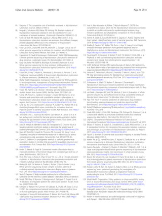
![Table 1 Antitubercular drug-resistance mechanismsa
WHO
category
Drug or drug class Resistance
genes
Rv number Gene function Mechanism of drug
resistance
Reference(s)
First-line
agents
Rifamycins (for
example, rifampicin)
rpoB Rv0667 RNA polymerase Target modification [6]
ponA1 Rv0050 Probable bifunctional penicillin-binding
protein
Unknown [7]
Isoniazid katG Rv1908c Catalase-peroxidase enzyme Decreased drug
activation
[8]
inhA Rv1484 NADH-dependent enoyl-acyl carrier protein Target amplification
or modification
[9, 10]
Pyrazinamideb
pncA Rv2043c Pyrazinamidase Decreased drug
activation
[11, 12]
panD Rv3601c Aspartate decarboxylase Unknown [13]
rpsA RRv1630 Ribosomal protein S1 Target modification [14]
Ethambutolb
embCAB
operon
Rv3793-5 Arabinosyltransferase Target modification [15, 16]
ubiA Rv3806c Arabinogalactan synthesis Gain-of-function [15]
Group A Levofloxacin
Moxifloxacin
gyrA Rv0006 DNA gyrase A Target modification [17, 18]
gyrB Rv0005 DNA gyrase B Target modification [18]
Bedaquiline atpE Rv1305 ATP synthase Target modification [19]
pepQ Rv2535c Putative Xaa-Pro aminopeptidase Unknown [20]
Rv0678 Rv0678 Transcriptional regulator of mmpL5 Drug efflux [21, 22]
Linezolid Rrl NA 23S rRNA Target modification [23]
rplC Rv0701 50S ribosomal protein L3 Target modification [24]
Group B Clofazimine pepQ Rv2535c Putative Xaa-Pro aminopeptidase Drug efflux [20]
Rv0678 Rv0678 Transcriptional regulator of mmpL5 Drug efflux [21]
Cycloserine
Terizidone
Ald Rv2780 L-alanine dehydrogenase Substrate shunting [25]
alr Rv3423c Alanine racemase Target modification [26, 27]
ddl Rv2981c D-alanine-D-alanine ligase Target modification [27]
cycA Rv1704c Bacterial D-serine/L-and D-alanine/glycine/D-
cycloserine proton symporter
Mechanism not
confirmed
[28]
Group C Delamanid
Pretomanid
ddn Rv3547 Oxidative stress Decreased drug
activation
[29]
fgd1 Rv0407 Glucose-6-phosphate oxidation Decreased drug
activation
[29]
Imipenem/cilastatin crfA Rv2421c-Rv2422
intergenic
Unknown Drug inactivation [30]
Amikacin,
Capreomycin,
Kanamycinc
Rrs NA 16S rRNA Target modification [31]
Streptomycin rpsL Rv0682 12S ribosomal protein Target modification [32–35]
rrs NA 16S rRNA Target modification [36]
gidB Rv3919c 7-Methylguanosine methyltransferase Target modification [37]
Ethionamide
Prothionamide
ethA Rv3854c Mono-oxygenase Decreased drug
activation
[38, 39]
ethR Rv3855 Transcriptional regulatory repressor protein
(TetR)
Decreased drug
activation
[39]
inhA Rv1484 NADH-dependent enoyl-acyl carrier protein Target amplification
or modification
[10]
Para-aminosalicylic
acid (PAS)
folC Rv2447c Folate pathway Decreased drug
activation
[40]
dfrA Rv2763c Dihydrofolate reductase Target amplification [40]
Cohen et al. Genome Medicine (2019) 11:45 Page 3 of 18](https://image.slidesharecdn.com/decipheringdrugresistanceinmtbusingwgs-211208101108/85/Deciphering-drug-resistance-in-mtb-using-wgs-3-320.jpg)
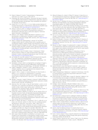
![containing media, and selecting for mutants that are able
to grow in the presence of the drug. Sequencing
laboratory-derived resistant mutants has played a critical
role in identifying both the mechanism of action of new
TB drug classes, including diarylquinolines (for example,
bedaquiline) [19] and nitroimidazopyrans (for example,
PA-824) [19, 29], and rare resistance mechanisms for
established antitubercular drugs, including ethambutol
[15], pyrazinamide [13], carbapenems [30], cycloserine
[87], clofazimine, and bedaquiline [20]. For example,
WGS of laboratory mutants identified drug efflux as a
mechanism of resistance to clofazimine and bedaquiline
[20–22], and this approach continues to be a mainstay
for identifying the mechanism of action of compounds
that are in development for TB [88].
Although laboratory-derived mutants are helpful in
elucidating novel resistance mechanisms, mutations that
have evolved in laboratory settings do not always match
those in clinical isolates of drug-resistant M. tuberculosis
[89, 90], for reasons that are largely unknown. Studies by
Ford et al. [91, 92] suggested that these mismatches
could not be explained by differences in the mutation
rate in these settings, because the in vitro mutation rate
of M. tuberculosis correlates well with the in vivo muta-
tion rate in both humans and in a macaque model. Dif-
ferences in the relative fitness of specific mutants grown
in in vitro compared to in vivo conditions are probably
responsible for these mismatches, but more work is
needed. Regardless of the reason, if the goal is to identify
a full complement of resistance mutations on which to
base molecular diagnostics, isolates from clinical collec-
tions must be studied as these bacteria have evolved
their resistance within the host.
Quantifying and identifying resistance determinants in
clinical strains
Among the larger studies exploring resistance in natural
populations, Walker et al. [58] analyzed the genomes of
3651 drug-resistant and -susceptible M. tuberculosis iso-
lates for associations between phenotypic resistance to
eight first- and second-line drugs, and then predicted
genotypic resistance on the basis of a compiled catalog
of 232 resistance mutations in 23 candidate resistance
genes. Resistance to most drugs could be predicted ac-
curately, with a mean sensitivity of 92% and specificity
of 98%, suggesting that the majority of resistance—par-
ticularly for first-line drugs—is explained by known
mechanisms and mutations (Table 1). Numerous other
studies have found similar results using smaller datasets
[7, 25, 57, 69, 93, 94]. This result was echoed in a more
recent study by the Comprehensive Resistance Predic-
tion for Tuberculosis (CRYPTIC) Consortium and the
100,000 Genomes Project that focused solely on first-
line drugs, which included analysis of 10,209 globally di-
verse M. tuberculosis isolate genomes against a database
of mutations identified in a literature search [60]. Not-
ably, predictions for mutations that are associated with
resistance to pyrazinamide were greatly improved over
earlier predictions; this study achieved 91.3% sensitivity
in predicting resistance to this drug compared to 57%
sensitivity in Walker et al. [58]. Although the news has
been encouraging with respect to completing the catalog
of mutations that cause resistance to first-line drugs, few
studies have attempted to predict resistance to second-
line drugs [95]. Some of these drugs, such as D-
cycloserine, pyrazinamide, and para-aminosalicylic acid
(PAS), are more difficult to assay because they have been
reported to have variable drug phenotypes in clinical
microbiology laboratories [96] (discussed later).
To fill gaps in the catalog of drug-resistance mecha-
nisms, genome-wide association study (GWAS) ap-
proaches, originally designed for use on human genomic
data, have been adapted for non-recombining microbes
such as M. tuberculosis and used to predict novel resist-
ance mechanisms [97, 98] (Table 3). The majority of
GWAS predictions remain experimentally unverified,
Table 1 Antitubercular drug-resistance mechanismsa
(Continued)
WHO
category
Drug or drug class Resistance
genes
Rv number Gene function Mechanism of drug
resistance
Reference(s)
thyA Rv2764c Thymidylate synthase Target modification [41, 42]
thyX Rv2754c Catalyzes dTMP and tetrahydrofolate Mitigating target
inhibition
[43]
ribD Rv2671 Enzyme in riboflavin biosynthesis Mitigating target
inhibition
[40, 44]
Other
medicinesc
Kanamycin Eis Rv2416c Aminoglycoside acetyltransferase Inactivating mutation [45]
Capreomycin tlyA Rv1694 rRNA methyltransferase Target modification [46]
Abbreviations: MDR-TB multidrug-resistant tuberculosis, NA not applicable, RR-TB rifampicin-resistant tuberculosis, WHO World Health Organization
a
Antitubercular drugs are listed by the 2018 WHO grouping of medicines recommended for use in longer, individualized MDR-TB regimens [47]. For each drug or
drug class, the specific genes in which drug-resistance mutations are commonly identified are listed with their gene name, gene number (Rv number), gene
function, and the confirmed or putative mechanisms of resistance. b
Pyrazinamide and ethambutol are first-line TB drugs that also are categorized as Group C
medicines for the treatment of longer MDR-TB regimens. c
Kanamycin and capreomycin are no longer recommended to be included in longer, individualized
MDR/RR-TB regimens
Cohen et al. Genome Medicine (2019) 11:45 Page 4 of 18](https://image.slidesharecdn.com/decipheringdrugresistanceinmtbusingwgs-211208101108/85/Deciphering-drug-resistance-in-mtb-using-wgs-4-320.jpg)
![Table 2 Spotlight on whole-genome sequencing studies of drug-resistant M. tuberculosis
Reference Description Advances
Identifying M. tuberculosis drug-resistance determinants
Farhat et al. 2013 [7] Large-scale WGS project: sequencing of 116 genomes from
around the globe
Developed a phylogenetic convergence test, PhyC, to
identify resistance associations; validated ponA1 mutations
that increase MIC for rifampicin
Zhang et al. 2013 [57] Large-scale WGS project: sequencing of 161 genomes from
China
Identified genes that are under positive selection and have
increased mutation frequencies in drug-resistant isolates
Walker et al. 2015 [58] Analysis of 23 candidate resistance genes from 3651 clinical
isolates
Demonstrated that drug-resistance in M. tuberculosis can be
predicted with high sensitivity and specificity
Desjardins et al. 2016
[25]
Use of a combination of the correlated evolution test and a
GWAS framework to identify drug-resistance-associated mu-
tations in 498 genomes from China and South Africa
Identified ald loss-of-function as a novel mechanism of D-
cycloserine resistance
Coll et al. 2018 [59] GWAS study of 6465 M. tuberculosis clinical isolates from
more than 30 countries
Identified new resistance-associated mutations in ethA and
the thyX promoter
The Cryptic Consortium
and the 100,000
Genomes Project [60]
Prediction of first-line-drug susceptibility in a dataset of 10,
209 clinical isolates from 16 countries
Predicted drug-susceptibility phenotypes with high sensitiv-
ity and specificity using WGS in a large global dataset
Within-patient evolution of resistance
Eldholm et al. 2014 [61] WGS of nine serial isolates cultured from a single patient
over a 42-month period
First documented case of the evolution of susceptible TB
into XDR-TB in a single patient in response to selective drug
pressure
Trauner et al. 2017 [62] Very deep WGS of serial sputum specimens from patients
receiving treatment for TB
Demonstrated that the combination of multiple active
drugs prevented fixing and dominance of transient
mutants. The fewer drugs used, the more likely it was that
resistance would develop and become fixed
Transmission versus de novo evolution of resistance
Nikolayevskyy et al.
2016 [63]
Literature review including meta-analysis of 12 studies pub-
lished between 2005 and 2014
Showed that WGS studies have higher discriminatory
power than fingerprinting techniques and can more
sensitively detect transmission events
Ioerger et al. 2010 [64] WGS of 14 phenotypically diverse strains within the Beijing
lineage in South Africa
Showed that resistance mutations arose independently
multiple times, and that XDR-TB isolates may be less fit and
less able to transmit
Shah et al. 2017 [65] Sequencing of more than 400 strains from South Africa The majority of cases of XDR-TB in KwaZulu-Natal were due
to transmission rather than de novo evolution
Manson et al. 2017 [66] WGS of a set of 5310 isolates, with diverse geographical
origin, genetic background, and drug-resistance profiles
Demonstrated that both de novo evolution and
transmission contribute to drug-resistance worldwide
Geographic spread of multidrug-resistance
Cohen et al. 2019 [67] Further analysis of geographic trends in MDR strains within
the set of 5310 strains from Manson et al. [66]
Revealed extensive worldwide spread of MDR-TB clades be-
tween countries of varying TB burden
Nelson et al. 2018 [68] Sequencing of 344 patients with XDR-TB, combined with
global positioning system coordinates
Identified many cases of probable person-to-person trans-
mission (≤ 5 SNPs) between people living a median of 108
km apart, suggesting that drivers of XDR-TB transmission in-
clude migration between urban and rural areas
Order of acquisition of resistance mutations
Cohen et al. 2015 [69] WGS and drug-susceptibility testing on 337 clinical isolates
collected in Kwazulu-Natal, South Africa
Showed that stepwise accumulation of mutations leading
to XDR-TB in Kwazulu-Natal occurred over decades. Estab-
lished the order of acquisition of drug-resistance mutations
leading to XDR-TB, showing that isoniazid resistance almost
always evolved prior to rifampicin resistance
Eldholm et al. 2015 [70] WGS of all 252 available clinical isolates from an outbreak
in Argentina
Showed stepwise accumulation of mutations leading to the
development of MDR-TB in Argentina
Manson et al. 2017 [66] WGS of 5310 isolates with diverse geographical origin,
genetic background, and drug-resistance profiles
Established that a clear order of acquisition of resistance
mutations holds globally: isoniazid resistance
overwhelmingly evolves prior to rifampicin resistance across
all geographies, lineages, and all time periods (including
decades after rifampicin introduction)
Evolution of compensatory and stepping-stone mutations
Fonseca et al. 2015 [71] Review paper Discussed the evolution of compensatory mutations that
can ease fitness effects caused by resistance
Comas et al. 2012 [72] Comparison of the genome sequences of ten clinical
rifampicin-resistant isolates to those of the corresponding
rifampicin-susceptible isolates from the same individual at
an earlier timepoint
Identified compensatory mutations in rpoB that conferred
high competitive fitness in vitro and were also found
frequently in clinical populations
Casali et al. 2014 [73] Large-scale analysis of 1000 strains from Russia Examined strains with primary rifampicin-resistance muta-
tions in rpoB, and identified accompanying compensatory
mutations in rpoA and rpoC
Cohen et al. 2015 [69] WGS and drug-susceptibility testing of 337 clinical isolates
collected in Kwazulu-Natal, South Africa
Identified putative rifampicin compensatory mutations in
rpoA, rpoB, and rpoC
Merker et al. 2018 [74] Sequencing of highly resistant TB strains from Central Asia Showed that the presence of rifampicin compensatory
mutations are associated with transmission success and
Cohen et al. Genome Medicine (2019) 11:45 Page 5 of 18](https://image.slidesharecdn.com/decipheringdrugresistanceinmtbusingwgs-211208101108/85/Deciphering-drug-resistance-in-mtb-using-wgs-5-320.jpg)
![but several new resistance-associated genotypes have
been validated. Farhat et al. [7] sequenced 116 M. tuber-
culosis genomes and developed a phylogenetic conver-
gence test, ‘PhyC’, to identify resistance associations.
They identified a mutation in ponA1 (c.1095G>T) and
showed that it conferred a minimum inhibitory concen-
tration (MIC) to rifampicin that was twofold higher than
that of wild-type bacteria. Zhang et al. [57] sequenced
161 genomes from China and searched for genes that
appeared to be under positive selection and more fre-
quently mutated in drug-resistant isolates. Resistance-
associated polymorphisms in two intergenic regions up-
stream of the known resistance genes thyA-Rv2765 and
thyX-hsdS.1 were shown to cause increased gene expres-
sion of a lacZ construct in Mycobacterium smegmatis,
suggesting that these mutations may mediate PAS resist-
ance through the overexpression of downstream genes.
Desjardins et al. [25] used a combination of the corre-
lated evolution test [104] (to test for correlated evolution
of genotype and phenotype) and a simple GWAS frame-
work to search for novel drug-resistance mechanisms in
498 genomes from South Africa and China. Of note,
they combined all variants within each gene that were
predicted to inactivate gene function, and used these
combinations as the input into the association test to
increase statistical power in the detection of genomic
features that are associated with resistance. They found
that loss-of-function mutations in ald (Rv2780), which is
predicted to encode an alanine dehydrogenase, corre-
lated with unexplained resistance [25]. They also con-
firmed experimentally that these mutations conferred
increased resistance of laboratory and clinical isolates to
D-cycloserine [25], a key drug in MDR- and XDR-TB
regimens that has severe psychiatric and central nervous
system toxicities.
Hicks et al. [105] used the algorithm phyOverlap to
perform a GWAS on 549 clinical isolates from China, in
which they identified mutations that disproportionately
occurred in isoniazid-resistant isolates. In addition to
known resistance and compensatory mutations for first-
and second-line drugs, they identified an association
with prpR (Rv1129c). They then went on to characterize
prpR as a transcriptional regulator of propionate metab-
olism which, instead of drug resistance, confers tolerance
to multiple antibiotics in a macrophage model of
infection.
In one of the largest GWAS published to date, Coll
et al. [59] combined PhyC with a GWAS approach
within a mixed-regression framework to detect determi-
nants of resistance to 14 drugs in a large dataset of 6465
Table 2 Spotlight on whole-genome sequencing studies of drug-resistant M. tuberculosis (Continued)
Reference Description Advances
higher drug-resistance rates
Coll et al. 2018 [59] GWAS study of 6465 M. tuberculosis clinical isolates from
more than 30 countries
Identified putative compensatory mutations for
pyrazinamide and PAS resistance
Safi et al. 2018 [15] Genetically and biochemically characterized strains selected
in vitro for ethambutol resistance
Showed that multi-step selection is required to achieve the
highest levels of ethambutol resistance
Understanding mixed infections and spatial heterogeneity within a patient
Köser et al. 2013 [75] WGS for rapid drug-susceptibility testing of a patient with
XDR-TB
Determined that the patient carried two different XDR-TB
Beijing strains with differing resistance mutations
Liu et al. 2015 [76] Deep WGS of serial sputum isolates within a patient Identified three dominant subclones differing by 10–14
SNPs within a single patient, with different resistance
patterns and probably different anatomical distributions
Lieberman et al. 2016
[77]
Sequencing of samples from post-mortem biopsies from
different body sites
Observed sublineages evolving within a patient, as well as
distinct strains from mixed infections that were differentially
distributed across body sites
Dheda et al. 2018 [78] Sequencing of samples biopsied from seven different body
sites, as well as pre-treatment and serial sputum samples
Showed that drug concentrations at different sites were
inversely correlated with bacterial MICs. Sequencing and
comparison to sputum samples suggested ongoing
acquired resistance
Sobkowiak et al. 2018
[79]
Assessed methods for detecting mixed infections using
WGS data from in vitro and in silico artificially mixed M.
tuberculosis samples
Frequency of mixed infections in the Karonga Study in Mali
is approximately 10% and only associated with year of
diagnosis, not with age, sex, HIV or prior TB infection.
Computational methods can identify mixed infections using
WGS data
Bench to bedside with WGS
Pankhurst et al. 2016
[80]
Prospective study evaluating the use of WGS for diagnosis Compared WGS of positive liquid cultures to routine
laboratory workflows. Illumina MiSeq-based bioinformatics
classification of species and drug resistance was faster (by a
median of 21 days) and cheaper (by 7%), yet offered similar
accuracy to routine techniques
Doughty et al. 2014
[81]
Sequencing-based detection without culturing Proof-of-concept culture-free metagenomics detection of
M. tuberculosis from sputum samples using Illumina MiSeq
Votintseva et al. 2017
[82]
Evaluation of Oxford Nanopore sequencing for diagnostic
or surveillance purposes
Proof-of-concept detection of M. tuberculosis DNA in
sputum samples using a portable sequencer
Abbreviations: GWAS genome-wide association study, MDR multidrug-resistant, MIC minimum inhibitory concentration, PAS para-aminosalicylic
acid, SNP single nucleotide polymorphism, TB tuberculosis, XDR extensively drug-resistant
Cohen et al. Genome Medicine (2019) 11:45 Page 6 of 18](https://image.slidesharecdn.com/decipheringdrugresistanceinmtbusingwgs-211208101108/85/Deciphering-drug-resistance-in-mtb-using-wgs-6-320.jpg)
![global M. tuberculosis clinical isolates. Although no
functional experiments were performed to validate the
predictions, new resistance-associated mutations were
identified, including new codons in ethA (a gene known
to activate ethionamide, which is a prodrug) that are as-
sociated with ethionamide resistance, and mutations in
the thyX promoter associated with PAS resistance. Mu-
tations in the promoter of thyX have been previously
shown to upregulate thyX [43, 57, 106].
Predicting susceptibility and drug resistance in M.
tuberculosis
As the list of suspected resistance determinants grows,
there has been a need to establish well-curated databases
of drug-resistance single nucleotide polymorphisms
(SNPs) [107]. Initially, SNP databases, including TBDB
[108] and PATRIC [109], were created to bring together
genome annotation data and other functional data. Un-
fortunately, some of the pioneering databases of drug-
resistance-associated mutations in M. tuberculosis, in-
cluding TBDReamDB [110], have not been maintained
to include emerging data.
Software and web-based tools have also been devel-
oped to enable the community to infer TB drug resist-
ance from WGS data. These tools include CASTB [111],
KVarQ [112], MyKrobe Predictor TB [113], PhyResSE
[114], TBProfiler [115], and TGS-TB [116]. Studies have
compared the sensitivity and specificity of these tools in
predicting drug resistance [117–119], and have found
that they tend to perform quite well for first-line drugs
but less optimally for second-line drugs. In addition to
tools, there have been improvements to databases, in-
cluding the creation of the Relational Sequencing TB
Database Platform (ReSeqTB) [120, 121] and efforts
from the CRyPTIC Consortium [122], which seeks to
develop a curated database of clinically relevant drug-
resistance mutations.
Continued refinement of these drug-resistance data-
bases and prediction tools is necessary. Miotto et al.
[123] performed a systematic review in which they
assigned a confidence level to associations of individual
and groups of mutations with phenotypic drug resist-
ance. Importantly, they identified that certain mutations
that are included in current commercial diagnostic tests,
including eis c-2a, do not have a convincing association
with drug resistance. Input from ongoing large sequen-
cing projects will be needed to optimize the inference of
resistance phenotypes from sequence data, especially for
mutations that are present at low frequency in natural
populations.
Challenges in uncovering the remaining resistance
elements
Although WGS approaches have been successful in
identifying resistance mechanisms, there are computa-
tional and experimental challenges that hamper efforts
to complete the catalog of TB drug resistance. For ex-
ample, for non-recombining organisms such as M. tu-
berculosis, interpretation of GWAS output can be
complicated because non-causal variation can be tightly
Table 3 Publicly available software packages implementing microbial GWAS methods for identifying drug-resistance-associated
genetic variants in bacteria
Method Details of approach Key recent studies and advances achieved in
identifying drug-resistance-associated genetic
variants
Availability Reference(s)
bugwas Uses linear mixed models with a correction for
population stratification. Uses SNPs identified through
mapping to a reference
Applied to identify resistance to 17 drugs across
3144 isolates from four diverse species of bacteria,
including M. tuberculosis [99]. Confirmed that some
major known resistance determinants could be
recovered. The method was recently extended in a
kmer-based method based on bugwas [100]
https://github.
com/sgearle/
bugwas
[99, 100]
SEER Uses logistic and linear regression with a correction
for population stratification. Uses SNPs identified
through mapping to a reference
Initially applied to Streptococcus. To date, has not
been applied to M. tuberculosis
https://github.
com/johnlees/
seer/wiki
[101]
treeWAS Uses a phylogenetic test to identify convergent
evolution using kmers, which can detect both
individual variants and gene presence or absence
agnostic of a reference
Initially applied to Neisseria meningitidis. Has not yet
been applied to M. tuberculosis
https://github.
com/
caitiecollins/
treeWAS
[102, 103]
phyC Uses phylogenetic tests to identify convergent
evolution, using SNPs identified through mapping to a
reference
Identified 39 genomic regions that are potentially
involved in resistance, and confirmed a rifampicin-
conferring mutation in ponA1 [7]. Used within a
mixed-regression framework to detect resistance
determinants to 14 drugs in a dataset of 6465 glo-
bal clinical isolates. Identified new ethionamide-
resistance codons in ethA and PAS-resistance muta-
tions in the thyX promoter [59]
https://
bitbucket.org/
rpetit3/visa-
gwas
[7, 59, 102]
Abbreviation: GWAS genome-wide association study, SNP single nucleotide polymorphism
Cohen et al. Genome Medicine (2019) 11:45 Page 7 of 18](https://image.slidesharecdn.com/decipheringdrugresistanceinmtbusingwgs-211208101108/85/Deciphering-drug-resistance-in-mtb-using-wgs-7-320.jpg)
![linked to causal variation [124]. Furthermore, as a result
of frequent multidrug resistance, resistance mutations
for one drug can appear to be highly associated with
phenotypic resistance to multiple drugs [25], and con-
firmatory wet lab studies, which are non-trivial in M. tu-
berculosis, are often necessary to identify causal
resistance mutations correctly. In addition, genotype–
phenotype associations are largely dependent on accur-
ate phylogenies, and phylogenetic reconstruction can be
challenging in M. tuberculosis because of its slow rate of
evolution [92, 125–128], which gives rise to relatively
few SNPs in clinical isolates.
When defining phenotypic resistance, different studies
often use different drug concentration cutoffs and test in
different media, complicating the meta-analysis of mul-
tiple datasets. In addition, phenotypic resistance testing
of some antitubercular drugs, including pyrazinamide
and D-cycloserine, is notoriously challenging and unreli-
able [129], introducing phenotypic inaccuracies that can
confound analyses. Furthermore, the dichotomous clas-
sification of phenotypic resistance as ‘resistant’ or ‘sus-
ceptible’ will fail to identify drug-resistance mutations
that result only in minimal increases in MIC, and there
is emerging evidence that such mutations may be clinic-
ally relevant. TB relapse following treatment has been
found to occur more commonly in individuals who har-
bored M. tuberculosis isolates that were susceptible to,
yet had minimally increased MIC values for, either iso-
niazid or rifampicin [130]. Future study designs that ad-
dress phenotypic resistance as a spectrum, rather than a
binary value, will be needed to identify such mutations.
Heteroresistance, defined as the coexistence of patho-
gen populations that have differing nucleotides at a spe-
cific drug-resistance locus [131], can also confound
genotype–phenotype comparisons [132–134]. A bacter-
ial culture in which only a small fraction of the popula-
tion is resistant can appear to be resistant when tested
on media containing a drug, yet when grown on drug-
free media for genome sequencing, the sensitive frac-
tion can dominate, resulting in a genotypic prediction
of sensitivity [132]. The problem of heteroresistance
seems to be particularly common with fluoroquinolone
resistance [135].
Last, innate characteristics of the M. tuberculosis gen-
ome—namely, highly repetitive DNA sequences and the
high guanine-cytosine (GC) content of the genome
(65.6%) [53]—present technical difficulties for both
WGS and bioinformatic analyses. GC-rich regions can
be troublesome for library PCR amplification and se-
quencing, and reads that represent highly repetitive re-
gions of the genome can confound alignments by
mapping to multiple regions of the genome and hamper-
ing accurate de novo assemblies. In addition, approxi-
mately 10% of the coding regions in M. tuberculosis are
dedicated to two repetitive protein families that are
unique to mycobacteria (the PE and PPE families), which
have conserved Pro-Glu (PE) and Pro-Pro-Glu (PPE)
motifs [53]. Even with WGS investigation [136], the
function of the PE and PPE genes has remained elusive,
although recent studies have suggested that they may
play a role in virulence [137]. Their association with
drug resistance remains largely unexplored because bio-
informatic studies of M. tuberculosis often exclude these
genes [138, 139]. In the future, long-read sequencing
technology may allow these regions to be sequenced
successfully in order to assess if they have a role in drug
resistance.
Understanding the evolution and spread of drug
resistance in M. tuberculosis
Prior to WGS, the diversity and epidemiology of resist-
ant M. tuberculosis were examined using DNA finger-
printing techniques, like IS6110 restriction fragment
length polymorphism (RFLP) typing [140], spoligotyping
(spacer oligonucleotide typing, a method of typing
strains according to the distinct hybridization patterns of
their spacer oligonucleotides) [141], and mycobacterial
interspersed repetitive units-variable number of tandem
repeats (MIRU-VNTR) typing [142–145]. These tech-
niques enabled assessments of the diversity of resistant
strains in specific geographic regions [146–149] and,
when combined with the genetic profiling of resistance
mutations, allowed strain-level monitoring of patients
on TB therapy [150].
The dramatic increase in resolution afforded by WGS
has extended the sensitivity and resolution with which
the diversity and evolution of drug-resistant M. tubercu-
losis can be assessed. This has resulted in the more
confident identification of cases of recent transmission
[151] and re-infection [152], and has provided insights
into the evolution of resistance within individual patients
and across populations. WGS has also enabled more
sensitive differentiation of de novo acquisition of resist-
ance (where resistance mutations emerge within a host)
from person-to-person transmission of resistance, a crit-
ical capability given that these two scenarios require dif-
ferent health-system responses in order to stem
resistance.
Within patient evolution of drug resistance
Despite the slow evolutionary rate of M. tuberculosis, es-
timated at 0.3–0.6 SNPs/genome/year [69, 92, 125–128],
experimental data suggest that drug resistance can
evolve within an individual patient during TB treatment.
Eldholm et al. [61] described the first documented case
of XDR evolution of M. tuberculosis from a fully suscep-
tible ancestor within a single patient, by sequencing nine
serial isolates collected over a 42-month period. During
Cohen et al. Genome Medicine (2019) 11:45 Page 8 of 18](https://image.slidesharecdn.com/decipheringdrugresistanceinmtbusingwgs-211208101108/85/Deciphering-drug-resistance-in-mtb-using-wgs-8-320.jpg)
![this time, seven known resistance mutations emerged in
a stepwise fashion after the clinical use of each corre-
sponding drug, revealing how TB drug pressures can
rapidly shape M. tuberculosis populations in vivo.
However, the evolution of drug resistance within a host
is not always linear, and instead can involve a complex
interplay of heterogeneous M. tuberculosis populations
[153, 154]. In particular, transient genetic diversity can
exist before a dominant clone emerges. In addition, as the
size of the transmission bottleneck (the number of bac-
teria transmitted during an infection event) in M. tubercu-
losis is not well understood [155], it is difficult to estimate
the relative contribution of diversity that is transmitted to
the patient versus diversity that evolves within the patient.
Numerous WGS studies, performed either on isolates or
directly on DNA extracted from serially collected sputum
samples, have revealed substantial transient genetic diver-
sity in pathogen populations within patients, particularly
within resistance genes [61, 62, 106, 156–159]. This diver-
sity was observed to endure months before a single variant
became fixed in the population (the situation when only a
single variant remains). In the study by Eldholm et al. [61]
mentioned above, the seven resistance-conferring muta-
tions that eventually dominated were from amongst 35
mutations observed in total throughout the sampling
period [61, 160]. They joined eight other mutations that
were not resistance-associated but that also became fixed
in the population, probably as the result of a phenomenon
called ‘hitchhiking’ in which non-adaptive mutations are
selected because of their linkage and physical proximity to
consequential mutations.
The relative fitness cost of drug-resistance mutations
often determines which mutations become fixed within a
host. While multiple mutations that confer resistance to a
specific drug can evolve repeatedly, mutations conferring
no or little fitness cost are typically selected, resulting in
fixed dominant mutations [61, 156]. Compensatory muta-
tions (discussed in more detail later), which serve to coun-
terbalance the deleterious effects of acquired resistance,
have also been shown to emerge during treatment [156].
WGS has also revealed how combination chemother-
apy effectively prevents the emergence of drug resistance
during treatment for TB. In a study of very deep WGS
of serial sputum specimens from patients receiving treat-
ment for TB, Trauner et al. [62] demonstrated that the
combined action of multiple active drugs prevented
transient mutants from fixing within a population and
becoming dominant. The fewer the drugs that were ap-
plied, the more likely it was that resistance would de-
velop and become fixed.
Population views of drug-resistance evolution
A number of careful WGS studies have empirically
established SNP-based criteria to discriminate cases of
recent transmission from unrelated infections—usually
using the criterion that recently transmitted strains differ
by fewer than 6–12 total SNPs across the M. tuberculosis
genome [63, 125, 126, 161]. In a 2016 review, Niko-
layevskyy and colleagues [63] systematically compared
WGS to fingerprinting techniques for detecting trans-
mission, including a meta-analysis of 12 studies pub-
lished between 2005 and 2014. They concluded that
results from WGS studies not only have higher discrim-
inatory power, but they also enable more sensitive detec-
tion of transmission events that may have been missed
by epidemiologic methods.
Although traditional spoligotyping analyses suggested
that drug-resistant strains were diverse, WGS of clinical
isolates began to reveal the full breadth of diversity in
resistant M. tuberculosis. The TB epidemic in South Af-
rica over the past two decades has been well-studied in
this regard. In an early WGS investigation, Ioerger et al.
[64] examined 14 phenotypically diverse strains within
the Beijing lineage and showed that resistance mutations
arose independently multiple times, and that XDR iso-
lates may be less fit and less able to transmit. WGS stud-
ies across larger sets of strains from the same region in
South Africa suggested that, although de novo resistance
is indeed common, highly resistant strains (including
MDR and XDR strains) have the ability to spread
broadly by person-to-person transmission. This includes
the ongoing transmission of a circulating XDR clone in
South Africa that is linked to the infamous Tugela Ferry
XDR outbreak [162] that brought XDR-TB to the world
stage in 2005. A more recent large-scale study confirmed
that XDR strains have been broadly transmitted person-
to-person in KwaZulu-Natal [65].
The patterns observed in South Africa hold for many
other parts of the world. Recent studies have shown that
patterns of both de novo evolution and person-to-
person spread of drug resistance in M. tuberculosis also
occur in Belarus, Russia, England, and Malawi [73, 139,
159, 163, 164]. In a composite analysis of over 5000 M.
tuberculosis isolates from patients from around the
globe, Manson et al. [66] confirmed that both de novo
evolution and person-to-person transmission are import-
ant factors for the rise and spread of drug-resistant TB
worldwide. The emergence of MDR and XDR M. tuber-
culosis was found to be a frequent occurrence that is dis-
tributed fairly evenly across the globe [66]. This analysis
also predicted that 37% of MDR isolates in this study
had spread person-to-person, which is probably a vast
underestimate of how frequently MDR is transmitted
once evolved [66].
Geographic movement of people is also an important
consideration with regard to person-to-person transmis-
sion. Further examination of the MDR clades from Man-
son et al. [66] revealed that they included widespread
Cohen et al. Genome Medicine (2019) 11:45 Page 9 of 18](https://image.slidesharecdn.com/decipheringdrugresistanceinmtbusingwgs-211208101108/85/Deciphering-drug-resistance-in-mtb-using-wgs-9-320.jpg)
![international, and even intercontinental, dissemination
of strains that were separated by as few as four SNPs,
probably due to spread via international travel [67]. Even
within a single province in South Africa, Nelson et al.
[68] showed, using genomic sequence data and global
positioning system coordinates, that many cases of
person-to-person transmission (with ≤ 5 SNPs) of XDR-
TB occur between people living a median of 108 km
apart, pointing to migration between urban and rural
areas as a driver of TB spread. Collectively, these studies
reinforce the idea that the geographic movement of
people must be taken into consideration in any strategy
for controlling the spread of TB resistance.
Ordering of the acquisition of resistance and
compensatory mutations
Recent WGS studies have helped to illuminate the steps
or ‘fitness landscape’ through which M. tuberculosis de-
velops and compensates for drug resistance. Several
studies [66, 69, 70] have shown that the order of acquisi-
tion of drug-resistance mutations in complex resistance
cases is partly constrained in clinical M. tuberculosis. For
example, in MDR-TB, isoniazid resistance (most often
involving a katG S315T mutation) overwhelmingly
evolves prior to resistance to rifampicin and second-line
drugs. This was first shown using regional datasets from
South Africa [69] and Argentina [70], and recently con-
firmed by Manson et al. [66] using a global dataset of
5310 strains. In the study by Manson et al. [66], this or-
dering was shown to hold true over 95% of the time,
even for distinct global regions and time frames, includ-
ing times when both rifampicin and isoniazid were in
use, suggesting that the earlier introduction of isoniazid
in the 1950s was not the major contributor to this effect.
It was also shown that inhA promoter mutations that
confer isoniazid resistance (such as those observed by
Perdigão et al. [165] in Portugal) were acquired earlier
than rifampicin mutations, although the number of sam-
ples harboring these mutations was much smaller. Fur-
ther studies are necessary to determine whether
isoniazid preventive monotherapy, which is one of the
treatments for latent tuberculosis, may account for some
of this effect, as this could result in a background level
of increased isoniazid monoresistance.
Compensatory mutations that potentially ease fitness
effects caused by resistance often occur after the evolu-
tion of primary resistance. This phenomenon was
reviewed by Fonseca et al. [71], and examples include
mutations in the ahpC promoter region and the rpoC/
rpoA genes, which act as compensatory mutations for
isoniazid and rifampicin resistance, respectively. Newer
WGS work has pointed to several novel compensatory
mutations in M. tuberculosis, particularly for rifampicin
resistance. Comas et al. [72] identified a set of
compensatory mutations in the rpoB gene that conferred
high competitive fitness in vitro and were also found fre-
quently in clinical populations. In a large-scale analysis
of 1000 strains from Russia, Casali et al. [73] examined
strains with primary resistance mutations in rpoB and
identified accompanying compensatory mutations in
rpoA and rpoC. Cohen et al. [69] identified putative ri-
fampicin compensatory mutations that are present in
South African strains by searching for rpoA, rpoB, and
rpoC mutations that evolved only after or concurrent
with rifampicin resistance-conferring mutations. A re-
cent study of highly resistant M. tuberculosis strains
from Central Asia confirmed that the presence of com-
pensatory mutations, particularly those compensating
for the fitness cost of mutations that confer rifampicin
resistance, is associated with transmission success and
higher drug-resistance rates [74]. Beyond rifampicin re-
sistance compensation, Coll et al. [59] identified muta-
tions in pncB2 that may compensate for pyrazinamide
resistance conferred by pncA, and similarly, mutations in
thyX-hsdS.1 (the thyX promoter) that may compensate
for PAS resistance conferred by thyA; however, experi-
mental validation of these potential compensatory rela-
tionships is needed. Even fewer studies have identified
stepping-stone mutations in M. tuberculosis, which
emerge prior to higher-level resistance mutations. Cohen
et al. [69] found that ubiA mutations emerge in a
stepping-stone fashion prior to more classic embB muta-
tions that confer ethambutol resistance. Safi et al. [15]
also showed in vitro that multi-step selection involving
ubiA, aftA, embB, and embC is required to achieve the
highest levels of ethambutol resistance.
The challenge of mixed infections
Although WGS approaches have great sensitivity in de-
tecting cases of recent transmission, reconstructing the
details of transmission networks with WGS [166–168]
can be difficult. Transmission network mapping is highly
dependent on sampling density and studies rarely, if
ever, comprehensively sample an outbreak or the extent
of within-host diversity. It is also becoming clear, from
the prevalence of very close relationships between iso-
lates from patients who have no other direct epidemio-
logical connections, that transmission may largely result
from casual contact in community settings [169]. In
addition, the phylogenetic reconstruction of transmis-
sion networks can be especially challenging, particularly
because of the very close relationships between strains
and the slow rate of evolution of M. tuberculosis [92,
125–128].
Mixed infections represent a major challenge for un-
derstanding drug-resistance evolution within patients
[153, 158, 159]. It can be straightforward to disambigu-
ate co-infections of strains from different lineages, but
Cohen et al. Genome Medicine (2019) 11:45 Page 10 of 18](https://image.slidesharecdn.com/decipheringdrugresistanceinmtbusingwgs-211208101108/85/Deciphering-drug-resistance-in-mtb-using-wgs-10-320.jpg)
![mixed infections involving strains that have few genetic
differences can also occur, making these strains difficult
to distinguish. Köser et al. [75] used WGS for rapid
drug-susceptibility testing of a patient with XDR-TB,
and determined that the patient carried two different
XDR-TB Beijing strains with differing resistance muta-
tions. In a study by Liu et al. [76], three dominant sub-
clones differing by 10–14 SNPs were detected within a
single patient, each with different resistance patterns and
probably different anatomical distributions. Also, co-
infection by strains with differing resistance patterns
may yield misleading composite views of resistance; for
example, co-infection with two MDR-TB strains—one
with quinolone resistance and the other with aminogly-
coside resistance—may be mistaken for infection with a
single XDR-TB strain.
Furthermore, newer data suggest that there can be
genetic heterogeneity among M. tuberculosis isolates
from different parts of the body, potentially leading to
incomplete views of drug resistance within a patient
(Fig. 1). In a study by Lieberman et al. [77], the authors
observed evidence for both within-host evolution and
mixed infection by piecing together the genetic variation
observed among M. tuberculosis isolates from multiple
post-mortem biopsies from the same patient. Another
recent study by Dheda et al. [78] showed that drug con-
centrations at seven body sites were inversely correlated
with the MIC of the bacteria isolated from these sites.
Sequencing and comparison to pre-treatment and serial
sputum samples suggested ongoing acquired resistance
and differential evolution across sites [78]. These find-
ings underscore the limitations of diagnosing or studying
the evolution of drug-resistant M. tuberculosis using a
single patient specimen. However, they also show the
promise of WGS for informing interventions related to
drug delivery, dosing, and diagnostics, thereby helping
to prevent the development of acquired resistance within
a patient. More research in this area is needed to deter-
mine the breadth and scope of mixed infections among
patients with active TB, their contribution to changing
drug-resistance patterns over time, and the role of
spatial heterogeneity in the evolution of drug resistance.
From bench to bedside: promise and challenges
Given that the failure to identify and treat patients who
have drug-resistant TB leads to increased mortality,
spread of resistant strains, and gain of additional drug
resistance [171], there is a critical need to diagnose re-
sistant M. tuberculosis in patients rapidly. Several im-
portant molecular diagnostic platforms have been
established for the identification of M. tuberculosis and
drug resistance within this organism, but they are lim-
ited to the identification of a defined subset of resistance
mutations [172], do not always include the earliest-
arising mutations that precede MDR [66], and do not
provide knowledge that is useful in determining whether
a patient has been re-infected, whether the patient has a
recurrent or mixed infection, or whether a particular in-
fection represents a transmission event. WGS holds sig-
nificant potential to modernize the TB laboratory and
improve upon TB management [173], and this topic has
been reviewed previously [173, 174]. To date, WGS has
been primarily applied as a clinical tool to achieve two
goals: first, to detect M. tuberculosis within a clinical
sample, and second, to detect resistance mutations and
predict resistance patterns so that appropriate treatment
can be provided. In order to provide clinically useful
information, a diagnostic platform must be rapid.
Historically, WGS has relied upon an input of pure
mycobacterial cultures, which is time-consuming (re-
quiring multiple weeks) and therefore of less clinical
utility. Several investigations have attempted to address
this issue by using earlier culture inputs or by attempt-
ing culture-independent, direct sequencing from clinical
specimens [80, 82, 175]. In a rapid, yet still culture-
dependent method, Pankhurst et al. [80] prospectively
compared real-time WGS of “early positive liquid cul-
tures” to routine M. tuberculosis diagnostics, and found
Fig. 1 Challenges to predicting drug resistance accurately from clinical specimens using current culture-dependent molecular diagnostics. The
left panel depicts an expectorated sputum sample, which may not accurately represent the microbiologic diversity within the source patient.
Culturing this sample (center panel) introduces further biases between faster- and slower-growing strains, such that faster-growing strains are
over-represented within the cultured sample. Genomic DNA that is isolated and sequenced is input to computer algorithms that determine the
genomic content, including the identification of drug-resistance mutations. However, disambiguating samples that contain mixed strains or
detecting heteroresistance remains a computational challenge. The left panel was adapted from Ford et al. [170], with permission from Elsevier
Cohen et al. Genome Medicine (2019) 11:45 Page 11 of 18](https://image.slidesharecdn.com/decipheringdrugresistanceinmtbusingwgs-211208101108/85/Deciphering-drug-resistance-in-mtb-using-wgs-11-320.jpg)
![that WGS achieved a faster time to diagnosis at a lower
cost.
Although the advances achieved using WGS are prom-
ising, several hurdles must be overcome before it can be
put into practical use in the clinic (Fig. 1). Requirements
for costly equipment, technical expertise, and substantial
computational resources present challenges to imple-
mentation [173]. Direct sequencing of patient samples
has revealed that the vast majority of DNA present is
from the patient or from non-mycobacterial prokaryotes,
with variable quantities of mycobacterial DNA present.
Doughty et al. [81] performed a pilot study demonstrat-
ing the feasibility of direct sequencing using a benchtop
sequencer (Illumina MiSeq, San Diego, CA) and sputum
samples from eight patients. Although they were able to
identify the presence of M. tuberculosis, the low depth of
sequencing coverage of the genome (0.002 to 0.7x) pre-
vented drug-susceptibility prediction. Separately, Brown
et al. [176] performed an enrichment step with biotinyl-
ated RNA baits prior to direct sequencing of sputum,
resulting in higher quality data (> 20x depth and > 90%
coverage) that allowed the identification of resistance
mutations.
Using a targeted DNA enrichment strategy to study 43
individuals with active pulmonary TB, Doyle et al. [177]
compared WGS directly from sputum with mycobacter-
ial growth indicator tube (MGIT) WGS. Although direct
sputum sequencing was able to identify drug resistance
much more rapidly than MGIT WGS, only 74% of spu-
tum samples yielded interpretable WGS data (vs 100%
from MGIT); thus, additional optimization of these
methods is needed to increase the sensitivity of this ap-
proach. Similarly, in a recent study, the use of pyrose-
quencing of a concentrated sputum sediment (rather
than from sputum directly), dramatically shortened the
time to initiation of an MDR-treatment regimen [178].
One promising technology that could change clinical
WGS is long-read sequencing using the Oxford Nano-
pore Technologies (ONT; Oxford, UK) platform. An ad-
vantage of ONT is the ability to allow sequencing to
continue until sufficient coverage of the genome has
been obtained, potentially solving the problem of low or
variable amounts of M. tuberculosis in clinical samples
[82]. Early ONT studies have shown promise in identify-
ing antimicrobial-resistance genes in different bacterial
species [179]. Unfortunately, at present, both the high
error rate of ONT MinION and potential difficulties
with GC-rich regions limit the utility of this technology;
thus, improvements in accuracy are necessary to enable
the identification of resistance associated with point mu-
tations [179]. ONT metagenomic sequencing has been
successfully applied to improve pathogen detection and
antimicrobial-resistance testing in other clinical settings
[180]; however, to date, applications of this technology
to M. tuberculosis have been limited to pre-clinical re-
search [82].
Despite these challenges, WGS offers several advan-
tages over the technologies that are currently employed
for diagnosis and epidemiological monitoring of TB.
Using WGS directly on patient sputum could reduce the
turnaround time for diagnosis and determination of
antibiotic-resistance status from weeks to hours [61,
159], and would prevent the introduction of culture-
induced biases. The depth of information provided by
WGS could be used to identify whether an individual
harbors multiple co-infecting strains [106, 160] and to
distinguish recurrent infection as either relapse or re-
infection [174, 181]. In addition, WGS could provide
real-time epidemiological information that could be use-
ful for understanding patterns of drug resistance and for
establishing chains of transmission [174]. Encouragingly,
the high levels of concordance observed between the ge-
notypes and phenotypes of clinical samples indicate that
WGS can provide high accuracy for both diagnosing TB
and informing treatment options [113]. Finally, WGS of
patient samples would provide a high level of conveni-
ence, by combining diagnosis, resistance profiling, and
epidemiological analysis into a single test [85]. Given
these advantages, the WHO has recently published a
technical guide for the implementation of next-
generation sequencing (NGS) technologies for the detec-
tion of drug resistance in M. tuberculosis [182].
Routine whole-genome sequencing of mycobacterial
isolates
In 2017, England became the first nation to launch rou-
tine WGS of all prospectively identified M. tuberculosis
clinical isolates [183]. Sponsored by Public Health Eng-
land (PHE), prospective WGS is being performed on all
positive mycobacterial cultures. Within 5–7 days of re-
ceipt of the culture from the reference lab, data will be
provided on the mycobacterial species, the predicted
drug susceptibility, and the molecular epidemiology of
the strains. If, from the sequence analysis, a strain is pre-
dicted to be fully susceptible to first-line antitubercular
drugs, phenotypic drug-susceptibility testing (DST) will
no longer be performed routinely. However, if drug re-
sistance to any first-line drug is identified, then pheno-
typic DST will follow. Beyond drug-susceptibility
prediction, these efforts will have profound implications
for TB control because WGS data can be used for real-
time molecular epidemiology in this context.
Given the high sensitivity of WGS in detecting drug
resistance to first-line TB drugs [60], similar algorithms
utilizing WGS to predict susceptibility (rather than to
identify drug resistance) for first-line drugs, in lieu of
phenotypic DST, have been endorsed in the Netherlands
and in New York [60]. It seems highly likely that these
Cohen et al. Genome Medicine (2019) 11:45 Page 12 of 18](https://image.slidesharecdn.com/decipheringdrugresistanceinmtbusingwgs-211208101108/85/Deciphering-drug-resistance-in-mtb-using-wgs-12-320.jpg)
![kinds of efforts would be helpful in higher-burden TB
settings than those mentioned here, but the feasibility of
this approach has not yet been established, from either a
practical or economic standpoint, in settings where the
numbers of drug-resistant TB cases are high.
Conclusions and future directions
Since the first applications of WGS to M. tuberculosis in
1998, WGS techniques have greatly accelerated our un-
derstanding of drug-resistance mechanisms in this
pathogen. Importantly, WGS studies now indicate that,
for many drugs, the vast majority of resistance is ex-
plained by known mutations. The increasing availability
of whole-genome sequences from phenotypically diverse
M. tuberculosis, combined with improved GWAS algo-
rithms, is enabling the discovery of the remaining deter-
minants of unexplained resistance. In addition, WGS has
provided valuable insight into how resistance mutations
evolve and spread. It is clear that both de novo acquisi-
tion of resistance mutations and clonal transmission are
critical factors in the spread of drug-resistant TB.
Furthermore, WGS investigations have revealed that
there is a specific order in which drug-resistance muta-
tions are acquired: isoniazid resistance is almost always
acquired before rifampicin resistance, which has signifi-
cant implications for the design of diagnostic tests. Within
individual patients, WGS studies have highlighted that
mixed infections are common, and often represent im-
portant intermediates in the evolution of drug resistance.
WGS also holds great promise for revolutionizing the
rapid clinical diagnosis of TB in the future. Although
there are still substantial technical hurdles, WGS can be
used to diagnose the presence of M. tuberculosis rapidly,
as well as to pinpoint appropriate antibiotic treatment
regimens by identifying the complement of M. tubercu-
losis drug-resistance mutations that are present within a
clinical sample. Indeed, improvements in the prediction
of drug susceptibility with WGS may obviate the need
for phenotypic culture methods, especially for first-line
drugs.
Although WGS offers many benefits, targeted NGS, in
which sequence data are obtained from only a focused
panel of genes or genetic regions rather than from the
entire genome, is gaining momentum [184]. One of the
advantages of targeted NGS over WGS is that it can be
performed directly on clinical specimens and is, there-
fore, faster than culture-based WGS. Other advantages
include reduction in both labor and computational
efforts and reduced costs. The potential offered by the
application of targeted NGS to the prediction of drug re-
sistance from genomic data is self-evident. Nevertheless,
it seems that WGS would have greater discriminatory
power than targeted NGS for molecular epidemiology
purposes.
Ultimately, the use of WGS is expected to continue to
advance our understanding of M. tuberculosis drug re-
sistance. Furthermore, its practical use in clinical settings
holds great potential to improve public health through
real-time molecular epidemiology tracking, to identify
global hotspots of drug-resistance emergence, and to fa-
cilitate the development of improved approaches for the
diagnosis and treatment of drug-resistant TB.
Abbreviations
DST: Drug-susceptibility testing; GWAS: Genome-wide association study;
MDR: Multidrug-resistant; MIC: Minimum inhibitory concentration; NGS: Next
generation sequencing; ONT: Oxford Nanopore Technologies; PAS: Para-
aminosalicylic acid; RR-TB: Rifampicin-resistant-TB; SNP: Single nucleotide
polymorphism; TB: Tuberculosis; WGS: Whole-genome sequencing;
WHO: World Health Organization (WHO); XDR: Extensively drug-resistant
Acknowledgements
The authors thank Arlin Keo for her assistance with the creation of Fig. 1.
Authors’ contributions
KAC, ALM, CAD, TA, and AME wrote the manuscript. All authors read and
approved the final manuscript.
Authors’ information
Keira A. Cohen, MD is a pulmonary and critical care medicine specialist at
Johns Hopkins University School of Medicine. Abigail L. Manson, PhD is a
senior computational biologist and computational group leader for Bacterial
Genomics Group in the Infectious Disease and Microbiome Program at the
Broad Institute of MIT and Harvard. Christopher Desjardins, PhD is a senior
computational biologist working in the Infectious Disease and Microbiome
Program at the Broad Institute of MIT and Harvard. Thomas Abeel, PhD is an
assistant professor of bioinformatics at Delft University of Technology and a
visiting scientist at the Broad Institute of MIT and Harvard. Ashlee M. Earl,
PhD is a research scientist and senior group leader for the Bacterial
Genomics Group in the Infectious Disease and Microbiome Program at the
Broad Institute of MIT and Harvard.
Funding
The authors’ work has been funded by the National Institute of Allergy and
Infectious Diseases (grant number U19AI110818 to AME) and by The
National Heart Lung and Blood Institute (K08 HL139994 to KAC) and the
Burroughs Wellcome Fund CAMS (to KAC). The funders had no role in
preparation of the manuscript.
Competing interests
The authors declare that they have no competing interests.
Author details
1
Division of Pulmonary and Critical Care Medicine, Johns Hopkins University
School of Medicine, Baltimore, MA 21205, USA. 2
Broad Institute of Harvard
and Massachusetts Institute of Technology, 415 Main Street, Cambridge, MA
02142, USA. 3
Delft Bioinformatics Lab, Delft University of Technology, 2628
XE Delft, The Netherlands.
References
1. World Health Organization. Global Tuberculosis Report 2018. Geneva: WHO;
2018. http://apps.who.int/iris/bitstream/handle/10665/274453/
9789241565646-eng.pdf?ua=1. Accessed 9 July 2019
2. Streptomycin in Tuberculosis Trials Committee. Streptomycin treatment
for pulmonary tuberculosis. Br Med J. 1948;2:769–82 https://www.ncbi.
nlm.nih.gov/pmc/articles/PMC2091872/pdf/brmedj03701-0007.pdf.
Accessed 9 July 2019.
3. World Health Organization. Global Tuberculosis Report 2016. Geneva: WHO;
2016. https://apps.who.int/iris/bitstream/handle/10665/250441/97892415653
94-eng.pdf?sequence=1. Accessed 9 July 2019
Cohen et al. Genome Medicine (2019) 11:45 Page 13 of 18](https://image.slidesharecdn.com/decipheringdrugresistanceinmtbusingwgs-211208101108/85/Deciphering-drug-resistance-in-mtb-using-wgs-13-320.jpg)