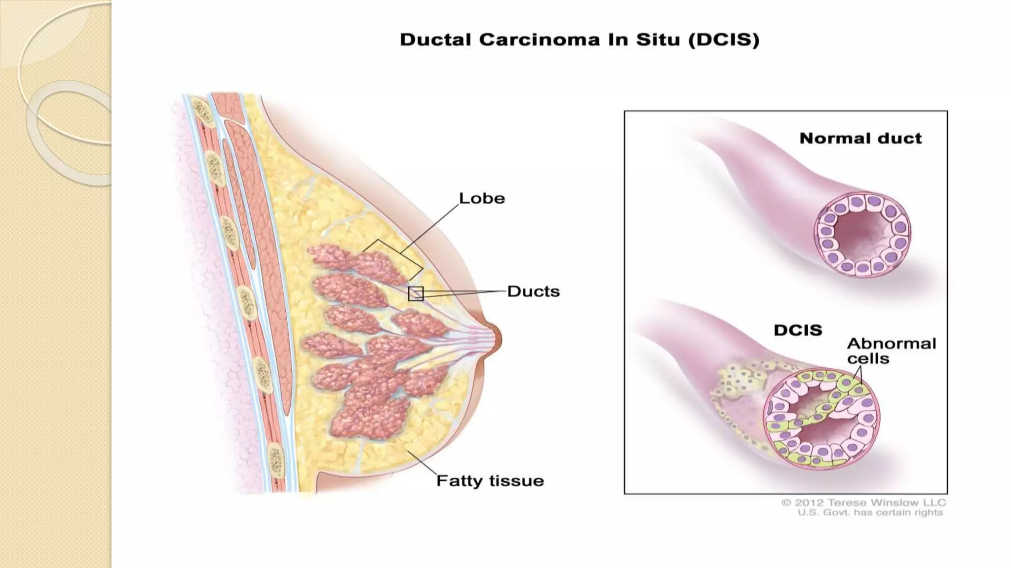
Ductal Carcinoma In
Situ (DCIS)
Saleh Yassin
Supervisor: Dr. Ibrahim Majjad
Introduction
Neoplastic process that is confined to the breast ductal system and lacks
histopathologic evidence of invasion
These cells neither disrupt the basement membrane nor involve the
surrounding breast tissues
Lacks the ability to metastasize and is confined to the breast
Earliest form of cancer is DCIS then it progresses to invasive ductal carcinoma
Approximately 25% of newly diagnosed breast cancers are DCIS
DCIS is less common than invasive breast cancer, the risk increases with age.
DCIS is uncommon in women younger than 30 years of age
More than 90% of all cases of DCIS are detected only on imaging studies
Risk factors are similar to that of invasive breast cancer:
Family history, increased breast density, obesity, nulliparity, mutations in
BRCA1 and BRCA2 genes
Pathology
Classification has followed its
architectural or morphological
appearance
Subtypes:
 Comedo
 Solid
 Cribriform
 Micropapillary
 Papillary
Less common subtypes:
 Apocrine
 Neuroendocrine
 Signet –cell cystic hypersecretory carcinoma
 Clinging DCIS
It’s common to encounter a mixture of subtypes within the same specimen
Clinical presentation
There are no specific clinical manifestations for patients with DCIS.
Before the use of screening mammography, DCIS typically presented as a
palpable mass or nipple discharge
An invasive component commonly was found, and pure DCIS rarely was
encountered
The widespread of mammography now routinely detects DCIS >1cm in
diameter.
Results in breast cancer-free survival rates that approach 100%
95% of new cases of DCIS present with mammographic abnormalities
Microcalcifications are most typical. 90% of women with DCIS have suspicious
microcalcifications on mammography
Asymmetric densities identified in 10%
Dominant masses in 8%
Abnormal galactograms (performed for evaluation of nipple discharge) in 6%
Linear and branching calcifications frequently are associated with high-grade
DCIS and necrosis, whereas fine and granular calcifications are associated more
commonly with low-grade
Initial evaluation should include magnification views that allow for complete
characterization of mammographic findings and determination of the need for
biopsy
The extent of the lesion as determined mammographically may be used as
guide for excision
Ultrasonography, digital mammography, and MRI all have the potential to be
helpful in the management of DCIS
MRI is not routinely indicated in the evaluation of newly diagnosed DCIS
Has high sensitivity but low specificity in the imaging of breast cancer
MRI appears to be no better than mammography for distinguishing DCIS from
benign, atypical lesions or microinvasions
Diagnosis
The diagnosis of DCIS is confirmed by pathologic examination of breast biopsy,
such as a core or excisional biopsy, which is typically performed for suspicious
calcifications detected by a screening mammogram
DCIS is characterized by proliferation of neoplastic epithelial cells within the
mammary ductal system, with no evidence of invasion
FNA is inadequate to distinguish between invasive and in situ disease
Treatment
The goal of the treatment with DCIS is to eradicate the initial cancer,
prevention of local recurrence, with particular emphasis on the prevention of
invasive breast cancer
The recommended workup and staging of DCIS includes:
 History and physical exam
 Bilateral diagnostic mammography
 MRI (optional)
 Pathology review
Options:
Mastectomy
Breast conserving therapy(BCT)
BCT consists of lumpectomy (breast-conserving surgery (BCS)) followed in
most cases by adjuvant radiation
A sentinel lymph node biopsy (SLNB) can be avoided in most women.
However, it should be obtained in women with high risk features for whom
resection may compromise the ability to perform a future SLNB
Mastectomy VS BCT
Mastectomy was the standard treatment of DCIS through the first four decades
Mastectomy is highly effective treatment for DCIS, with a locoregional control
rate of 98 to 100%
Cancer-specific mortality rates of 2% or less
Many retrospective studies suggest that the rates of local or regional recurrence
are significantly lower after mastectomy than after breast conserving therapy.
But there have been no significant differences in overall survival
No prospective randomized trials comparing mastectomy to BCT for DCIS
BCS ± RT
Studies have shown that the addition of whole breast irradiation to a margin-
free excision of pure DCIS, decrease the rate of in-breast disease recurrence,
but doesn’t affect survival, or distant metastasis-free survival
Whole breast irradiation after BCS reduces the relative risk of a local failure by
approximately one half.
However, treating all women who undergo BCS for DCIS with adjuvant RT may
be overtreatment for some
The current challenge is to identify women with DCIS whose risk of an
ipsilateral tumor recurrence (primarily invasive) with BCS, with or without
radiation
Systemic treatment
The primary role of systemic treatment is to reduce the risk of invasive breast
cancer in the ipsilateral and/or contralateral breast.
Chemotherapy plays no role in the management of these patients
Endocrine therapy:
Approximately 75% of DCIS lesions express estrogen receptor (ER) and/or
progesterone receptor (PR)
For women with ER-positive DCIS who have not undergo a bilateral
mastectomy, we suggest endocrine therapy rather than observation, by either
tamoxifen or aromatase inhibitors
In women having bilateral mastectomies for DCIS, the risks of adverse effects of
tamoxifen outweigh any potential benefit for risk reduction
Studies have shown that women treated with tamoxifen after BCT had a 3.4%
absolute reduction in ipsilateral in-breast tumor recurrence risk.
No differences in overall survival were noted
Factors predicting recurrence
Approximately one-half of all locoregional recurrences are invasive, regardless
the treatment approach
Age: increasing age is associated with decrease risk of ipsilateral breast tumor
recurrence
Size: increasing clinical size was associated with an increased risk of ipsilateral
tumor recurrence in patients treated with BCS alone, but not those treated with
conservative surgery and radiation
Multifocality: multifocal DCIS has been associated with an increased risk of
ipsilateral breast tumor recurrence. When compared with unifocal disease in
patients treated with conservative surgery alone or conservative surgery and
radiation
Resection margin status: positive margins of resection have been
associated with an increased risk of ipsilateral breast tumor recurrence
Critical margin >1mm and <10mm
Breast density
High nuclear grade and the presence of necrosis have been associated
with an increased risk of ipsilateral tumor recurrence in patients undergoing
conservative surgery
These factors have had less of an impact on recurrence rates in patients
undergoing conservative surgery and radiation
Prognosis
The prognosis for patient with DCIS is excellent. Advancements in screening,
standardized pathologic review and reporting of margins, and adjuvant
endocrine therapy have improved outcomes
20-year breast cancer mortality among DCIS patients is 3.3%
The risk of ipsilateral invasive recurrence at 20 years is 5.9%, and of
contralateral invasive recurrence is 6.2%
Follow up
The goals of follow up are early recognition and treatment of recurrences and
second primary breast cancer, evaluation of therapy related complications, and
detection of metastatic symptoms
History and physical examination every 6 to 12 months for 5 years and then
annually
As well as yearly diagnostic mammography
Thank you

DCIS

  • 1.
    Ductal Carcinoma In Situ(DCIS) Saleh Yassin Supervisor: Dr. Ibrahim Majjad
  • 2.
    Introduction Neoplastic process thatis confined to the breast ductal system and lacks histopathologic evidence of invasion These cells neither disrupt the basement membrane nor involve the surrounding breast tissues Lacks the ability to metastasize and is confined to the breast Earliest form of cancer is DCIS then it progresses to invasive ductal carcinoma
  • 4.
    Approximately 25% ofnewly diagnosed breast cancers are DCIS DCIS is less common than invasive breast cancer, the risk increases with age. DCIS is uncommon in women younger than 30 years of age More than 90% of all cases of DCIS are detected only on imaging studies Risk factors are similar to that of invasive breast cancer: Family history, increased breast density, obesity, nulliparity, mutations in BRCA1 and BRCA2 genes
  • 5.
    Pathology Classification has followedits architectural or morphological appearance Subtypes:  Comedo  Solid  Cribriform  Micropapillary  Papillary
  • 6.
    Less common subtypes: Apocrine  Neuroendocrine  Signet –cell cystic hypersecretory carcinoma  Clinging DCIS It’s common to encounter a mixture of subtypes within the same specimen
  • 7.
    Clinical presentation There areno specific clinical manifestations for patients with DCIS. Before the use of screening mammography, DCIS typically presented as a palpable mass or nipple discharge An invasive component commonly was found, and pure DCIS rarely was encountered The widespread of mammography now routinely detects DCIS >1cm in diameter. Results in breast cancer-free survival rates that approach 100%
  • 8.
    95% of newcases of DCIS present with mammographic abnormalities Microcalcifications are most typical. 90% of women with DCIS have suspicious microcalcifications on mammography Asymmetric densities identified in 10% Dominant masses in 8% Abnormal galactograms (performed for evaluation of nipple discharge) in 6%
  • 10.
    Linear and branchingcalcifications frequently are associated with high-grade DCIS and necrosis, whereas fine and granular calcifications are associated more commonly with low-grade Initial evaluation should include magnification views that allow for complete characterization of mammographic findings and determination of the need for biopsy The extent of the lesion as determined mammographically may be used as guide for excision Ultrasonography, digital mammography, and MRI all have the potential to be helpful in the management of DCIS
  • 11.
    MRI is notroutinely indicated in the evaluation of newly diagnosed DCIS Has high sensitivity but low specificity in the imaging of breast cancer MRI appears to be no better than mammography for distinguishing DCIS from benign, atypical lesions or microinvasions
  • 12.
    Diagnosis The diagnosis ofDCIS is confirmed by pathologic examination of breast biopsy, such as a core or excisional biopsy, which is typically performed for suspicious calcifications detected by a screening mammogram DCIS is characterized by proliferation of neoplastic epithelial cells within the mammary ductal system, with no evidence of invasion FNA is inadequate to distinguish between invasive and in situ disease
  • 13.
    Treatment The goal ofthe treatment with DCIS is to eradicate the initial cancer, prevention of local recurrence, with particular emphasis on the prevention of invasive breast cancer The recommended workup and staging of DCIS includes:  History and physical exam  Bilateral diagnostic mammography  MRI (optional)  Pathology review
  • 14.
    Options: Mastectomy Breast conserving therapy(BCT) BCTconsists of lumpectomy (breast-conserving surgery (BCS)) followed in most cases by adjuvant radiation A sentinel lymph node biopsy (SLNB) can be avoided in most women. However, it should be obtained in women with high risk features for whom resection may compromise the ability to perform a future SLNB
  • 15.
    Mastectomy VS BCT Mastectomywas the standard treatment of DCIS through the first four decades Mastectomy is highly effective treatment for DCIS, with a locoregional control rate of 98 to 100% Cancer-specific mortality rates of 2% or less Many retrospective studies suggest that the rates of local or regional recurrence are significantly lower after mastectomy than after breast conserving therapy. But there have been no significant differences in overall survival No prospective randomized trials comparing mastectomy to BCT for DCIS
  • 16.
    BCS ± RT Studieshave shown that the addition of whole breast irradiation to a margin- free excision of pure DCIS, decrease the rate of in-breast disease recurrence, but doesn’t affect survival, or distant metastasis-free survival Whole breast irradiation after BCS reduces the relative risk of a local failure by approximately one half. However, treating all women who undergo BCS for DCIS with adjuvant RT may be overtreatment for some The current challenge is to identify women with DCIS whose risk of an ipsilateral tumor recurrence (primarily invasive) with BCS, with or without radiation
  • 17.
    Systemic treatment The primaryrole of systemic treatment is to reduce the risk of invasive breast cancer in the ipsilateral and/or contralateral breast. Chemotherapy plays no role in the management of these patients Endocrine therapy: Approximately 75% of DCIS lesions express estrogen receptor (ER) and/or progesterone receptor (PR) For women with ER-positive DCIS who have not undergo a bilateral mastectomy, we suggest endocrine therapy rather than observation, by either tamoxifen or aromatase inhibitors
  • 18.
    In women havingbilateral mastectomies for DCIS, the risks of adverse effects of tamoxifen outweigh any potential benefit for risk reduction Studies have shown that women treated with tamoxifen after BCT had a 3.4% absolute reduction in ipsilateral in-breast tumor recurrence risk. No differences in overall survival were noted
  • 19.
    Factors predicting recurrence Approximatelyone-half of all locoregional recurrences are invasive, regardless the treatment approach Age: increasing age is associated with decrease risk of ipsilateral breast tumor recurrence Size: increasing clinical size was associated with an increased risk of ipsilateral tumor recurrence in patients treated with BCS alone, but not those treated with conservative surgery and radiation
  • 20.
    Multifocality: multifocal DCIShas been associated with an increased risk of ipsilateral breast tumor recurrence. When compared with unifocal disease in patients treated with conservative surgery alone or conservative surgery and radiation Resection margin status: positive margins of resection have been associated with an increased risk of ipsilateral breast tumor recurrence Critical margin >1mm and <10mm Breast density
  • 21.
    High nuclear gradeand the presence of necrosis have been associated with an increased risk of ipsilateral tumor recurrence in patients undergoing conservative surgery These factors have had less of an impact on recurrence rates in patients undergoing conservative surgery and radiation
  • 22.
    Prognosis The prognosis forpatient with DCIS is excellent. Advancements in screening, standardized pathologic review and reporting of margins, and adjuvant endocrine therapy have improved outcomes 20-year breast cancer mortality among DCIS patients is 3.3% The risk of ipsilateral invasive recurrence at 20 years is 5.9%, and of contralateral invasive recurrence is 6.2%
  • 23.
    Follow up The goalsof follow up are early recognition and treatment of recurrences and second primary breast cancer, evaluation of therapy related complications, and detection of metastatic symptoms History and physical examination every 6 to 12 months for 5 years and then annually As well as yearly diagnostic mammography
  • 24.