Cytation 3 single sheet english
•
0 likes•104 views

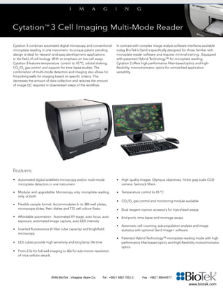
Cytation 3 combines automated digital microscopy and conventional microplate reading in one instrument. It features temperature control, gas control, orbital shaking, and support for time lapse studies, making it ideal for live-cell assays and research applications in cell biology. The instrument allows for hit-picking wells for imaging based on criteria to reduce data collection and image quality control. In contrast to complex interfaces, BioTek's Gen5 software is specifically designed for those familiar with microplate software and requires minimal training. Cytation 3 offers both high performance filter-based optics and high-flexibility monochromator optics for application versatility.

Recommended
Recommended
More Related Content
What's hot
What's hot (18)
Viewers also liked
Viewers also liked (18)
Similar to Cytation 3 single sheet english
Similar to Cytation 3 single sheet english (20)
More from Maziar Yari
More from Maziar Yari (20)
Recently uploaded
Recently uploaded (20)
Cytation 3 single sheet english
- 1. Cytation 3 combines automated digital microscopy and conventional microplate reading in one instrument. Its unique patent pending design is ideal for research and assay development applications in the field of cell biology. With an emphasis on live-cell assays, Cytation 3 features temperature control to 45 °C, orbital shaking, CO2 /O2 gas control and support for time lapse studies. The combination of multi-mode detection and imaging also allows for hit-picking wells for imaging based on specific criteria. This decreases the amount of data collection and reduces the amount of image QC required in downstream steps of the workflow. In contrast with complex image analysis software interfaces available today, BioTek’s Gen5 is specifically designed for those familiar with microplate reader software and requires minimal training. Equipped with patented Hybrid Technology™ for microplate reading, Cytation 3 offers high performance filter-based optics and high- flexibility monochromator optics for unmatched application versatility. • High quality images: Olympus objectives, 16-bit gray scale CCD camera, Semrock filters • Temperature control to 45 °C • CO2 /O2 gas control and monitoring module available • Dual reagent injector accessory for inject/read assays • End point, time-lapse and montage assays • Automatic cell counting, sub-population analysis and image statistics with optional Gen5 Image+ software • Patented Hybrid Technology™ microplate reading mode with high performance filter-based optics and high-flexibility monochromator optics Features: • Automated digital widefield microscopy and/or multi-mode microplate detection in one instrument • Modular and upgradable: Microscopy only, microplate reading only, or both • Flexible sample format: Accommodates 6- to 384-well plates, microscope slides, Petri dishes and T25 cell culture flasks • Affordable automation: Automated XY stage, auto focus, auto exposure, automated image capture, auto LED intensity • Inverted fluorescence (4 filter cube capacity) and brightfield microscopy • LED cubes provide high sensitivity and long lamp life time • From 2.5x for full-well imaging to 60x for sub-micron resolution of intra-cellular details www.biotek.com IRAN BioTek , Viragene Akam Co. Tel : +9821 88611552-3 Fax : +9821 88044577
- 2. Specifications: General Imaging modes: Fluorescence and brightfield Detection method: Monochromators: FL, Lum., UV-Vis Abs. Filters: FL, TRF, FP, Lum. Read method: End point, time-lapse, kinetic, well mode, montage Labware type: 6- to 384-well plates, microscope slides, Petri dishes, cell culture flasks (T25) Compatible with Take3™ Micro-Volume Plates with 2 µL microspots Temperature control: To 45 °C; ±0.2 °C at 37 °C Independent top and bottom temperature control Shaking: Linear, orbital, double orbital Automation: Compatible with BioStack™ and 3rd party automation CO2 and O2 control: 0 – 20% CO2 control and 1 – 19% O2 control, with optional Gas Controller Software: Gen5™ Data Analysis Software; optional Gen5 Image+ Imaging Camera: 16-bit gray scale, Sony CCD, 1.1 megapixel Imaging filter cube capacity: 4 onboard, user-replaceable filter cubes Imaging filter cubes available: DAPI, CFP, GFP, YFP, RFP, Texas Red, CY5 and CY7, Propidium Iodide, Acridine Orange, CYP-YFP FRET, Chlorophyll, Phycoerythrin, CY5.5, TagBFP Imaging LED cubes available: 365 nm, 390 nm, 465 nm, 505 nm, 523 nm, 590 nm, 623 nm, 655 nm, 740 nm Objective capacity: 2 onboard, user-replaceable objectives Available objectives: 2.5x (2.25x eff), 2.5x (2.75x eff), 4x, 10x, 20x, 40x, 60x Image collection rate: 96 wells, 1 color (DAPI), 4x, 6 minutes 96 wells, 3 colors, 4x, 12 minutes Fluorescence Intensity Sensitivity: Monochromators: Top: Fluorescein 2.5 pM (0.25 fmol/well 384-well plate) Bottom: Fluorescein 4 pM (0.4 fmol/well 384-well plate) Filters/mirrors: Fluorescein 0.25 pM (0.025 fmol/well 384-well plate) Light source: Xenon flash lamp Wavelength selection: Double grating monochromators (top and bottom) Deep blocking bandpass filters/dichroic mirrors (top) Wavelength range: Monochromators: 250 – 700 nm; Filters: 200 – 700 nm (850 nm option) Dynamic range: 5 decades Detection system: Two PMT detectors: one for monochromator system, one for filter system Fluorescence Polarization Sensitivity: 1.2 mP standard deviation at 1nM fluorescein Wavelength range: 280 – 700 nm (850 nm option) Time-Resolved Fluorescence Sensitivity: Europium 40 fM with filters (4 amol/well in 384-well plate) Europium 1200 fM with monos (120 amol/well in 384-well plate) Light source: Xenon flash lamp Wavelength range: Monos: 250 – 700 nm Filters: 200 – 700 nm (850 nm option) Luminescence Sensitivity: Monochromators: <20 amol ATP (flash) Filters: <10 amol ATP (flash) Wavelength range: 300 – 700 nm Dynamic range: >6 decades Absorbance Light source: Xenon flash lamp Wavelength selection: Monochromator Wavelength range: 230 – 999 nm, 1 nm increment Bandpass: 4 nm (230 – 285 nm), 8 nm (>285 nm) Dynamic range: 0 – 4.0 OD Resolution: 0.0001 OD Reagent Dispensers Number: 2 syringe pumps Dispense volume: 5 – 1000 µL in 1 µL increment Dead volume: 1 mL, 100 µL with back flush Plate geometry: 6- to 384-well microplates Dispense precision: <2% at 50 – 200 μL Dispense accuracy: ±1 μL or 2% Performance values represent the average observed factory test values. BioTek Instruments, Inc. Highland Park, P.O. Box 998 Winooski, Vermont 05404-0998, USA Phone: 802-655-4040 • Toll-Free: 888-451-5171 Outside the USA: 802-655-4740 www.biotek.com Optional Accessories: • CO2 /O2 Gas Controller Module • Dual Reagent Injector Module • BioStack™ Microplate Stacker • Take3™ Micro-Volume Plate • Gen5™ Secure for 21 CFR part 11 compliance • Luminescence, Fluorescence and Absorbance Test Plates • Gen5 Image+ Software Configurations: CYT3V: Cytation 3 w/imaging CYT3FV: Cytation 3 w/filter optics and imaging CYT3MV: Cytation 3 w/mono optics and imaging CYT3MFV: Cytation 3 w/mono and filter optics and imaging CYT3MF: Cytation 3 w/mono and filter optics CYT3M: Cytation 3 w/mono optics CYT3F: Cytation 3 w/filter optics Typical Applications: • Cell imaging and analysis • Cell proliferation • Cytotoxicity • Protein expression • Biomarker quantification • Drug discovery • Genetic analysis • Drug absorption and metabolism • Biologics drug discovery and development • Environmental testing • Food safety • Nucleic acid quantification • Protein quantification 3T3 cells, 4x magnification. BPAE cells, 20x, from FluoCells® prepared slide #2. HepG2 cells, Caspase3, 20x magnification. FluoCells® is a registered trademark of Molecular Probes, Inc. BioTek’s Hybrid Technolgy™ is protected under US Patent 8,218,141. *Specifications subject to change. Rev. 01/21/15 IRAN BioTek , Viragene Akam Co. Tel : +9821 88611552-3 Fax : +9821 88044577