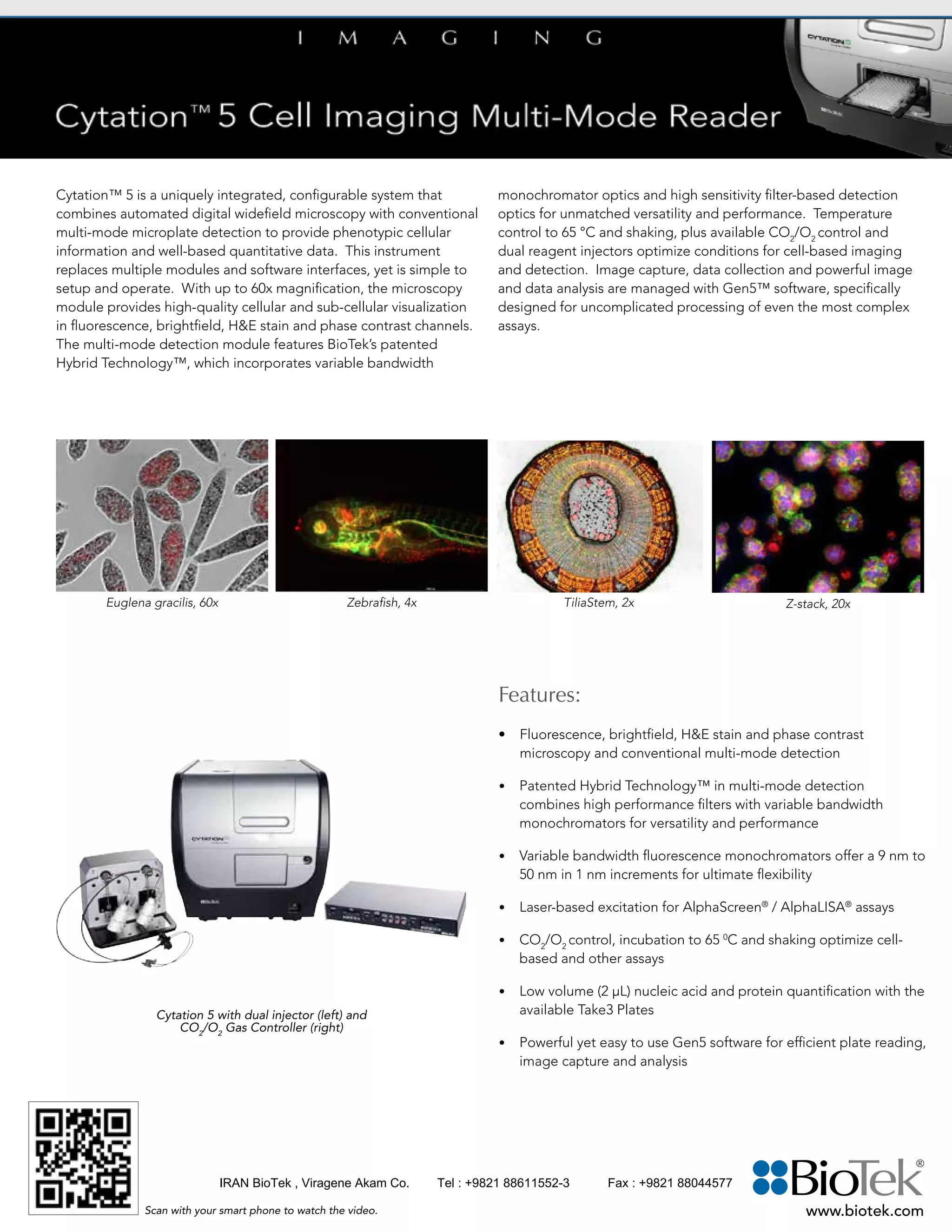
Cytation 5 is an integrated microscope and microplate detection system that combines automated digital widefield microscopy with multi-mode microplate detection. It provides cellular imaging and quantitative data from wells. The system replaces multiple modules and software with a simple, configurable setup. It features fluorescence, brightfield, H&E stain, and phase contrast microscopy at up to 60x magnification, as well as multi-mode detection including monochromators and filters. Temperature control, shaking, and optional gas control optimize cell-based imaging and detection assays. Powerful yet easy-to-use Gen5 software manages image capture, data collection, and analysis.
The first AND ONLY human SPECT/CT/PET imager, a unique solution in the Nuclear Medicine world.
AnyScan SPECT-CT-PET Multi Modality System delivers
the current and future promise of nuclear medicine by
maximizing molecular information in combination with
precise anatomical detail. This technology will help to spot the exact location, size, nature and extent of malignancy anywhere in the body.
2019-06-07 Characterization and research of semiconductors with an FTIR spect...LeonidBovkun
2019-06-07 Educational seminar at EP-3 University of Wuerzburg
I will present particular experiments and related results with FTIR spectrometer, so one may consider these experiments complimentary for you research.
(June 22nd, 2021) Introducing Vilber’s Product Line of Gel Imaging SystemsScintica Instrumentation
Overview:
In this webinar, Alexis will introduce the Fusion fluorescence and chemiluminescence systems manufactured by Vilber. The Fusion systems are the ideal multimodal imaging platform designed for molecular biology laboratories. They cover the spectrum of chemiluminescence, multiplexing fluorescence and chemifluorescence applications. The system offers the ultimate speed and sensitivity across all DNA, RNA and protein-based applications. The Fusion is advanced in the technology it uses to take images and the ease at which users can process and analyze them. The Fusion system is ideal for publication, quantification, and documentation grade imaging.
The Fusion systems are ideal for quantification grade imaging. Chemiluminescent Western blot data poses distinct challenges in producing quantifiable and reproducible data. Most of these issues are a result of low-dynamic range of detection and difficulty in accurately determining the limit of detection. Fusion systems eliminate all these issues thanks to a High Sensitivity Reading (HSR) camera technology that delivers reliable dynamic range, linearity, and unsurpassed sensitivity for the lowest limit of detection. The advanced camera technology reduces noise allowing faint signals to stand out from the surrounding background.
Further the Fusion systems can deliver consistent and reproducible data, independently of a chemiluminescence time course. The chemiluminescence intensity/time profile consists of an initial rise period up to a prolonged emission at a pseudo-plateau level and a decline. VILBER Fusions Automatic imaging mode compensates the time course of the chemiluminescence reaction by adjusting the exposure time while maintaining the larger possible image dynamic.
VILBER is a leading life science company developing and manufacturing fluorescence and chemiluminescence imaging systems for applications ranging from small animal to cell biology research. Founded in 1954, Vilber is a leader in the molecular imaging sector, equipping more than 20,000 laboratories worldwide. An estimated 60,000 researchers use their products every day in over 100 countries.
Learning Objectives:
Gel imaging standard workflow
Chemiluminescence vs. fluorescence imaging
What makes the Vilber systems unique?
Multiplexing capabilities
Image analysis
20160219 - M. Agostini - Nuove tecnologie per lo studio del DNA tumorale libe...Roberto Scarafia
Nano Inspired Biomedicine Laboratory
1 Department of Surgical, Oncological and Gastroenterological Sciences, University of Padua, Italy.
2 Istituto di Ricerca Pediatrica- Città della Speranza, Padova, Italy.
Decoding Turbulent Chemical Plumes With Improved Signal Processing and Machin...InsideScientific
During this webinar sponsored by Aurora Scientific, Dr. Burgués will discuss how he and his team are leveraging various signal processing and machine learning techniques in order to decode the fine-scale structure of turbulent chemical plumes using low-cost chemical sensors. Specifically, he will discuss three signal processing methods they developed to improve MOX sensor dynamics, and share the experimental setups they used to test their theories. Finally, he will share data from recent experiments and elaborate on the conclusions of their studies and how robotic plume tracking technology might apply to industrial and air quality monitoring, research and more.
For more information, please visit: https://insidescientific.com/webinar/decoding-turbulent-chemical-plumes-improved-signal-processing-machine-learning-aurora-scientific
From Jisc's student experience experts group meeting in Birmingham on 21 April 2016.
https://www.jisc.ac.uk/events/student-experience-experts-group-meeting-20-apr-2016
Implementing the Digital Capability Service in my college or university.James Clay
So how do you build digital capability within your institution? The proposed Jisc Digital Capability Service provides a framework, audit and diagnostic tools and an online offer of activities and CPD. However this is only part of the story, there are key challenges and potential enablers within every institution. This workshop will enable participants to work together to identify the potential barriers, blockers and challenges an organisation will face in building digital capability and think about the potential enablers that will allow them to maximise the impact of the Jisc Digital Capability Service to improve the skills and effectiveness of staff across their organisation.
The SuperArgus state-of-the-art preclinical PET/CT system: An overview of the...Scintica Instrumentation
These systems are ideally suited for pre-clinical imaging of small animals such as mice and rats, all the way up to medium sized animals such as rabbits, non-human primates and other similarly sized animals. Some of the unique imaging capabilities include real-time imaging of awake animals, as well as multiplexed PET imaging of standard and non-standard isotopes. Key research applications and example images were reviewed.
Positron Emission Tomography (PET) is the gold standard in metabolic imaging, providing high sensitivity to specific radiotracer used to detect specific metabolic activity or biomarkers in vivo. The most common uses for PET imaging in pre-clinical research include oncology, neurobiology, cardiology, as well as dynamic imaging.
These systems are considered to be best in class imaging system with state of the art detectors and electronics. The systems have been designed to be self-shielded, requiring no additional shielding at the location selected for installation. The systems come in a three different bore sizes allowing for imaging of animals such as mice all the way up to rabbits and even non-human primates. The CT component of these systems has been optimized for reduced radiation exposure, rapid acquisition times, and high resolution images; all ideal for the longitudinal studies so commonly performed in pre-clinical research.
The SuperArgus system is uniquely designed to provide consistent resolution across the entire field of view, while maintaining sensitivity and system performance. Reconstruction algorithms have also been implemented to rapidly process and display the acquired images. The system performs very well for standard imaging applications such as oncology, cardiology, etc. Additionally, the system has some unique features which allow for some unique imaging capabilities such as real-time awake animal imaging, self-gated cardiac imaging, as well as multiplex imaging of standard and non-standard isotopes.
Gas chromatography LZ-GC-A100: Column Oven Temperature Range Ambient temperature: +7 °C to 400 °C (in 1 °C increments) For more information, visit www.Labozon.com.
Spectrolytic was founded with the aim of making spectroscopy solutions available to the masses and to broaden the view of usability.
Spectrolytic GmbH is a developer and supplier of infrared spectrometers and related solutions for a wide range of applications and markets. It also provides development and manufacturing services to third parties for both systems and solutions.
Spectrolytic GmbH has its registered office in Wernberg-Köblitz, Germany, and its operating HQ in Wackersdorf, Germany. The Companies design, manufacture (internally and/or externally) a range of spectroscopy systems for a number of market sectors including but not limited to industrial and consumer applications.
The Company also provide technology consulting, system design and development services, plus related manufacturing to customers on a contract basis.
We believe we have some of the greatest minds in our business and will utilize this capability to expand our business to the benefits of the team and all stakeholders.
The RA802 Pharmaceutical Analyser combines Renishaw's proprietary LiveTrackTM and StreamLineTM technologies to generate chemical images up to 150 times faster than conventional methods, whilst maintaining focus ? ensuring high quality pharmaceutical tablet imaging.
It offers high throughput with 3 shaking modes, fast reading speed of 5 seconds and
malfunction alarm system along with 96-well multimode and bichromatic multi-channel
optical system. Different detection modes like comprehensive qualitative and quantitative
data-evaluation functions with cut-offs, curve-fits and transformation formulas are
available. It is a basic tool in drug detection and life science research, bioassay
testing and quality control pharmaceutical processes.
FTIR Spectrometer is a benchtop unit designed with multilayer Ge coated KBr beam splitter, offering a spectral range of 7800 cm-1 to 350 cm-1. Equipped with FTIR
software workstation, Cube-corner Michelson interferometer, auto software updating system, high intensity and long-lasting air-cooled IR source. Intelligent
system with real-time monitoring of parameters.
NIR Spectrophotometer is a spectrophotometer with grating monochromator. The wavelength range is 900 nm to 2500 nm and temperature ranges from
5 to 35℃. Single channel whole spectrum scanning offers low dark noise and high sensitivity. It features built-in high-quality PTFE reference module
and polystyrene wavelength standard filter. It combines the data with the corresponding NIR data model, various components of the tested sample can be
obtained at the same time.
Are You Ready for Harmful Algal Bloom Season? | Preparing for HAB MonitoringXylem Inc.
This webinar presentation provides a foundational understanding of YSI Total Algae sensors, including how to calibrate them, which units to use, and how to interpret data gathered with the sensors. This webinar will be especially useful for new users and users transitioning from our legacy 6-series to our EXO and ProDSS platforms.
With the webinar presentation, and you’ll learn:
• Using algal pigments for early HAB detection
• 6-series chlorophyll and BGA vs. the new Total Algae (TAL) sensors
• Calibration with Rhadamine WT
• Choosing the right units
• The new cells/mL tool in KorEXO software
• Real-world data examples and challenges
Interested in total algae sampling? Check out the total algae sampling package at: https://www.ysi.com/prodss/tap-pc
There are many other visible spectrophotometer, you can visit our official website if you are interested. Also you can contact us when you got any problem, the contact information can be found on our websites.
شرکت ویراژن نماینده فروش و خدمات پس از فروش فریزرهای منفی 86 درجه ایلشین کره جنوبی در حجمهای مختلف از 50 لیتر تا 500 لیتر
Viragene Akam co. is Ilshin distributor in Iran for sales and after sales services .
Ozempic: Preoperative Management of Patients on GLP-1 Receptor Agonists Saeid Safari
Preoperative Management of Patients on GLP-1 Receptor Agonists like Ozempic and Semiglutide
ASA GUIDELINE
NYSORA Guideline
2 Case Reports of Gastric Ultrasound
Tom Selleck Health: A Comprehensive Look at the Iconic Actor’s Wellness Journeygreendigital
Tom Selleck, an enduring figure in Hollywood. has captivated audiences for decades with his rugged charm, iconic moustache. and memorable roles in television and film. From his breakout role as Thomas Magnum in Magnum P.I. to his current portrayal of Frank Reagan in Blue Bloods. Selleck's career has spanned over 50 years. But beyond his professional achievements. fans have often been curious about Tom Selleck Health. especially as he has aged in the public eye.
Follow us on: Pinterest
Introduction
Many have been interested in Tom Selleck health. not only because of his enduring presence on screen but also because of the challenges. and lifestyle choices he has faced and made over the years. This article delves into the various aspects of Tom Selleck health. exploring his fitness regimen, diet, mental health. and the challenges he has encountered as he ages. We'll look at how he maintains his well-being. the health issues he has faced, and his approach to ageing .
Early Life and Career
Childhood and Athletic Beginnings
Tom Selleck was born on January 29, 1945, in Detroit, Michigan, and grew up in Sherman Oaks, California. From an early age, he was involved in sports, particularly basketball. which played a significant role in his physical development. His athletic pursuits continued into college. where he attended the University of Southern California (USC) on a basketball scholarship. This early involvement in sports laid a strong foundation for his physical health and disciplined lifestyle.
Transition to Acting
Selleck's transition from an athlete to an actor came with its physical demands. His first significant role in "Magnum P.I." required him to perform various stunts and maintain a fit appearance. This role, which he played from 1980 to 1988. necessitated a rigorous fitness routine to meet the show's demands. setting the stage for his long-term commitment to health and wellness.
Fitness Regimen
Workout Routine
Tom Selleck health and fitness regimen has evolved. adapting to his changing roles and age. During his "Magnum, P.I." days. Selleck's workouts were intense and focused on building and maintaining muscle mass. His routine included weightlifting, cardiovascular exercises. and specific training for the stunts he performed on the show.
Selleck adjusted his fitness routine as he aged to suit his body's needs. Today, his workouts focus on maintaining flexibility, strength, and cardiovascular health. He incorporates low-impact exercises such as swimming, walking, and light weightlifting. This balanced approach helps him stay fit without putting undue strain on his joints and muscles.
Importance of Flexibility and Mobility
In recent years, Selleck has emphasized the importance of flexibility and mobility in his fitness regimen. Understanding the natural decline in muscle mass and joint flexibility with age. he includes stretching and yoga in his routine. These practices help prevent injuries, improve posture, and maintain mobilit
Acute scrotum is a general term referring to an emergency condition affecting the contents or the wall of the scrotum.
There are a number of conditions that present acutely, predominantly with pain and/or swelling
A careful and detailed history and examination, and in some cases, investigations allow differentiation between these diagnoses. A prompt diagnosis is essential as the patient may require urgent surgical intervention
Testicular torsion refers to twisting of the spermatic cord, causing ischaemia of the testicle.
Testicular torsion results from inadequate fixation of the testis to the tunica vaginalis producing ischemia from reduced arterial inflow and venous outflow obstruction.
The prevalence of testicular torsion in adult patients hospitalized with acute scrotal pain is approximately 25 to 50 percent
New Drug Discovery and Development .....NEHA GUPTA
The "New Drug Discovery and Development" process involves the identification, design, testing, and manufacturing of novel pharmaceutical compounds with the aim of introducing new and improved treatments for various medical conditions. This comprehensive endeavor encompasses various stages, including target identification, preclinical studies, clinical trials, regulatory approval, and post-market surveillance. It involves multidisciplinary collaboration among scientists, researchers, clinicians, regulatory experts, and pharmaceutical companies to bring innovative therapies to market and address unmet medical needs.
Explore natural remedies for syphilis treatment in Singapore. Discover alternative therapies, herbal remedies, and lifestyle changes that may complement conventional treatments. Learn about holistic approaches to managing syphilis symptoms and supporting overall health.
Flu Vaccine Alert in Bangalore Karnatakaaddon Scans
As flu season approaches, health officials in Bangalore, Karnataka, are urging residents to get their flu vaccinations. The seasonal flu, while common, can lead to severe health complications, particularly for vulnerable populations such as young children, the elderly, and those with underlying health conditions.
Dr. Vidisha Kumari, a leading epidemiologist in Bangalore, emphasizes the importance of getting vaccinated. "The flu vaccine is our best defense against the influenza virus. It not only protects individuals but also helps prevent the spread of the virus in our communities," he says.
This year, the flu season is expected to coincide with a potential increase in other respiratory illnesses. The Karnataka Health Department has launched an awareness campaign highlighting the significance of flu vaccinations. They have set up multiple vaccination centers across Bangalore, making it convenient for residents to receive their shots.
To encourage widespread vaccination, the government is also collaborating with local schools, workplaces, and community centers to facilitate vaccination drives. Special attention is being given to ensuring that the vaccine is accessible to all, including marginalized communities who may have limited access to healthcare.
Residents are reminded that the flu vaccine is safe and effective. Common side effects are mild and may include soreness at the injection site, mild fever, or muscle aches. These side effects are generally short-lived and far less severe than the flu itself.
Healthcare providers are also stressing the importance of continuing COVID-19 precautions. Wearing masks, practicing good hand hygiene, and maintaining social distancing are still crucial, especially in crowded places.
Protect yourself and your loved ones by getting vaccinated. Together, we can help keep Bangalore healthy and safe this flu season. For more information on vaccination centers and schedules, residents can visit the Karnataka Health Department’s official website or follow their social media pages.
Stay informed, stay safe, and get your flu shot today!
Title: Sense of Taste
Presenter: Dr. Faiza, Assistant Professor of Physiology
Qualifications:
MBBS (Best Graduate, AIMC Lahore)
FCPS Physiology
ICMT, CHPE, DHPE (STMU)
MPH (GC University, Faisalabad)
MBA (Virtual University of Pakistan)
Learning Objectives:
Describe the structure and function of taste buds.
Describe the relationship between the taste threshold and taste index of common substances.
Explain the chemical basis and signal transduction of taste perception for each type of primary taste sensation.
Recognize different abnormalities of taste perception and their causes.
Key Topics:
Significance of Taste Sensation:
Differentiation between pleasant and harmful food
Influence on behavior
Selection of food based on metabolic needs
Receptors of Taste:
Taste buds on the tongue
Influence of sense of smell, texture of food, and pain stimulation (e.g., by pepper)
Primary and Secondary Taste Sensations:
Primary taste sensations: Sweet, Sour, Salty, Bitter, Umami
Chemical basis and signal transduction mechanisms for each taste
Taste Threshold and Index:
Taste threshold values for Sweet (sucrose), Salty (NaCl), Sour (HCl), and Bitter (Quinine)
Taste index relationship: Inversely proportional to taste threshold
Taste Blindness:
Inability to taste certain substances, particularly thiourea compounds
Example: Phenylthiocarbamide
Structure and Function of Taste Buds:
Composition: Epithelial cells, Sustentacular/Supporting cells, Taste cells, Basal cells
Features: Taste pores, Taste hairs/microvilli, and Taste nerve fibers
Location of Taste Buds:
Found in papillae of the tongue (Fungiform, Circumvallate, Foliate)
Also present on the palate, tonsillar pillars, epiglottis, and proximal esophagus
Mechanism of Taste Stimulation:
Interaction of taste substances with receptors on microvilli
Signal transduction pathways for Umami, Sweet, Bitter, Sour, and Salty tastes
Taste Sensitivity and Adaptation:
Decrease in sensitivity with age
Rapid adaptation of taste sensation
Role of Saliva in Taste:
Dissolution of tastants to reach receptors
Washing away the stimulus
Taste Preferences and Aversions:
Mechanisms behind taste preference and aversion
Influence of receptors and neural pathways
Impact of Sensory Nerve Damage:
Degeneration of taste buds if the sensory nerve fiber is cut
Abnormalities of Taste Detection:
Conditions: Ageusia, Hypogeusia, Dysgeusia (parageusia)
Causes: Nerve damage, neurological disorders, infections, poor oral hygiene, adverse drug effects, deficiencies, aging, tobacco use, altered neurotransmitter levels
Neurotransmitters and Taste Threshold:
Effects of serotonin (5-HT) and norepinephrine (NE) on taste sensitivity
Supertasters:
25% of the population with heightened sensitivity to taste, especially bitterness
Increased number of fungiform papillae
For Better Surat #ℂall #Girl Service ❤85270-49040❤ Surat #ℂall #Girls
Cytation 5 single sheet english version
1. Cytation™ 5 is a uniquely integrated, configurable system that
combines automated digital widefield microscopy with conventional
multi-mode microplate detection to provide phenotypic cellular
information and well-based quantitative data. This instrument
replaces multiple modules and software interfaces, yet is simple to
setup and operate. With up to 60x magnification, the microscopy
module provides high-quality cellular and sub-cellular visualization
in fluorescence, brightfield, H&E stain and phase contrast channels.
The multi-mode detection module features BioTek’s patented
Hybrid Technology™, which incorporates variable bandwidth
monochromator optics and high sensitivity filter-based detection
optics for unmatched versatility and performance. Temperature
control to 65 °C and shaking, plus available CO2
/O2
control and
dual reagent injectors optimize conditions for cell-based imaging
and detection. Image capture, data collection and powerful image
and data analysis are managed with Gen5™ software, specifically
designed for uncomplicated processing of even the most complex
assays.
Features:
• Fluorescence, brightfield, H&E stain and phase contrast
microscopy and conventional multi-mode detection
• Patented Hybrid Technology™ in multi-mode detection
combines high performance filters with variable bandwidth
monochromators for versatility and performance
• Variable bandwidth fluorescence monochromators offer a 9 nm to
50 nm in 1 nm increments for ultimate flexibility
• Laser-based excitation for AlphaScreen®
/ AlphaLISA®
assays
• CO2
/O2
control, incubation to 65 0
C and shaking optimize cell-
based and other assays
• Low volume (2 µL) nucleic acid and protein quantification with the
available Take3 Plates
• Powerful yet easy to use Gen5 software for efficient plate reading,
image capture and analysis
www.biotek.com
Euglena gracilis, 60x TiliaStem, 2x Z-stack, 20xZebrafish, 4x
Cytation 5 with dual injector (left) and
CO2
/O2
Gas Controller (right)
Scan with your smart phone to watch the video.
IRAN BioTek , Viragene Akam Co. Tel : +9821 88611552-3 Fax : +9821 88044577
2. Specifications:
General
Imaging modes: Fluorescence, brightfield, phase contrast, H&E stain
Detection mode: Monochromators: FL, Lum., UV-Vis Abs., TRF (secondary)
Filters: FL, TRF, FP, Lum., Alpha
Read method: End point, kinetic, well mode, time-lapse, montage
Labware type: 6- to 384-well plates, microscope slides, Petri dishes, cell culture
flasks (T25)
Take3™ Micro-Volume Plates
Temperature control: 4-Zone™ incubation to 65 °C with Condensation Control™
Variation: ±0.2 °C at 37 °C
Shaking: Linear, orbital, double orbital
Automation: Compatible with BioStack™ and 3rd party automation
CO2
and O2
control: Optional Gas Controller available
Software: Gen5™ Data Analysis Software included; Gen5 Image+ software
available for full image analysis
Imaging
Light source: High power LEDs (available wavelengths: 365 nm, 405 nm, 465 nm,
590 nm, 523 nm, 505 nm, 623 nm, 740 nm)
Camera: 16-bit gray scale, Sony CCD, 1.1 megapixel
Filter cube capacity: 4 onboard, user-replaceable filter cubes
Objective capacity: 6 objectives turret
Available objectives: Fluorescence: 2.5x (2.25x eff), 2.5x (2.75x eff), 4x, 10x, 20x, 40x, 60x
Phase contrast: 4x, 10x, 20x, 40x
Image collection rate: 96 wells, 1 color (DAPI), 4x, 6 minutes
96 wells, 3 colors, 4x, 12 minutes
Resolution: 0.3µm/pixel at 20x
Automated functions: Autofocus, autoexposure, auto-LED intensity
Fluorescence Intensity
Sensitivity: Monochromators:
Top: Fluorescein 2.5 pM (0.25 fmol/well 384-well plate)
Bottom: Fluorescein 4 pM (0.4 fmol/well 384-well plate)
Filters/mirrors: Fluorescein 0.25 pM (0.025 fmol/well 384-well plate)
Light source: Xenon flash lamp
Wavelength selection: Double grating monochromators (top and bottom)
Deep blocking bandpass filters/dichroic mirrors (top)
Wavelength range: Monochromators: 250 – 700 nm (850 nm option)
Filters: 200 – 700 nm (850 nm option)
Dynamic range: 7 decades
Detection system: Two PMTs: (1) for monochromator system, (1) for filter system
Luminescence
Sensitivity: Monochromators: 20 amol ATP (flash); Filters: 10 amol ATP (flash)
Wavelength range: 300 – 700 nm
Dynamic range: >6 decades
Fluorescence Polarization
Sensitivity: 1.2 mP standard deviation at 1nM fluorescein
Wavelength range: 320 – 700 nm (850 nm option)
Time-Resolved Fluorescence
Sensitivity: Europium 40 fM with filters (4 amol/well in 384-well plate)
Europium 1200 fM with monos (120 amol/well in 384-well plate)
Light source: Xenon flash lamp
Wavelength range: Monos: 250 – 700 nm (850 nm option)
Filters: 200 – 700 nm (850 nm option)
Absorbance
Light source: Xenon flash lamp
Wavelength selection: Monochromator
Wavelength range: 230 – 999 nm, 1 nm increment
Bandpass: 4 nm (230 – 285 nm), 8 nm (>285 nm)
Dynamic range: 0 – 4.0 OD
Alpha Detection
Light source: 680 nm laser, 100 mW +/-10%
Wavelength selection: Filter (top only)
Sensitivity: 100 amol LCK peptide (384-well low volume plate)
Reagent Dispensers
Number: 2 syringe pumps
Dispense volume: 5 – 1000 µL in 1 µL increment
Dead volume: 1 mL, 100 µL with back flush
Plate geometry: 6- to 384-well microplates
Dispense precision: <2% at 50 – 200 μL
Dispense accuracy: ±1 μL or 2%
Performance values represent the average observed factory test values.
BioTek Instruments, Inc.
Highland Park, P.O. Box 998
Winooski, Vermont 05404-0998, USA
Phone: 802-655-4040 • Toll-Free: 888-451-5171
Outside the USA: 802-655-4740
www.biotek.com
Optional Accessories:
• Joystick Controller
• CO2
/O2
Gas Controller Module
• Dual Reagent Injector Module
• BioStack™ Microplate Stacker
• Take3™ Micro-Volume Plate
• Gen5™ Secure for 21 CFR part 11 compliance
• Luminescence, Fluorescence and Absorbance Test Plates
• Gen5 Image+ Software
Configurations:
CYT5PV: Cytation 5 w/phase contrast imaging and fluorescence,
brightfield and H&E imaging
CYT5MFV: Cytation 5 w/monochromator fluorescence, monochromator
UV-Vis aborbance, filtered luminescence, luminescence,
time-resolved fluorescence, filter / dichroic fluorescence,
fluorescence polarization. Fluorescence, brightfield and
H&E imaging
CYT5MV: Cytation 5 w/monochromator fluorescence, monochromator
UV-Vis aborbance, luminescence, time-resolved fluorescence
(2° mode). Fluorescence, brightfield and H&E imaging
CYT5MFA: Cytation 5 w/monochromator fluorescence, monochromator
UV-Vis aborbance, time-resolved fluorescence, filter / dichroic
fluorescence, fluorescence polarization, luminescence, filtered
luminescence, and laser AlphaScreen®
/AlphaLISA®
CYT5M: Cytation 5 w/monochromator fluorescence, monochromator
UV-Vis aborbance, luminescence, and time-resolved fluores-
cence (2° mode)
Note: Several other configurations are available. Ask your BioTek Sales
Representative or visit www.biotek.com for details.
*Specifications subject to change. Rev. 10/23/14
Typical Applications:
• 2D and 3D cell imaging and analysis
• Cell proliferation
• Cytotoxicity
• Protein expression
• Biomarker quantification
• Drug discovery
• Genetic analysis
• Drug absorption and metabolism
• Biologics drug discovery and development
• Environmental testing
• Food safety
• Nucleic acid quantification
• Protein quantification
IRAN BioTek , Viragene Akam Co. Tel : +9821 88611552-3 Fax : +9821 88044577