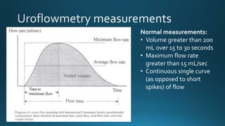
This document discusses normal bladder function measurements including a volume greater than 200 mL over 15 to 30 seconds, a maximum flow rate greater than 15 mL/sec, and a continuous single curve of flow as opposed to short spikes.
1. Urodynamics describes physiological tests used to investigate lower urinary tract function, with cystometry being the most important test. Cystometry measures pressure-volume relationships during bladder filling and voiding.
2. Urodynamics tests the storage and evacuation of urine to reproduce a patient's symptoms and determine their underlying cause. Tests include cystometry, uroflowmetry, and pressure-flow studies.
3. Urodynamics is indicated for incontinence, suspected outflow obstruction, neurogenic bladder dysfunction, and children with voiding issues. It helps characterize detrusor and bladder outlet function and diagnose neuropathies.
This document provides an overview of urodynamics, including the physiology of micturition, urodynamics equipment, cystometry, uroflowmetry, pressure flow studies, and abnormal findings. Key points include:
1. Urodynamics evaluates bladder filling, storage, and emptying through tests like cystometry, uroflowmetry, and pressure flow studies.
2. Cystometry measures bladder pressure during filling and identifies abnormalities like impaired compliance, detrusor overactivity, and high leak point pressures.
3. Pressure flow studies quantify voiding and help differentiate obstruction from poor contractility.
4. Abnormal findings on urodynamics like high pressures, impaired compliance,
This document provides an overview of urodynamic studies (UDS), which are used to evaluate bladder storage and voiding functions. It describes various UDS techniques including uroflowmetry, post-void residual measurement, cystometry, and pressure-flow studies. Cystometry involves bladder filling while measuring pressures, and is used to assess capacity, compliance, and for detecting detrusor overactivity. Pressure-flow studies performed during voiding provide information about bladder contractility and outflow obstruction. Together these invasive UDS techniques provide valuable information to characterize lower urinary tract dysfunction.
This document discusses urodynamics, which involves testing to evaluate lower urinary tract symptoms. It describes the components of a urodynamic study including uroflowmetry, cystometrography, and pressure flow study. Common indications for urodynamics include evaluating young males with untreated LUTS, neurogenic bladder issues, and mixed urinary incontinence. The document outlines the procedure, parameters evaluated, normal values, and how urodynamics can help guide appropriate treatment.
Urodynamics tests measure how the urinary bladder functions and provide objective evidence about any dysfunction. Common tests include uroflowmetry to measure urine flow, cystometry to evaluate bladder capacity and pressure, and pressure flow studies to identify bladder outlet blockage. Symptoms like urine leakage, frequent urination, and incomplete emptying may indicate the need for urodynamics to inform treatment for conditions like prostate enlargement or spinal injuries.
The document provides information about urodynamics testing performed at the Department of Urology, Government Royapettah Hospital and Kilpauk Medical College in Chennai. It discusses the professors and assistant professors who moderate the tests. It then describes the purpose and components of urodynamics testing, which involves a series of tests to evaluate urine storage and evacuation. The key components reviewed include uroflowmetry, measurement of post-void residual urine, cystometrogram, pressure flow studies, and videourodynamics. The document provides details on performing each test and interpreting the results.
This document provides an overview of overactive bladder (OAB). It defines OAB and its main symptoms of urgency, frequency, and nocturia. It discusses the prevalence of OAB increasing with age and being similar between genders. The document outlines the bladder anatomy and physiology, as well as theories around the etiology and pathophysiology of OAB. It describes the diagnosis and clinical evaluation of OAB through medical history, physical exam, urinalysis, and other tests. Finally, it covers treatment approaches for OAB including behavioral modifications, medications, injections, and surgeries.
Urodynamic studies provide physiological measurements of the lower urinary tract to assess function and dysfunction. The main types of urodynamic studies include cystometry, uroflowmetry, pressure flow studies, and video-urodynamics. Cystometry measures bladder pressure during filling and provides information on bladder sensation, capacity, compliance, and detrusor activity. Uroflowmetry measures urine flow rate during voiding to assess voiding effectiveness. Pressure flow studies simultaneously measure bladder pressure and flow rate throughout voiding to quantitatively analyze voiding function. Together, these tests can help diagnose incontinence, obstruction, and neurogenic bladder issues.
Urodynamic evaluation assesses normal and abnormal lower urinary tract function. It involves measuring bladder filling and emptying through cystometry and pressure-flow studies. Key measurements include bladder capacity, compliance, and identifying any detrusor overactivity. Abnormalities can indicate storage or voiding dysfunction. Urodynamic studies are useful for evaluating incontinence, neurogenic bladder, and outflow obstruction when other tests are insufficient for diagnosis or treatment planning. The test aims to reproduce symptoms and determine their cause through urodynamic measurements.
This document contains information about the Department of Urology at the Government Royapettah Hospital and Kilpauk Medical College in Chennai, India. It lists the professors and assistant professors in the department and provides an overview of the neurogenic control of micturition, including the roles of the brain, brainstem, spinal cord, and peripheral nerves. It also discusses the neuroanatomy and physiology of storage and emptying of the bladder, as well as changes in bladder function throughout life and various pathophysiological conditions.
Urodynamics tests the storage and voiding functions of the lower urinary tract through various tests like cystometry, uroflowmetry, and pressure-flow studies. Cystometry measures pressure-volume relationships during bladder filling and looks at parameters like compliance, capacity, and detrusor activity. Uroflowmetry analyzes urine flow over time through variables like maximum flow rate and voiding time. Together these tests can characterize detrusor and outlet functions, diagnose neuropathies, and determine the cause of lower urinary tract symptoms when other evaluations are inconclusive. The tests are indicated for incontinence, outflow obstruction, neurogenic bladder dysfunction, and voiding problems in children.
This document discusses female urinary incontinence. It begins by describing the functions of the urinary system and micturition cycle. It then discusses the different types of urinary incontinence including stress, urge, overflow and mixed incontinence. The document outlines the anatomy involved in maintaining continence like the urethral mechanism and Delancey's level II support. It also discusses various theories of continence. Treatment options covered include conservative treatments like pelvic floor exercises as well as surgical procedures for stress incontinence like vaginal slings and urethrocystopexy.
The document describes urodynamic evaluation (UDE) performed in the Department of Urology at Govt Royapettah Hospital and Kilpauk Medical College in Chennai. It lists the professors and assistant professors in the department and provides an introduction to UDE. It then describes the various components of UDE including uroflowmetry, cystometry, pressure flow studies and videourodynamics. It outlines the procedure for setting up and performing UDE, and analyzes storage and voiding phases and parameters measured.
This document provides an overview of overactive bladder, including its definition, etiology, pathophysiology, symptoms, diagnosis, and treatment. It defines overactive bladder as a symptom complex of urgency, usually with frequency and nocturia, with or without incontinence, in the absence of infection or other obvious pathology. The pathophysiology involves detrusor overactivity due to various hypotheses like outflow obstruction, neurogenic mechanisms, and myogenic and urothelial mechanisms. Treatment involves behavioral therapy, drug therapy using antimuscarinics, neuromodulation techniques, surgery for refractory cases, and newer developments in drug delivery and mechanisms of action.
This document describes uroflowmetry - a noninvasive test used to evaluate urine flow. It discusses the normal and abnormal flow patterns seen in uroflowmetry and their clinical significance. Uroflowmetry provides parameters like maximum flow rate, average flow rate and voided volume. It can detect bladder outlet obstruction, detrusor underactivity or overactivity. However, pressure-flow studies are needed to precisely define lower urinary tract function. Uroflowmetry is useful for screening and monitoring treatment response, though invasive therapy should not be based on uroflowmetry alone per AUA guidelines.
- Hypospadias is a congenital condition where the urethral opening is on the underside of the penis rather than at the tip. It can range from mild to severe forms.
- The cause is usually unknown but may involve genetic factors or exposure to chemicals during pregnancy. Risk increases with family history, maternal age over 35, or in vitro fertilization.
- Treatment involves surgery to reposition the urethral opening, which is usually done between ages 6-18 months. The goal is to create a normal appearing penis and allow for normal urination and sexual function.
- Surgical techniques vary depending on the severity but aim to lengthen and tubularize the urethra
The prostate is a gland located below the bladder. It helps produce fluid for semen. Common prostate issues include benign prostatic hyperplasia (BPH), prostatitis, and prostate cancer. BPH involves non-cancerous growth of the prostate and affects most men as they age. Prostatitis is prostate inflammation that can be acute or chronic. Prostate cancer is the most common cancer in men over 65. Early prostate cancer may have no symptoms, while advanced cases can spread to bones.
Uroflowmetry is a test to measure urine flow and assess for bladder outlet obstruction. It is performed by having a patient urinate into a special toilet that measures parameters like voided volume, average flow rate, maximum flow rate, and time to maximum flow. Abnormal results can indicate conditions like benign prostatic hyperplasia, bladder outlet obstruction, or a neurogenic bladder.
This document discusses urodynamic testing procedures like uroflowmetry and cystometrogram that are used to evaluate lower urinary tract function and diagnose conditions like overactive bladder. It provides details on parameters measured and what different tests can reveal. Common urodynamic findings are defined, like detrusor overactivity and poor bladder compliance. Neurogenic causes of lower urinary tract dysfunction are outlined for different spinal cord injury levels. A step-wise approach to managing the neurogenic bladder is proposed starting with self-voiding and progressing to clean intermittent catheterization or other options if needed.
Recent trends in management of undescended testesAwaneesh Katiyar
This document discusses recent trends in the management of undescended testes. It begins with definitions and prevalence rates of undescended testes. It then covers the development of the testes, the phases of testicular descent, and factors that can affect descent. Risk factors for undescended testes like genetics, syndromes, and environmental exposures are outlined. Complications include reduced fertility, torsion, and testicular cancer. Diagnostic tools like ultrasound, CT, MRI, and laparoscopy are discussed. Management includes hormonal therapy or surgical orchiopexy depending on factors like age and palpability. Surgical techniques for orchiopexy like inguinal and trans-scrotal approaches are described.
The document discusses the physiology of micturition (urination). It describes the functional anatomy of the bladder, its innervation, and the physiological processes of filling and emptying. Filling is controlled by sympathetic nerves, while emptying involves a parasympathetic reflex initiated once the bladder reaches a certain volume. This reflex can be inhibited by higher brain centers until a convenient time. Abnormalities like atonic, automatic, and neurogenic bladders are also discussed.
Urodynamic techniques involve dynamic studies of urine storage and voiding to help diagnose lower urinary tract dysfunction. Simple techniques include voiding diaries while complex techniques involve pressure-flow studies and cystometry. Cystometry measures the pressure-volume relationship of the bladder during filling and voiding to describe normal and abnormal bladder and urethral function. It aims to reproduce the patient's symptoms and provide a physiological explanation by correlating findings to symptoms. Interpretation of cystometry evaluates detrusor activity, bladder sensation, compliance, capacity and other factors. Together, urodynamic studies can help accurately diagnose conditions, predict treatment outcomes, and assess the progression of lower urinary tract problems.
This document discusses ureteroceles, which are cystic dilations of the distal ureter that may be associated with defects in ureteral maturation. Ureteroceles can be intravesical, extending into the bladder, or extravesical/ectopic, extending beyond the bladder neck. They are usually associated with the upper renal moiety in a duplex system. Clinical presentations include infections, incontinence, pain, or being found incidentally. Diagnostic imaging includes ultrasound, IVU, VCUG, nuclear medicine scans, and cystoscopy. Management goals are preserving renal function, eliminating obstruction/reflux, and continence. Treatment depends on individual factors and may include observation, acute decomp
Pathophysiology of urinary incontinencedr.hafsa asim
Urinary incontinence is defined as the involuntary loss of urine, which is both a social and hygienic problem that can be objectively demonstrated. It occurs when the internal sphincter and urethral closure pressure are unable to withstand increases in intra-abdominal pressure due to reductions in the differences between intraurethral and intravesical pressures.
This document provides guidelines for the evaluation and management of hematuria from the Department of Urology at Govt Royapettah Hospital and Kilpauk Medical College in Chennai. It discusses the classification, timing, differential diagnosis, and evaluation of hematuria. The evaluation includes history, physical exam, urinalysis, urine culture if indicated, renal function testing, cystoscopy, and imaging such as CT urogram. The goal of evaluation is to identify any underlying causes of hematuria such as infection or malignancy. Close follow up is recommended depending on the diagnosis and persistence of microscopic hematuria.
This document discusses the management of overactive bladder. It begins by describing the normal bladder functions of storage and emptying. It then discusses overactive bladder, noting it is defined by symptoms of urgency and frequency. Causes can be neurologic or myogenic defects. Evaluation involves history, exam, urodynamics and cystoscopy. Treatment begins with lifestyle changes and pharmacotherapy using antimuscarinic drugs to inhibit bladder contractions. For refractory cases, options include intravesical injections, botulinum toxin, neuromodulation or urinary reconstruction. Overall understanding of overactive bladder pathophysiology and treatment options has improved.
Urodynamics describes physiological tests used to investigate abnormalities of lower urinary tract function. The principles of urodynamics are simple - it involves studying the relationship between bladder pressure, volume, and flow during different stages of the micturition cycle. Both non-invasive tests like flowmetry and invasive tests like cystometry are used to evaluate patients and make a urodynamic diagnosis. Special considerations are needed when performing urodynamics on certain patient populations like those with spinal cord injuries.
Making sense of Urodynamic studies for women with Urinary Incontinence and ...GLUP2010
This document discusses urodynamic studies (UDS) for women undergoing surgery for pelvic organ prolapse or stress urinary incontinence. It notes that while UDS can identify conditions like intrinsic sphincter deficiency or detrusor overactivity, it may not always change treatment plans. UDS is still recommended preoperatively to identify issues, but its ability to predict surgical outcomes is limited as current treatments are non-specific. The document questions if UDS is testing the right parameters given the complex nature of incontinence.
The document discusses the benefits of exercise for mental health. Regular physical activity can help reduce anxiety and depression and improve mood and cognitive functioning. Exercise causes chemical changes in the brain that may help protect against mental illness and improve symptoms.
This document discusses detrusor sphincter dyssynergia (DSD), an impairment of coordination between the detrusor and urethral sphincter muscles during urination. It defines DSD and provides details on the micturition cycle, neural control of the lower urinary tract, causes of DSD including spinal cord injury and multiple sclerosis, classification systems for DSD, clinical presentation, diagnostic evaluation including urodynamics, and treatment options. DSD is a common cause of urinary obstruction in patients with neurological conditions that interrupt signaling between the brain and sacral spinal cord controlling micturition.
Overzicht van urologische (plas-, ophoud- EN seksuele) problemen die kunnen optreden bij Multiple Sclerose en hoe daar mee om te gaan, steeds gespiegeld aan de multidisciplinaire richtlijn.
IoT Standardisatie werkgroepbijeenkomst 4 juni 2014Erik Van Der Zee
Als onderdeel van de 2e plenaire bijeenkomst van de Geonovum Pilot Making Sense for Society namens de IoT Standaardisatie werkgroep een presentatie gehouden over het belang van standaardisatie en de beschikbaarheid en mogelijkheden van de OGC Sensor Web Enablement (SWE) standaardenset.
Standardization of Terminology of Lower Urinary Tract Function in Children an...TC İÜ İTF Üroloji AD
Standardization of Terminology of Lower Urinary Tract Function in Children and Adolescents :Update Report from the Standardization Committee of International Children's Continence Society. Evidence based journal club by Yaşar Pazır
The document summarizes the structure and functions of the digestive system. It describes the layers of the gastrointestinal tract and the roles of organs like the liver, gallbladder and pancreas. Key functions include motility, secretion, digestion, absorption, storage and elimination of waste. Hormones and neural pathways regulate digestive processes and secretions throughout the gastrointestinal tract.
This document summarizes the key points regarding urodynamics studies for assessing urinary incontinence. It discusses the general aspects of urodynamics, including informing patients, test conditions, the role of the investigator, use of catheters and transducers, and definitions of various pressure measurements. The document emphasizes that urodynamics aims to objectively observe lower urinary tract function and dysfunction in order to appropriately manage incontinence and its underlying causes.
Este documento contiene los resultados de pruebas realizadas a una paciente, incluyendo una flujometría, una cistometría y un estudio de presión-flujo. La cistometría mostró escapes durante la fase de llenado y al final debido a contracciones, y la capacidad cistométrica máxima fue de 338 ml. El estudio de presión-flujo documentó los valores de presión abdominal, vesical y detrusor durante la micción.
The document describes a computer software program for semi-automated diagnosis of urodynamic studies. It outlines the rationale for developing the software to minimize incorrect diagnoses and improve standardization. The software utilizes published guidelines and literature on urodynamic terminology, techniques, tracing interpretation and diagnosis of lower urinary tract conditions. Validation studies of the software's accuracy are still underway.
El documento describe la anatomía, fisiología, factores de riesgo, síntomas, diagnóstico y tratamientos de la hiperplasia prostática benigna. La HPB es el agrandamiento benigno de la próstata causado por un aumento del tejido prostático y es más común en hombres mayores de 50 años. Los síntomas incluyen disuria, nicturia y flujo urinario débil. El diagnóstico incluye examen rectal, PSA y uroflujometría. Los tratamientos son farmacológicos con blo
The document discusses various urological diseases, procedures, and treatments. It provides details on:
1) Benign prostatic hyperplasia (BPH), prostate cancer, urinary incontinence, and stones - including causes, symptoms, and long term effects.
2) Common procedures for BPH include watchful waiting, drug therapy, and minimally invasive surgeries like TURP, TUIP, and stents. Prostate cancer treatments discussed include brachytherapy, cryotherapy, and external beam radiation therapy.
3) The market for urological devices and procedures is large, with the highest values seen for BPH and stones. Procedures for urinary in
Neuro Urology...Fantastic presentation by Prof Drake of Southmeadmeducationdotnet
- The document discusses the initial assessment and management of neurogenic urinary incontinence. It covers taking a detailed history, performing a neurological examination, investigating with urinalysis and urodynamics, and classifying the neurological lesion. Special considerations for safety include assessing for detrusor sphincter dyssynergia, poor bladder compliance, autonomic dysreflexia, and latex allergies before managing symptoms. Urodynamics helps evaluate the bladder and sphincter function to guide appropriate treatment and ensure renal safety.
O documento descreve o processo de micção, incluindo a anatomia e fisiologia da bexiga, esfíncteres e controle nervoso. A micção envolve o armazenamento de urina na bexiga através do relaxamento do músculo detrusor e contração do esfíncter, e esvaziamento da bexiga através da contração do detrusor e relaxamento dos esfíncteres. O sistema nervoso central e autônomo controlam este processo através de vias parasimpáticas e simpáticas.
O documento descreve a anatomia, histologia, irrigação, drenagem e fisiologia da bexiga urinária. A bexiga é um órgão musculomembranoso localizado na cavidade pélvica que armazena e expulsa a urina. Ela possui quatro camadas (serosa, muscular, submucosa e mucosa) e é irrigada e drenada por vasos que se conectam à aorta e veia ilíaca. A fisiologia da micção envolve fases de enchimento e esvaziamento da bexiga através de
This document provides an overview of neurogenic bladder. It begins with an introduction defining neurogenic bladder as bladder dysfunction due to central nervous system or peripheral nerve disease. It then covers the relevant anatomy and physiology including the innervation of the bladder. The document discusses the central neural control of micturition and the normal voiding process. It also covers development of bladder control in adults and provides descriptions of terminology related to storage and emptying problems.
This document discusses the anatomy, physiology, and dysfunction of the urinary bladder and micturition process. It begins with an overview of bladder anatomy, including its parts and innervation. It then covers the normal physiology of bladder filling and emptying, controlled by the brain, spinal cord, and peripheral nerves. Different types of bladder dysfunction are described, such as overactive bladder, dysfunctional voiding, underactive bladder, and detrusor-sphincter dyssynergia. Evaluation of voiding disorders and urodynamic studies are also mentioned. In summary, the document provides a comprehensive review of the structure and function of the urinary bladder and the potential sites of dysfunction.
Adhd Medication Shortage Uk - trinexpharmacy.comreignlana06
The UK is currently facing a Adhd Medication Shortage Uk, which has left many patients and their families grappling with uncertainty and frustration. ADHD, or Attention Deficit Hyperactivity Disorder, is a chronic condition that requires consistent medication to manage effectively. This shortage has highlighted the critical role these medications play in the daily lives of those affected by ADHD. Contact : +1 (747) 209 – 3649 E-mail : sales@trinexpharmacy.com
share - Lions, tigers, AI and health misinformation, oh my!.pptxTina Purnat
• Pitfalls and pivots needed to use AI effectively in public health
• Evidence-based strategies to address health misinformation effectively
• Building trust with communities online and offline
• Equipping health professionals to address questions, concerns and health misinformation
• Assessing risk and mitigating harm from adverse health narratives in communities, health workforce and health system
These lecture slides, by Dr Sidra Arshad, offer a quick overview of the physiological basis of a normal electrocardiogram.
Learning objectives:
1. Define an electrocardiogram (ECG) and electrocardiography
2. Describe how dipoles generated by the heart produce the waveforms of the ECG
3. Describe the components of a normal electrocardiogram of a typical bipolar lead (limb II)
4. Differentiate between intervals and segments
5. Enlist some common indications for obtaining an ECG
6. Describe the flow of current around the heart during the cardiac cycle
7. Discuss the placement and polarity of the leads of electrocardiograph
8. Describe the normal electrocardiograms recorded from the limb leads and explain the physiological basis of the different records that are obtained
9. Define mean electrical vector (axis) of the heart and give the normal range
10. Define the mean QRS vector
11. Describe the axes of leads (hexagonal reference system)
12. Comprehend the vectorial analysis of the normal ECG
13. Determine the mean electrical axis of the ventricular QRS and appreciate the mean axis deviation
14. Explain the concepts of current of injury, J point, and their significance
Study Resources:
1. Chapter 11, Guyton and Hall Textbook of Medical Physiology, 14th edition
2. Chapter 9, Human Physiology - From Cells to Systems, Lauralee Sherwood, 9th edition
3. Chapter 29, Ganong’s Review of Medical Physiology, 26th edition
4. Electrocardiogram, StatPearls - https://www.ncbi.nlm.nih.gov/books/NBK549803/
5. ECG in Medical Practice by ABM Abdullah, 4th edition
6. Chapter 3, Cardiology Explained, https://www.ncbi.nlm.nih.gov/books/NBK2214/
7. ECG Basics, http://www.nataliescasebook.com/tag/e-c-g-basics
Histololgy of Female Reproductive System.pptxAyeshaZaid1
Dive into an in-depth exploration of the histological structure of female reproductive system with this comprehensive lecture. Presented by Dr. Ayesha Irfan, Assistant Professor of Anatomy, this presentation covers the Gross anatomy and functional histology of the female reproductive organs. Ideal for students, educators, and anyone interested in medical science, this lecture provides clear explanations, detailed diagrams, and valuable insights into female reproductive system. Enhance your knowledge and understanding of this essential aspect of human biology.
Cell Therapy Expansion and Challenges in Autoimmune DiseaseHealth Advances
There is increasing confidence that cell therapies will soon play a role in the treatment of autoimmune disorders, but the extent of this impact remains to be seen. Early readouts on autologous CAR-Ts in lupus are encouraging, but manufacturing and cost limitations are likely to restrict access to highly refractory patients. Allogeneic CAR-Ts have the potential to broaden access to earlier lines of treatment due to their inherent cost benefits, however they will need to demonstrate comparable or improved efficacy to established modalities.
In addition to infrastructure and capacity constraints, CAR-Ts face a very different risk-benefit dynamic in autoimmune compared to oncology, highlighting the need for tolerable therapies with low adverse event risk. CAR-NK and Treg-based therapies are also being developed in certain autoimmune disorders and may demonstrate favorable safety profiles. Several novel non-cell therapies such as bispecific antibodies, nanobodies, and RNAi drugs, may also offer future alternative competitive solutions with variable value propositions.
Widespread adoption of cell therapies will not only require strong efficacy and safety data, but also adapted pricing and access strategies. At oncology-based price points, CAR-Ts are unlikely to achieve broad market access in autoimmune disorders, with eligible patient populations that are potentially orders of magnitude greater than the number of currently addressable cancer patients. Developers have made strides towards reducing cell therapy COGS while improving manufacturing efficiency, but payors will inevitably restrict access until more sustainable pricing is achieved.
Despite these headwinds, industry leaders and investors remain confident that cell therapies are poised to address significant unmet need in patients suffering from autoimmune disorders. However, the extent of this impact on the treatment landscape remains to be seen, as the industry rapidly approaches an inflection point.
ABDOMINAL TRAUMA in pediatrics part one.drhasanrajab
Abdominal trauma in pediatrics refers to injuries or damage to the abdominal organs in children. It can occur due to various causes such as falls, motor vehicle accidents, sports-related injuries, and physical abuse. Children are more vulnerable to abdominal trauma due to their unique anatomical and physiological characteristics. Signs and symptoms include abdominal pain, tenderness, distension, vomiting, and signs of shock. Diagnosis involves physical examination, imaging studies, and laboratory tests. Management depends on the severity and may involve conservative treatment or surgical intervention. Prevention is crucial in reducing the incidence of abdominal trauma in children.
NVBDCP.pptx Nation vector borne disease control programSapna Thakur
NVBDCP was launched in 2003-2004 . Vector-Borne Disease: Disease that results from an infection transmitted to humans and other animals by blood-feeding arthropods, such as mosquitoes, ticks, and fleas. Examples of vector-borne diseases include Dengue fever, West Nile Virus, Lyme disease, and malaria.
Osteoporosis - Definition , Evaluation and Management .pdfJim Jacob Roy
Osteoporosis is an increasing cause of morbidity among the elderly.
In this document , a brief outline of osteoporosis is given , including the risk factors of osteoporosis fractures , the indications for testing bone mineral density and the management of osteoporosis
Does Over-Masturbation Contribute to Chronic Prostatitis.pptxwalterHu5
In some case, your chronic prostatitis may be related to over-masturbation. Generally, natural medicine Diuretic and Anti-inflammatory Pill can help mee get a cure.
Does Over-Masturbation Contribute to Chronic Prostatitis.pptx
Cystometry and uroflowmetry
1.
2.
3.
4.
5.
6. FD = First Desire toVoid,
ND = Normal desire to void,
SD = Strong desire to void,
U = Urgency,
L = leakage,
MCC = Maximum Cystometric
Capacity.
7.
8.
9. Normal measurements:
• Volume greater than 200
mL over 15 to 30 seconds
• Maximum flow rate
greater than 15 mL/sec
• Continuous single curve
(as opposed to short
spikes) of flow
Editor's Notes
Urodynamic stress incontinence: insufficient strength of the pelvic floor muscles to prevent the passage of urine, especially during activities that increase intra-abdominal pressure, such as coughing, sneezing, or bearing down. (around 50% of all patients). Common causes are trauma and stretching during vaginal delivery, hysterectomy, menopause causing hormonal changes, pelvic denervation. It can also be caused by intrinsic sphincter dysfunction due to multiple previous operation, trauma, radiation and atrophic changes due to lack of estrogen
Urge incontinence: involuntary loss of urine occurring for no apparent reason while suddenly feeling the need or urge to urinate, can be due to primary causes (detrusor hyperactivity) or secondary causes that leads to irritation of the bladder wall (e.g. UTI) – among 20% of all patients
Mixed = ~30%
Overflow incontinence: Patient doesn’t have the urge of urination but the urine flows out involuntarilty – causes include obstructive uropathies and DM causing neuropathy to the bladder – more common in men due to more diversity of causes of obstruction, comparatively less common in female
Stress urinary incontinence is characterized by leakage that occurs with an increase in abdominal pressure, such as coughing or Valsalva, without a rise in true detrusor pressure.
International continence society (ICS definitions)
First desire to void – The patient would like to pass urine at the next convenient moment, but voiding can be delayed if necessary
●Strong desire to void – A persistent desire to void, but without fear of leakage
●Urgency – a sudden compelling desire to void
●Maximum cystometric capacity – The patient feels she can no longer delay micturition
Detrusor overactivity (overactive bladder) can be diagnosed if there is urgency or leakage with a detrusor contraction that the patient cannot suppress. The involuntary detrusor contractions during the filling phase may be spontaneous or provoked by maneuvers such as posture change from prone to standing, toe raises, running water, or hand washing. In women with neurologic disease, uninhibited detrusor contractions are termed neurogenic detrusor overactivity. When there is no defined cause, detrusor overactivity is labeled as “idiopathic.”
●Phasic detrusor overactivity – A characteristic wave form which may or may not lead to urinary incontinence
●Terminal detrusor overactivity – A single, involuntary detrusor contraction, occurring at cystometric capacity, which cannot be suppressed and results in incontinence usually with bladder emptying (voiding)
●Detrusor overactivity incontinence – Incontinence due to an involuntary detrusor contraction
Ensure patient is not stressed by:
Explain the simple nature of the test to patient to let them have a normal urge to urinate
Should be allowed to void in private – avoid tension to the patient
Ask the patient is the test results are representative – if not, test should be repeated
●Frequency, urgency, and urgency incontinence as some of these patients have outlet obstruction.
●Voiding difficulty, hesitancy, or difficulty maintaining the urine stream, which can also be due to outlet obstruction (from previous pelvic surgery or urethral kinking with anterior vaginal wall prolapse) or weak detrusor (as in neurologic diseases).
●Planned pelvic surgery because poor uroflow may be a predictor of postoperative voiding difficulty after incontinence surgery or radical pelvic surgery.
Flow rates less than 15 mL/sec may indicate outlet obstruction, detrusor weakness, or significant Valsalva effort during voiding. An acontractile detrusor is unable to initiate a contraction and will lead to overflow incontinence.
Urinary diary - a record of volume and frequency of fluid intake and voiding over one to seven days
Perineal pad test: to quantify leakage over a 1- to 24-hour period by measuring changes in pad weight
Residual urine: important in all incontinent women and distinguish between true incontinence (<50mL) and overflow incontinence (>100mL) – done by ultrasound or straight catheterization
Cystoscopy to rule out any organic lesions inside the bladder – do when suspicious