Downloaded 58 times


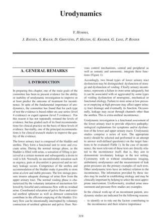
![- to obtain information about other aspects of the
lower urinary tract dysfunction
2. GENERAL
- to predict the consequences of the dysfunction for
the upper urinary tract
The patient should be informed of the procedures before the studies, preferably by written leaflets but in any
event by oral explanation. Any medication that may
affect the patientÕs consciousness or that has been prescribed for lower urinary tract dysfunction should be
avoided before the procedure, unless the test is specifically intended to study its effect or there is a clinical
reason for not stopping medication. The nature of any
such medication and the timing of its administration
(especially the last dose) should be noted. Medications
that affect lower urinary tract function but have been
prescribed for other reasons should be taken into
account when interpreting the findings.
CONDITIONS
AND
CIRCUM -
STANCES OF THE STUDY
- to predict the outcome, including undesirable side
effects, of a contemplated treatment.
- to confirm the effects of treatment or to understand
the mode of action of a particular type of treatment,
especially a new one
- to understand the reasons for failure of previous
treatments for incontinence
In short, urodynamic studies are indicated to objectively observe lower urinary tract function and dysfunction
with the idea of choosing an appropriate treatment for
the incontinence and its associated pathology. Basically, the urodynamic study should be performed and
reported in accordance with the standards of the Inter1
national Continence Society (ICS) [1], so as to optimize interpretation and facilitate comparison between different studies. This principle is applied hereafter;
however, the chapter is not intended to simply reproduce the ICS standardization report but rather to focus on
the clinical relevance of urodynamics to urinary incontinence. It includes recommendations for study procedures, interpretation of study results and the ability to
predict treatment. Electrophysiological studies are treated in more detail in chapter 4.
The subject should be awake and unanesthetized during
the study. In children, studies are sometimes performed
under mild sedation. However, this is not desirable and
can be avoided if the study is thoroughly explained to
them beforehand and if care is taken to distract and
calm them during the procedure (see section III.5.e).
The position of the patient during the examination
(supine, sitting, standing or ambulatory) needs to be
considered and should be specified in the report. In
general it may be better to perform bladder filling in the
sitting or standing position, or even to change the
patientÕs position, in order to facilitate demonstration of
the incontinence. If the position is changed, the pressure transducers (if external) must be repositioned at the
reference level (see section I.2.e). In some cases the
choice of position may be determined by the patientÕs
condition. For example, if incontinence is due to neurological disease, demonstration of leakage during the
examination is usually relatively easy, and it may be
simplest to examine the patient supine.
II. THE GENERAL ASPECTS OF
URODYNAMICS IN INCONTINENCE
ASSESSMENT
Urodynamic investigations must be carried out in a
safe, comfortable, and scientific manner, and should be
reproducible within the limits of physiological variability, if repeated. This section emphasizes points that are
pertinent to all urodynamic studies in the assessment of
2 3
incontinence [2, 3]. These points will be repeated in
other sections of this chapter where relevant to the discussion. Further details are available in textbooks [4-8].
4-8
3. THE INVESTIGATOR
The investigator plays a crucial role in the urodynamics. The tasks of the investigator include recognition
and minimization of artifacts (quality control), communication with the patient regarding sensation and intention, and direction of the whole examination. Quality
control requires careful observation of the data as it is
being collected. If data quality problems are identified
and corrected at this time, a valid examination may be
obtained. If not, the study may be uninterpretable. The
investigator should talk to patients in a polite and explicit way to facilitate good communication. This is essential so that the patient understands what the investigator
requires and the investigator knows how the patient
feels and whether the patient is consciously inhibiting
the leakage. Also he/she directs the investigation, for
1. INFORMATION PRIOR TO STUDY
Prior to the urodynamic investigation a medical history,
a physical examination and/or a voiding diary should be
taken. Such information is absolutely necessary to
select the appropriate studies and to anticipate what
events might take place during the urodynamic investigation.
321](https://image.slidesharecdn.com/urodin-131127082012-phpapp01/85/Urodinamia-5-320.jpg)
![example, by repeating a test if the result is unclear, or
introducing extra tests if needed to clarify the situation.
the detrusor pressure. The symbols for these pressures
are pves, pabd, pura and pdet, respectively.
Thus, these tasks require diligent scrutiny throughout
the progress of the study and understanding of the
results while the test is being carried out. Consequently
the person conducting the investigations must note and
record all relevant events as well as simultaneously
interpreting the findings. Simple inspection of traces
after the study is completed does not yield a satisfactory interpretation [9].
9
The measurement of pressure is the most important aim
of urodynamic tests; nevertheless it is prone to artifacts.
To monitor measurement validity, coughing at regular
intervals, e.g. every 60 seconds or every 50ml infused,
immediately before the examination, during the whole
storage phase and immediately after the examination, is
therefore essential. Coughing should consistently give
similar pressure changes in pves and pabd (Figure 2).
Formal qualifications for urodynamic investigators
have not been developed, but are being considered on a
national basis in the UK and in Germany. Provided the
person is experienced, the investigator conducting the
study may be a physician or a nurse, or a person with a
science, engineering or radiology background.
Currently, pressure is most frequently recorded by the
conversion of pressure changes to the electrical properties of a strain gauge transducer. When the strain gauge
is outside the body, the pressures that are generated
inside the body must be transferred to it. This is possible with fluid-filled catheters and external tubing.
Thus, the inserted catheter and connecting tube should
be short and flexible, and should not yield to pressure
change nor leak at any of connection points. All air
bubbles in the system should be meticulously removed.
4. CATHETERS AND TRANSDUCERS
Urethral catheters for bladder filling and for pressure
measurement should be as small as possible in diameter
so as not to interfere with observations of incontinence
(leakage) and voiding. However, with a small catheter it
may be difficult to drain the bladder when desired. A
catheter as small as 6 or 7 French gauge reduces the voiding flow rate in both men [10] and women [11]. Even a
10
11
5 French gauge catheter increases the voiding pressure in
males [12]. However, the obstructive effect of an 8 Fren12
13
ch gauge catheter is clinically acceptable in men [13],
while a 10 French catheter has a more significant effect
[14]. An 8 French gauge catheter tends to increase the
14
measured Valsalva leak point pressure [15]. Thus, for
15
adults, some authorities recommend a maximum catheter
size of 8 French gauge, although others permit 10 French gauge. If external pressure transducers are employed,
two small single-lumen catheters or a twin-lumen catheter should be used for bladder filling/drainage and intravesical pressure measurement, respectively. Such catheters can be left in place throughout the study so that it can
readily be repeated. Optionally, a single urethral catheter
with a third channel for simultaneous urethral pressure
measurement may be used. If catheter-mounted transducers are employed, the catheter size and the type of transducer (e.g., strain gauge or fibre-optic) are important for
interpretation and should be specified in the report. The
manufacturer of the catheter and the model number or
name should also be specified. Rectal catheters should be
similarly described and the name of the manufacturer
and the model should be specified as well.
Transducers to measure pressure can also be mounted
on a ÒmicrotipÓ or fiber-optic catheter that can be inserted into the body cavity. Problems related to the tubing
system are not important in this case. However, hydrostatic forces inside the abdomen influence the measurement in a variable way, because the pressure reference
level is not clearly defined (see Intravesical pressure,
16
below) [16]. Another undesirable property of cathetermounted transducers is that they respond not only to
pressures but also to forces exerted on them by solid
objects, for example, by contact with the bladder wall.
Consequently, intravesical pressures measured by
external transducers may differ from those measured
internally by 20 cm H2O or more [17].
17
Alternatively the pressure-measuring catheter can be
air-filled; the catheter is provided with a small air-filled
balloon to prevent entry of liquid from the bladder and
is connected to external transducers by an air-filled
connecting tube. As for catheter-mounted transducers,
the pressure reference level is not clearly defined. The
balloon must not be over-inflated (see Abdominal pressure, below).
In clinical urodynamic practice, absolute pressure
values sometimes seem less important than pressure
patterns. However, the reliability of the absolute value
plays an important role in the control of measurement
quality. In many clinical situations furthermore it is
essential to ensure that the measured pressures are correct. For instance, when comparisons with reference
values from the literature are used in clinical decisionmaking; or when cystometric values before and after
treatment are compared in outcome analysis based on
multicenter data; or when longitudinal observations on
5. PRESSURE MEASUREMENT
The principal pressures measured during urodynamic
studies are the intravesical pressure, the abdominal
pressure and the urethral pressure. The difference between the intravesical and abdominal pressures is called
322](https://image.slidesharecdn.com/urodin-131127082012-phpapp01/85/Urodinamia-6-320.jpg)
![cm H2O
Pves
50
Pabd
50
a
b
a
b
Pdet
50
0
0
100
200
300
Volume (ml)
Figure 2 : IIdeal cystometric traces with coughing at regular intervals of 50ml filling. Note coughs before starting (a) and after
ending the test (b).
b) Abdominal pressure
a single patient are compared. Specific examples include leak point pressure measurement and grading of
bladder outlet obstruction by pressure-flow analysis.
pabd represents the net effect of the forces exerted on
the bladder by surrounding organs. Measuring the pressure inside the rectum or the vagina [18, 19] approxi18 19
mates pabd.
a) Intravesical pressure
In the physical sense pves, which is the pressure in the
liquid contained within the bladder, is a true pressure.
This pressure is the height, above a given reference
level, to which the liquid would rise in an open catheter
puncturing the bladder. pves consists of 2 components:
a contribution from the internal forces in the bladder
wall (pdet), and a contribution from the organs surrounding the bladder (pabd):
If an external pressure transducer with water-filled
tubes and catheter is used to measure pabd, it should be
placed at the same level as the pves transducer and
zeroed in the same way, so that the same reference level
is employed. If a catheter-mounted transducer is used,
the reference level for pabdis at the position of the
transducer and is unlikely to be the same as for pves.
This in itself can be a source of artifact.
i.e., pves = pdet + pabd.
If a water-filled balloon is inserted in the rectum for
pressure measurement it is essential not to overinflate it
(to no more than 50% of nominal capacity), in order to
avoid an artificially elevated pabd. The balloon may be
punctured to prevent this possibility. Accurate measurement is not possible unless the vagina or the anal
sphincter forms a tight seal around the catheter. In this
regard intravaginal recording appears to be less reliable
[20] unless the catheter is high in the vaginal vault.
20
Whatever the means used to measure the abdominal
pressure, its accuracy should be monitored throughout
the study, by ensuring that transient pressure excursions
The standard reference level for all pressure recording
is defined as the upper border of the pubic symphysis.
If an external pressure transducer is used with waterfilled connecting tubes, it should be zeroed to atmospheric pressure and placed at this level during the procedure. If a catheter-mounted transducer or an air-filled
balloon catheter is used, the reference level for pves is
at the level of the transducer or the balloon. Thus its
relation to the pubic symphysis not known exactly. In
these cases the transducer or balloon should be zeroed
to the atmospheric pressure prior to insertion.
323](https://image.slidesharecdn.com/urodin-131127082012-phpapp01/85/Urodinamia-7-320.jpg)
![of the catheter sidehole or transducer within the urethra.
With some methods of measurement however the pressure reading does depend on the orientation (see section
II.2.a). This is also a sign that the urethral pressure is
impossible to measure correctly (by such methods) in
clinical practice. Choosing a very flexible catheter [22]
22
and a lateral orientation for the sidehole or cathetermounted transducer minimizes the systematic error.
The subtraction of pves from pura produces the urethral
closure pressure.
due to coughing are recorded equally in pves and pabd.
Even if these conditions are met, slow rectal contractions, or an elevated tone in the rectal wall, may occur
and lead to an artificially elevated and/or fluctuating
value for pabd.
c) Detrusor pressure
Rearranging the above equation shows that the detrusor
pressure is defined as
pdet = pves - pabd.
6. DETECTION OF LEAKAGE
Therefore the detrusor pressure can be calculated from
measurements of pves and pabd. It represents the effect
of the active and/or passive forces generated by the
detrusor muscle, separate from any external pressures
applied to the bladder wall. In other words it eliminates
the effects of coughing and straining and shows what
the detrusor itself is doing. It is difficult to distinguish
the effects of straining from detrusor contractions if
only pves is measured [21].
21
If recording is started with a nearly empty bladder, one
expects the forces in the bladder wall to be very small.
Consequently, provided the zeroing has been carried
out correctly, pves and pabd should be nearly equal and
pdet close to zero. With catheter-mounted transducers,
because the abdominal and intravesical pressures are
referenced differently and to unknown reference levels,
this may not be exactly correct [17]. The apparent ini17
tial value of pdet may be slightly greater than zero or
slightly negative.
Methods of detecting leakage from the bladder have not
been standardized despite their critical importance in
the evaluation of incontinent subjects. An electrical
method of detecting urine in the urethra by measuring
distal electric conductance has been shown to be a sensitive index of urine leakage [25]. Pads incorporating
25
wire grids or temperature-sensitive diodes have been
used to detect urinary leakage by the change of electric
resistance or temperature. If videourodynamics is available, fluoroscopy provides a way of detecting leakage
of X-ray contrast. A flowmeter placed below the patient
may record leakage. In most cases, however, demonstration of a leakage relies on naked-eye observation by
the investigator. If there is substantial loss, detection is
easy, but loss of a few drops may be overlooked. A dry
piece of cotton cloth, preferably dark-green in color, or
26
a simple paper towel applied to the orifice [26] may
help the investigator detect the urine loss.
d) Urethral pressure
7. EQUIPMENT FOR URODYNAMICS
The pressure in the urethra may be measured during
storage and/or during voiding, the former being the primary concern in this chapter. There are difficulties in
the definition and measurement of pura during storage,
because the urethra is collapsed during the storage
phase. It contains no fluid and so pura cannot represent
a true pressure in a physical sense.
Urodynamic instruments intended for pressure measurement should be equipped with at least 2 pressuremeasuring channels, for pves and pabd, and a means of
calculating and recording pdet. The pdet channel
should be capable of recording slightly negative as well
as positive values. A minimum sampling rate of 10 data
points per second is probably necessary, although
higher sampling rates have been recommended [27].
27
Depending on the complexity of the testing, urodynamic instruments may also have channels for infused
volume, urethral pressure, voiding flow rate, voided
volume, or EMG, and a means of displaying and recording these measurements together with simultaneous
images. Despite such sophistication some newer systems do not allow post-processing of ÒautomaticallyÓ
analyzed data, and use recording paper with a narrow
width and/or height, which can make the traces difficult
to read. To rule out artifacts the examiner should inspect the traces and compare them to the data that have
been automatically analyzed
Further consideration has shown that pura is the fluid
pressure that would hypothetically be required to force
open the collapsed urethra and so allow urine to flow
[22]. The method of measurement that conforms most
22
closely to this definition is the perfusion or BrownWickham method [23, 24]. In this method the liquid is
23 24
slowly infused into the urethra through the sidehole
port on the catheter. To accommodate the liquid, the
urethra has to be forced open very slightly. The fluid
pressure needed to do so is measured. The urethral pressure measured in this way varies from point to point
within the urethra. Thus, a graph of urethral pressure
against distance along the urethra can be drawn, the
urethral pressure profile (UPP). By definition, the urethral pressure should be independent of the orientation
The manufacturers provide data on accuracy of equipment but there is no external organization that monitors
324](https://image.slidesharecdn.com/urodin-131127082012-phpapp01/85/Urodinamia-8-320.jpg)
![the technical and clinical data quality. Studies of intrinsic
clinical and technical Ôrobustness,Õ determined by comparing different measuring techniques, are rare [28-30].
28 - 30
Consequently, it is not certain whether the data obtained
on urodynamic equipment from different manufacturers
are truly interchangeable [31]. It has also not been shown
31
whether the data from similar equipment used in different departments are interchangeable. The intrinsic technical quality of the urodynamic equipment on the market
is probably adequate; however, it should be remembered
that no objective quality control exists.
priate clinical decision. Different urodynamic findings
may be present with a given clinical presentation, and
the same urodynamic observations may be made in the
presence of different symptoms [32-37]. The results are
32 - 37
sometimes expressed in terms of values of selected
variables. In order to attain accurate interpretation at
the individual level, however, the whole chart should
always be taken into account.
a) Variability in urodynamic data
Lower urinary tract function has a certain physiological
variability. This variation and methodological inconsistency inevitably limit the reproducibility of urodynamic
investigation. For instance, uroflowmetry in symptomatic elderly men shows considerable variation in
maximum flow rate [38]. Inter- and intra- observer
38
variability in reading the maximum flow rate from a
given flow curve is typically 1 ml/s or more [39].
39
Repeated cystometries demonstrate a tendency for
40 capacity or volume to first contraction to increase [40,
41]. In a 3-way drug trial in women with Òdetrusor
41
instabilityÓ, 5/20 (25%) changed from unstable to
stable after 3 weeks on placebo [42]. When cystome42
trograms were repeated in girls (mean age 8 years),
10% changed from stable to unstable and 14% changed
from unstable to stable, from the second to the third
study [43]. The proportion showing detrusor overacti43
vity declined in successive filling cystometries, with
the results of the third study showing the strongest asso44
ciation with symptoms [44].
The users of equipment should carefully maintain the
machine in good condition, and the calibration of uroflow and pressure transducers should be checked periodically, for example, every month. If the filling volume
is derived from a weight transducer this too should be
checked regularly. If it is derived from counting the
revolutions of a peristaltic pump, it is probably necessary to recheck or recalibrate the pump for every test,
following the manufacturerÕs instructions.
8. DESCRIPTION
OF
URODYNAMIC
STUDY
CONDITIONS
The procedures for urodynamic studies are so variable
in their details that it is important to report the study
conditions, so as to allow others to judge the quality of
the investigations. Shown in Table 2 are the basic study
conditions that should be reported in scientific articles
dealing with urodynamic aspects of urinary incontinence.
b) Urodynamic classification of voiding dysfunction
9. INTERPRETATION OF STUDY RESULTS
When reporting or reviewing information about a urodynamic study, there is a certain minimum amount of
information about the storage and voiding phases that
should be described according to a well-defined terminology. The classification of lower urinary tract dysfunction shown in Table 3 is derived from the ICS, but
several others have been proposed [45]. In some cases
45
(e.g. pressure-flow studies in men) borderline (cut-off)
values have been established, but in others further
investigations will be needed to achieve this.
Urodynamic test results should be interpreted and integrated with other clinical findings to make an approTable 2 : Basic Study Conditions to be Documented in
Scientific Communications
¥ Investigator
¥ Circumstances during study
¥ Patient conditions
- sedation, medication, position, provocation
If the dysfunction observed during a urodynamic investigation is caused by an anatomical or neurological
abnormality, it is advisable to add Ôsecondary to...Õ followed by a description of the dysfunction. It is sometimes better to state that the dysfunction is Ôin combination with ...Õ, if the dysfunction is of an unexpected
type and/or if it is not clear whether it is attributable to
the presumed primary disease.
¥ Equipment
- type, calibration
¥ Pressure measurement
- reference level, transducer
¥ Catheter
- size, side-holes, type for microtip transducer
catheter, number of channels, manufacturer
¥ Fluid
- infusate, rate of infusion, temperature
¥ Method of leakage detection
325](https://image.slidesharecdn.com/urodin-131127082012-phpapp01/85/Urodinamia-9-320.jpg)
![Table 3 : Urodynamic Classification of Lower Urinary
Tract Dysfunction
STORAGE PHASE
VOIDING PHASE
¥ Detrusor activity
¥ Detrusor activity
- normal (stable)
- normal
- overactive
phasic
terminal
neurogenic
idiopathic
standard that other investigations can rely upon, because incontinence is a dysfunction of the lower urinary
tract and only urodynamics can describe the function or
dysfunction. For this reason an explicit and unambiguous urodynamic definition of urinary incontinence
and associated findings is needed.
- underactive
- acontractile
Stress incontinence denotes a symptom and a sign: the
patientÕs statement of involuntary loss of urine during
physical exertion and the observation of leakage from
the urethra synchronous with physical exertion (e.g.,
coughing), respectively. Urge incontinence is a symptom: an involuntary loss of urine associated with a
strong desire to void (urgency). Terms for the corresponding urodynamic observations are currently under
reconsideration. Urodynamic stress incontinence and
detrusor overactivity incontinence, respectively, are
used in this chapter. Urodynamic stress incontinence is
the urodynamic observation of involuntary leakage in
the absence of a detrusor contraction, with elevated
intravesical pressure. Detrusor overactivity incontinence is the urodynamic observation of urine loss caused
by an involuntary detrusor contraction. It may ultimately be necessary to further elaborate the terminology,
since different types of urge incontinence with different
etiologies exist [49-51]. Combinations of these types of
49 - 51
incontinence, mixed stress and urge incontinence, are
frequently encountered (Table 4).
¥ Bladder sensation
- normal
- increased (hypersensitive)
- reduced (hyposensitive)
- absent
¥ Bladder capacity*
¥ Compliance*
¥ Urethral function
¥ Urethral function
- normal
- normal
- incompetent
- abnormal
*) No classification terms are given (see text 2.1.3. and
2.1.4.)
Other types of incontinence are symptomatic descriptions. Various underlying urodynamic observations are
possible [52]. Previously, Òreflex incontinenceÓ was
52
defined as the loss of urine due to detrusor overactivity
and/or involuntary urethral relaxation in the absence of
the sensation of the desire to void. ÒOverflow incontinenceÓ was defined as any involuntary loss of urine
associated with over-distension of the bladder. These
terms are currently being reconsidered and will probably no longer be recommended.
c) Urodynamic definitions of the types of incontinence
The symptom of incontinence is usually the result of a
complex spectrum of anatomical and physiological
disorders of the lower urinary tract [46, 47]. Overactive
46 47
detrusor contractions vary in duration and amplitude
and may occur with or without concomitant urgency.
Bladder sensation may be aroused by involuntary
contraction of the detrusor or by other ill-defined factors. Urethral competence is maintained by urethral and
para-urethral factors that become deficient in stress
incontinence [48]. These deficiencies are reflected in a
48
low maximum urethral closure pressure (MUCP, section II.2.a), a low leak point pressure (LPP, section
II.3), a low pressure transmission ratio (PTR, section
II.2.b), or a reduced sphincter thickness, or in pronounced urethral hypermobility and bladder descent. There
is a continuous gradation of severity in these abnormalities, which is coupled with or confounded by related
functions and dysfunctions. Therefore for many urodynamic variables it is impossible to provide fixed cut-off
values on a clear scientific basis, so as to define any
specific pathologic feature.
Nocturnal enuresis means involuntary loss of urine
during sleep. It becomes clinically relevant only after
the age of, for example, 5-6 years, although it is not
uncommon for children to wet at night (with decreasing
prevalence) until puberty.
d) Association of symptoms and urodynamic finding
It has been argued that symptoms and urodynamic findings do not match. Jensen et al reviewed 29 articles
between 1975 and 1992 that addressed the clinical evaluation of urinary incontinence, and analysed the diagnostic performance of symptoms [53]. They found
53
that the sensitivity and specificity of symptoms suggestive of either of 3 final urodynamic findings (Urodynamic stress incontinence, detrusor overactivity incontinence, or both) were 0.48 to 0.91 and 0.51 to 0.66, res-
Nevertheless urodynamics must be taken as the gold
326](https://image.slidesharecdn.com/urodin-131127082012-phpapp01/85/Urodinamia-10-320.jpg)
![Table 4 : Terms related to urinary incontinence that either do not (A) or do (B) need urodynamic confirmation
A
Urinary incontinence as a symptom
the complaint of involuntary urine loss
Urinary incontinence as a sign
the objective demonstration of urine loss
Stress incontinence as a symptom
the complaint of involuntary loss of urine during coughing, sneezing, or
physical exertion
Stress incontinence as a sign
the observation of urine loss from the urethra synchronous with
coughing, sneezing, or physical exertion
Urge incontinence
the complaint of involuntary loss of urine associated with a sudden,
strong desire to void (urgency)
Mixed incontinence
the complaint of both stress and urge incontinence
B
Urodynamic stress incontinence
the involuntary leakage of urine during raised intravesical pressure secon
dary to increased abdominal pressure, in the absence of a detrusor
contraction
Detrusor overactivity incontinence
the involuntary leakage of urine during raised detrusor pressure resulting
from detrusor overactivity. In patients with sensation, urgency is
experienced before the leakage episode.
Urodynamic mixed incontinence
both urodynamic stress incontinence and detrusor overactivity
(± incontinence)
Detrusor overactivity
the involuntary detrusor contractions during the filling phase at any time
prior to "permission to void" being given. The contractions may be of any
size and may be spontaneous or provoked. If contraction is observed
without leakage, it may be only suggestive of detrusor overactivity
incontinence. The current terminology does not distinguish overactivity
accompanied by the sensation of urgency from sensation-free overactivity.
ve value for urodynamic stress incontinence of more
than 70% (Table 5). However, the positive predictive
value of overactive bladder syndrome (frequency,
urgency and/or urge incontinence) for detrusor overactivity was only 54% [64].
64
Thus, symptom-based diagnosis is misleading as a predictor of detrusor overactivity. It is felt, however, that
stress incontinence as the dominant symptom with
auxiliary evidence is specific to and predictive of urodynamic stress incontinence, especially in patients
without prior surgery [55, 57].
55 57
pectively, depending on the type of incontinence (Table
5). Analysis of selected or more homogenous populations did not significantly alter the validity. More recent
studies on this subject have given similar results [33,
33
54-59].
54 - 59 To improve prediction most of them utilised
non-urodynamic variables and a standard questionnaire
or frequency-volume chart to assess the symptoms.
Using voiding frequency and voided volume retrieved
from frequency-volume charts, one study gave a nomogram for the probability of detrusor overactivity [60].
60
However, the validity of the nomogram was not confirmed in a following study [61]. The mean voided volu61
me in frequency-volume recording was significantly
smaller in detrusor overactivity incontinence (151ml,
n=23) than in urodynamic stress incontinence (220ml,
n=73), but there was a substantial overlap [62]. The
62
symptom score for leakage associated with physical
activity was higher for stress incontinence but other
symptom scores addressing nocturia, frequency, urgency, urge incontinence or incomplete voiding did not dif63
fer between stress and urge incontinence [63]. In practice, positive predictive value and negative predictive
value are of more clinical significance. Stress incontinence as the dominant symptom has a positive predicti-
10. SUMMARY
¥ A urodynamic investigation is a functional assessment of the lower urinary tract, usually performed to
provide objective pathophysiological explanations
of symptoms and/or dysfunction
¥ Urodynamic investigations should be conducted
safely in a scientific and respectful manner.
¥ Urodynamic measurements are prone to artifacts.
They should be carefully identified and eliminated
during the study whenever possible. Accurate description of study conditions and methods is essential.
327](https://image.slidesharecdn.com/urodin-131127082012-phpapp01/85/Urodinamia-11-320.jpg)
![Table 5 : Value of patient history for predicting urodynamic findings
Author
year
Method Sample
size
Urodynamic stress
incontinence
Detrusor overactivity
incontinence
Mixed
incontinence
STV
SPT
PPV
STV
SPT
STV
SPT
0.91
0.51
0.75
0.74
0.55
0.48
0.66
0.68
0.48
Jensen
1994
Review
Handa*
1995
A
101
0.77
0.44
0.52
Handa*
1995
B
101
0.82
0.59
0.70
Haeusler
1995
C
1938
0.56
0.45
0.88
0.62
0.56
Cundiff
1997
D
535
0.44
0.87
0.87
0.71
0.41
Videla
1998
E
72
Diokno*
1999
F
76
James
1999
G
555
0.81
Lemack*
2000
H
174
0.92
0.82
0.83
1.0
1.0
Abbreviations: STV; sensitivity, SPT; specificity, PPV; positive predictive value
*Predictive value for type II stress incontinence
A: limited evaluation (no urodynamics required) by AHCPR criteria [128]
128
B: stress incontinence dominant, no prior surgery, positive stress test, hypermobility, residual < 50ml, age < 65, no prolapse
C: Gaudenz Incontinence questionnaire
D: stress incontinence dominant
E: stress incontinence dominant, positive stress test, residual < 50ml, maximum functional capacity > 400ml
F: stress incontinence dominant, no prior surgery, hypermobility, no grade 4 prolapse and residual < 200ml
G: stress incontinence without bladder filling symptoms H: stress incontinence dominant, no prior surgery
¥ Appropriate examinations should be selected so as to
achieve the best possible assessment of the patientÕs
condition.
B. URODYNAMIC STUDIES
¥ The investigator should be well versed in the procedures and interpretation of urodynamic studies, and
understand their clinical relevance in each patient.
I. CYSTOMETRY
¥ The limitations on the accuracy and the interchangeability of study results should be kept in mind
during interpretation.
The core test of a urodynamic investigation is cystometry. Cystometry is the continuous measurement of the
pressure/volume relationship of the bladder to assess
sensations, detrusor activity, bladder capacity and bladder compliance. In the context of this chapter, an important aim of cystometry is to reproduce the symptom of
incontinence. For this purpose, maneuvers intended to
provoke either urodynamic stress incontinence or
detrusor overactivity incontinence are important.
11. FUTURE STUDY AREAS
¥ Development of formal qualifications for investigators through certification of courses in the practice of
urodynamics.
¥ Comparative studies of different methods of detecting urine loss during a urodynamic study.
¥ Standardization of equipment, instrumentation, techniques, and documentation, and adoption of a standard file format to enable interchange of results
¥ Determination of physiological, technical and interpretational variability for urodynamic study results.
¥ Better definition of the pathophysiological conditions underlying the types of incontinence and the
corresponding urodynamic observations, and further
refinement of the recommended terminology to
reflect this.
1. TECHNICAL ASPECTS
The bladder is most commonly filled through a transurethral catheter. The bladder can be catheterized suprapubically or it can be filled solely via (forced) renal
excretion [65]. Bladder filling may be carried out with
65
or without preliminary drainage of residual urine; whether or not this is done should be stated. If catheters are
introduced using an anesthetic agent, the effect must be
taken into account in the interpretation. It is important
to keep in mind that any variations in technique may
affect study results.
328](https://image.slidesharecdn.com/urodin-131127082012-phpapp01/85/Urodinamia-12-320.jpg)
![Other provocative tests intend to demonstrate detrusor
overactivity or urge or stress incontinence, include coughing (Figure 3), change of position from supine or sitting to standing, filling in the standing position, running
water, handwashing, and waiting with a full bladder
(sometimes when sitting on a commode). In women
with incontinence, filling cystometry in the supine position without provocation demonstrates Òdetrusor instabilityÓ in only 38% of bladders shown ultimately to be
unstable. In a further 29%, Òdetrusor instabilityÓ is provoked by a change of posture, and in 33% it is provoked by coughing [81].
81
Historically, both liquid and gas have been used as the
filling medium. Gas (carbon dioxide) is usually infused
at a high rate (> 100 ml/min), allowing rapid and inexpensive performance of a study. However, it is unphysiologic and compressible, and easily provokes detrusor
66
overactivity (see below) [66]. Rapid filling may also
lead to erroneous diagnosis of reduced bladder compliance. It is not suitable for studying voiding, and leakage is very difficult to detect due to invisibility of the
gas. Gas cystometry is not reliable [67] and thus not
67
recommended.
The liquid filling medium may be physiologic saline,
water, or radiographic contrast. The physical properties
of the liquid, its acidity, the type of contrast medium
and the concentration of ions such as K+ and Ca++ may
affect detrusor overactivity [68-70]. The temperature of
68 - 70
the liquid is usually either room temperature or body
temperature. Traditionally, the filling rate is referred to
as ÔfastÕ (> 100 ml/min), ÔmediumÕ or ÔslowÕ (< 10
ml/min). Natural bladder filling is on average 1-2 ml
per minute, although diuresis at up to 15 ml/min is possible for short periods. Therefore, even ÔslowÕ urodynamic filling is already non-physiologic filling rates. For
children a rate above 10% of predicted or known bladder capacity per minute might be considered a ÔfastÔ
filling rate. ÔFastÕ filling is considered to be provocative of detrusor overactivity (see below) and any unphysiologically ÔfastÕ filling tends to produce lower bladder
capacity and lower compliance. Stepwise cystometry,
with ÔfastÕ intermittent volume increments, has been
used in research settings to determine the viscoelastic
properties of the detrusor [71-73]. Particularly if ÔslowÕ
71 - 73
filling is used, the volume of liquid in the bladder may
be considerably larger than the measured volume (i.e.,
the volume introduced) because of urine production
during the examination.
Handwashing is another potent provocation of Òdetrusor instabilityÓ [82]. In women with symptoms of urge
82
incontinence, sitting on a commode with a full bladder
for 1 minute was the most provocative maneuver for
Òdetrusor instabilityÓ, being about 27 times more provocative than remaining supine [83]; the second most
83
provocative maneuver was handwashing for 1 minute.
However, these two results were based on carbon dioxide cystometry, a non-recommended method. In children, ÒslowÓ bladder filling while distracting their
attention should help to evoke Òdetrusor instabilityÒ
[84]. Since over-provocation may reveal overactive
84
detrusor function of no clinical significance, as observed in symptom-free volunteers, the results of provocative testing must be judged in relation to symptoms.
Bladder sensation during cystometry is judged on the
basis of the volume in the bladder at patientÕs Ôfirst sensation of bladder fillingÕ, Ôfirst desire to voidÕ and
Ôstrong desire to voidÕ. Urgency is a compelling desire
to void. A strong desire to void or urgency Ñ depending on the patient and the investigator Ñ usually
defines the urodynamic bladder capacity (see section
II.1.c Description of study results below). Methods of
questioning the patient regarding these sensation parameters are only vaguely defined. Their reproducibility
is not well documented. However, one group found that
repeated bladder filling increased sensation intensity,
which was more consistently related to intravesical
pressure increase than to bladder volume [85]. Provi85
ding the patient with a push-button system to record
sensations appears a promising way of standardizing
the testing of sensation and increasing reproducibility
[86].
86
Some authors advise that ÔfastÕ filling rates should be
used if no detrusor contractions can be elicited in the
74
individual suffering from urge incontinence [74]. This
may conflict with the aim to reproduce the symptoms
experienced in daily life (see over-provocation, below).
Ice water testing can be used to demonstrate the existence of a temperature-sensitive reflex detrusor contraction mediated by afferent C-fibers, The reflex is interpreted as evidence for a neurogenic abnormality [75-75
79]. Instructing patients not to voluntarily inhibit the
79
urge to void, but merely to communicate sensations,
increases the efficacy for identifying detrusor contractions. In a prospective study of 42 patients referred for
irritative symptoms, a randomized double blind protocol asking patients to either inhibit or not inhibit micturition during cystometry showed a statistically significant increase in the presence of involuntary contractions when patients were instructed not to inhibit micturition [80].
80
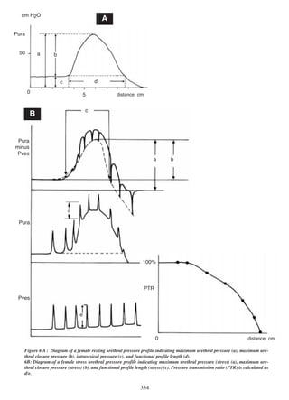
When no equipment is available or referral is not fea87
sible, Òsimple cystometryÓ (Figure 4) is an option [87,
88 For the detection of detrusor overactivity, taking
88].
multichannel cystometry as the standard, simple cystometry is reported to have specificity and sensitivity of
over 80% in elderly patients [89-91]. In both geriatric
89 - 91
and female populations, the rate of detection of detrusor overactivity was not substantially different in
90 91].
simple and multichannel investigations [90, 91
329](https://image.slidesharecdn.com/urodin-131127082012-phpapp01/85/Urodinamia-13-320.jpg)
![cm H2O
Stop filling
Pves
50
a
a
Pabd
50
Pdet
50
b
0
100
200
300
Volume (ml)
Figure 3 : Cough (a) provoked involuntary contraction (b) at 230ml.
2. SAFETY
of overactive detrusor function is described below. Cystometric capacity is determined differently in different
types of dysfunction. In the normal case it is the volume at which the patient states that he/she can no longer
delay micturition because of strong desire to void or
urgency. In urge incontinence it is the volume at which
involuntary voiding occurs. In the absence of sensation
cystometric capacity is the volume at which the investigator decides to terminate filling. Occasionally filling
has to be terminated because of patient discomfort. The
event that determines cessation of filling should be
reported. Compliance is defined as volume increment
per detrusor pressure increment (ml/cm H2O). Note
that all volumes should refer to the volume actually present in the bladder, not the volume introduced, and may
have to be estimated.
Screening of urine for bacteriuria at the time of the test
is important to rule out unrecognized infection. Antibiotics administered at or just after the study, are at the
discretion of the investigator.
The main risks of cystometry are those associated with
urethral catheterization. Dysuria (painful voiding)
occurs in some patients after urodynamic testing, but
usually disappears within 48 hours [92]. The technique
92
used for catheterization, and for handling of transducers
and connecting tubes, varies in different centers from
clean to sterile. It is not known whether these variations
have any effect on the infection rates. In any case,
appropriate aseptic techniques should be used.
3. DESCRIPTION OF STUDY RESULTS
4. INTERPRETATION OF ABNORMALITIES
The study results are expressed in terms of bladder
volume at various sensations or the amplitude and duration of detrusor pressure activity. Detrusor overactivity
is defined as an involuntary detrusor contraction during
the filling phase. It may be spontaneous or provoked
and of any magnitude and duration. Sub-classification
In the interpretation of detrusor activity during filling,
the amplitude and the duration of the contraction and
the intravesical volume at which the contraction occurs
should be taken into account. It should be remembered
that no method of monitoring attempted suppression or
330](https://image.slidesharecdn.com/urodin-131127082012-phpapp01/85/Urodinamia-14-320.jpg)
![inhibition has been standardized. If flow or leakage
occurs, the pressure attained does not fully represent
the strength of the contraction or its clinical significance.
Bladder compliance is influenced by infusion rates,
position of the patient, the volume of fluid in the bladder and the part of cystometrogram used for compliance calculation. There is insufficient data to precisely
define cut-off values between normal or abnormal compliance, but values in the range 12.5 to 30 ml/cm H2O
Observation of detrusor overactivity by itself is suggestive of underlying abnormality but is not conclusive,
because it is frequently observed in healthy volunteers
[65, 93], especially if observations are continued for
65 93
long periods, for example during ambulatory monitoring (see section II.8.c). Detrusor overactivity needs to
be interpreted in the light of symptoms and signs.
Observation of involuntary detrusor contraction that
leads to leakage (detrusor overactivity incontinence) is
more conclusive because it is clearly abnormal. However, it still requires interpretation in the light of the
patientÕs history.
have been suggested as the lower limit of normal [99].
99
Among healthy adults, compliance is higher in women
than in men [100]. Dynamic analysis of compliance,
100
taking into account the multiple phases of bladder
filling curve, has been suggested [101]. In children, the
101
compliance determined by urodynamic investigation is
an important outcome parameter [102-104]. If abnor102 - 104
mally low compliance is observed, cystometry at a
lower filling rate may lead to a different result.
Despite the differences of opinion mentioned above
about the interpretation of the cystometrogram, the
ÔarchetypalÕ cystometric patterns of a normal detrusor,
detrusor overactivity, and low compliance are straightforward and simple to understand. It is useful to judge
a cystometrogram according to these landmarks [105],
105
even though only approximate normal values are
known (Figures 4 and 5).
Traditionally, detrusor overactivity has been subdivided
into Òdetrusor hyperreflexiaÓ (overactivity with a relavant neurological condition) and Òdetrusor instabilityÒ
(overactivity with no definite cause) [65]. These terms
65
have been replaced by nerogenic detrusor overactivity
and idiopathic detrusor overactivity, respectively. A
more detailed classification of overactive bladder function, based mainly on observations of urodynamic patterns, has been proposed [94, 95]. In this classification
94 95
scheme phasic detrusor instability describes phasic
involuntary contractions of the detrusor during bladder
filling; it is found commonly in younger patients with
urge symptoms and no overt neurological disease.
Uninhibited overactive bladder describes the observation of a single involuntary detrusor contraction that
terminates bladder filling and causes leakage, often
accompanied by reduced sensation of bladder filling; it
is a common cause of urge incontinence among elderly
people and appears to be associated with cortical dysfunction [50, 51, 94, 95]. These conditions have been
50 51 94 95
adopted by the ICS as phasic detrusor overactivity and
terminal detrusor overactivity, respectively.
5. INDICATION
FOR CYSTOMETRY IN INCONTI-
NENT PATIENTS
Cystometry is the basic urodynamic evaluation for
incontinent patients. It may be indicated to evaluate
bladder function prior to therapeutic approaches, including medical and in particular surgical interventions.
Urodynamic assessment prior to surgery not only
allows an accurate diagnosis but also enables a discussion with the patient of any problems that might arise
Borderline (cut-off) values of volume or pressure for
the various sensation are at present undetermined.
However the sequence of sensations is fairly reproducible [96]. Similarly, exact reference values for normal
96
urodynamic capacity are not available, because they
depend on the technique of the investigator. The actual
capacity is the total volume of fluid a patient will hold
before voiding and is somewhat dependent on the rate
of infusion, but changes also with repeated filling [41].
41
As an approximate guide, a capacity of about 300-600
ml is normal in adults. For children 30 ml+30 ml x age
(in years) is an appropriate capacity [97]. As another
97
approximate guide, 60 ml + 60 ml x age (in years) for
children less than 2 years old and 180 ml+15 x age (in
years) for children over 2 years old have been recently
proposed [98].
98
Figures 4 : Schematic diagram of simple cystometry. The
height above the symphysis of the fluid meniscus in a syringe indicates intravesical pressure.
331](https://image.slidesharecdn.com/urodin-131127082012-phpapp01/85/Urodinamia-15-320.jpg)
![cm H2O
Pdet
50
a
Pdet
50
b
inhibition
voluntary micturition
inhibition
Pdet
50
c
0
100
Volume (ml)
200
Figure 5 : Diagrams to show low compliant bladder (a), overactive detrusor with phasic pressure waves that the patient can
suppress (b), and overactive detrusor with subsequent leakage that the patient cannot suppress (c).
after intervention because of other co-existing abnormalities.
From a health planning perspective, multichannel urodynamic assessment is more expensive than a cough
stress test plus simple cystometry, and it appears to
have a similar sensitivity for the diagnosis of urodynamic stress incontinence [106]. However, cost-effective106
ness, defined in terms of treatment outcome, was not
considered in this study.
¥ Filling rates should be chosen according to the aim
of the investigation
¥ The posture of the patient during the test is important; it is unlikely that incontinence will be consistently demonstrated in the supine position only
¥ To demonstrate incontinence, provocative maneuvers designed to elicit leakage should be performed
¥ The investigatorÕs awareness of the patientÕs sensations and the instructions given to the patient are critical parameters.
6. GOOD URODYNAMIC PRACTICE
¥ When filling cystometry is performed, intravesical
(pves) and abdominal pressures (pabd) should be
measured and detrusor pressure (pdet) should be calculated; all 3 pressures should be recorded
7. PREDICTIVE
VALUE OF FILLING CYSTOME-
TRY FOR TREATMENT OUTCOME
Investigations have repeatedly shown that the success
rate for surgery for stress incontinence is higher in
women without detrusor overactivity [107]. A recent
107
paper failed to show any difference in outcome in a
group receiving full urodynamics as opposed to simpler
methods [108]. Such investigations have usually inclu108
ded women both with and without symptoms of urgency and urge incontinence. An important question is therefore whether filling cystometry allows more precise
¥ Initial values of pves and padb should be plausible;
the initial value of pdet should be close to zero
¥ If catheter-mounted transducers are used, the initial
value of pdet may differ from zero by up to a few cm
H2O.
¥ pves and pabd should respond equally to coughs;
frequent cough checks are essential
332](https://image.slidesharecdn.com/urodin-131127082012-phpapp01/85/Urodinamia-16-320.jpg)
![selection of a group of stress-incontinent patients who
will respond particularly well to surgery in spite of
concurrent urge symptoms. Two papers suggest that
surgery successfully cured urge symptoms in 91% of
those with low-amplitude detrusor overactivity (peak
detrusor pressure < 15 cm H2O), but was less successful in those with no observable detrusor overactivity
(cure rate 39%) [109] or those with high-amplitude
109
110
overactivity (cure rate 28%) or low compliance [110].
These observations suggest an important place for
filling cystometry in women with stress incontinence
and urge symptoms.
II. URETHRAL PRESSURE
MEASUREMENT
Continence is dependent on the powers of urethral
resistance exceeding the forces of urinary expulsion
[112, 113]. In order to maintain continence the urethral
112 113
lumen should seal completely; this hermetic effect is
dependent upon the softness and compressibility of the
urethral wall [114]. Together, these properties determi114
ne the intraluminal urethral pressure. The urethral closure pressure represents the difference between the urethral pressure and the simultaneously recorded intravesical pressure, and conceptually, therefore, it represents
the ability of the urethra to prevent urine leakage.
Among men or patients with neuropathy detailed urodynamic examination is usually considered an essential
basis for rational management. Nevertheless few studies have been undertaken to evaluate the utility of cystometry. For men, preoperative filling cystometry was
unable to predict incontinence after radical prostatectomy [111] Treatment is usually conservative or medical
111
for the elderly.
Urethral pressure measurements may be taken from all
points along the urethra in steady-state conditions, and
are reproduced in the form of a profile, e.g. the resting
urethral pressure profile or the stress profile (provided
the stress is maintained at a constant level, see below)
(Figure 6 A, B). Alternatively the measurement may be
made at one or more points along the urethra over a period of time during which conditions may be changing;
the results may be presented in the form of a continuous
trace, e.g. continuous urethrocystometry, or as a profile, e.g. the micturitional pressure profile.
8. SUMMARY
¥ Filling cystometry is the basic test for examining the
aspects of bladder function concerned with the efficient storage of urine.
If the lumen of the urethra is filled with fluid, the intraluminal urethral pressure is a true fluid pressure that is
in equilibrium with the pressure exerted by the urethral
115
walls [115]. In practice, however, urethral pressures are
usually determined during the filling or storage phases
of the micturition cycle, when the urethra is empty and
collapsed; as a consequence difficulties arise in understanding, defining, and quantifying exactly what is being
measured.
¥ Good patient-observer communication throughout
the study is mandatory
¥ Analysis of the cystometrogram is based on pattern
recognition, and evidence-based quantification of
the cystometric observations is not yet possible.
¥ Artifacts produced by catheterization, infusion or
provocation should be taken into account in the
interpretation.
1. TECHNICAL ASPECTS
9. FUTURE STUDY AREAS
Urethral pressures may be measured by perfusion techniques, by catheter-mounted microtransducers, or by
catheter-mounted balloons connected to an external
transducer. All 3 methods have advantages and disadvantages. One problem is that, for any technique that
uses sideholes or a side-mounted transducer, the measured ÒpressureÓ is liable to show an artifactual dependence on the orientation of the sidehole(s) or transducer. This behavior depends on the stiffness of the catheter and may lead to gross artifacts. It can be minimized
by choosing a very flexible catheter or a catheter with
multiple sideholes or sensors.
¥ Quantification of observations during cystometric
investigation to achieve reliable, interchangeable
and clinically relevant information
¥ Assessment of the reproducibility of these observations in terms of clinical outcome measurements.
¥ Clinical significance of involuntary detrusor
contractions observed during bladder filling that do
not reproduce symptoms, e.g. because they are not
accompanied by sensation or leakage
¥ Improved methods of assessing bladder proprioception
a) Resting urethral pressure profile
In perfusion profilometry, catheters between 4 and 10
116
French gauge appear to give satisfactory results [116];
dimensions significantly greater than this may overestimate urethral pressure because of limited urethral dis-
¥ Confirmation of the ability of filling cystometry to
predict outcome of surgery among women with
stress incontinence and urge symptoms
333](https://image.slidesharecdn.com/urodin-131127082012-phpapp01/85/Urodinamia-17-320.jpg)
![timated. However, it is possible to overcome these problems.
24 117].
tensibility [24, 117 The number and location of side
holes should be specified; 2 opposed holes 5 cm from
the tip appear to be satisfactory, given the other limita24
tions of the technique[24]. Orientation dependence is
not usually important because the catheter is flexible,
and most systems use multiple sideholes.
Urethral pressure varies with bladder volume. In continent women urethral closure pressure tends to increase
with increasing volume, whereas in stress incontinent
women it tends to decrease with increasing volume[127, 128]. Urethral pressure also varies with posi127 128
tion. In continent women urethral closure pressure
usually increases on assuming the erect position, whereas in stress incontinent women there is either no change or a decrease in pressure on standing [127, 129,
127 129
130
130].
Perfusion is best achieved by syringe driver and not a
peristaltic pump. Rates of about 1-2 ml/min can give an
24
accurate measurement of urethral pressure [24],
although higher rates may have advantages.
Liquid perfusion systems have been shown to be
capable of recording a maximum rate of change of pressure of between 34 and 50 cm H2O/s, [24] depending
24
on the rate of perfusion, and the compliance of the perfusion system. The catheter may be withdrawn incrementally or continuously; the latter is preferred, and a
mechanical or electrical device should be used. The
24 118
optimal withdrawal speed is less than 7 mm/s [24, 118,
119
119]; typically, between 1 and 5 mm/s is chosen. Perfusion rate and withdrawal rate are important parameters which interact to determine the response time and
spatial resolution of the recording. They have to be
carefully chosen with this in mind.
Normal values are difficult to define. There is a dependence on age, which is discussed in section C.II.3
below.
In clinical practice the maximum value of the urethral
closure pressure (MUCP), determined from the urethral
pressure profile, has a standard deviation of approximately 5 cm H2O (95% confidence limits ± 10 cm H2O)
24 118
131
[24, 118] or ± 5% [131]. The microtransducer technique
has been shown to have greater repeatability and repro118 121 132
ducibility [118, 121, 132]: the standard deviation of
measurements made on a single occasion is approximately 3 cm H2O (4%) and for measurements on two separate occasions it is 3.5-5 cm H2O (depending on time
separation and menstrual status) [118, 132].
118 132
With microtransducer systems there is little smoothing
effect, and the high frequency response, estimated at
120
over 2000 Hz [120], is more than adequate to record
any physiological event in the lower urinary tract. The
method is however prone to several potential methodological artifacts.
b) Stress urethral pressure profiles
In the measurement of stress urethral pressure profiles
the ÔstressÕ may be provided by either repeated coughs
or Valsalva manoeuvres on the part of the patient. In
the latter case, asking the patient to blow into a manometer to maintain a constant level [133] can control the
133
degree of abdominal pressure rise. The amplitude of
abdominal pressure rise during coughing is more difficult to standardise. It is not clear whether the pressure
1
transmission ratio [1] (defined in the following subsection), whilst in itself inherently variable, is dependent
135
[134] or independent [135] of the extent of the rise in
134
abdominal pressure. It has been shown however that
the consistency of measurements is significantly greater during the Ôcough profileÕ than the Ôstrain profi136
leÕ[136]. The most possible flexible catheter is recommended for stress testing to prevent catheter movement
and to minimise orientation artifacts.
The profiles show a significant degree of directional
121 - 123
(orientation) dependence [121-123]. Resting urethral
pressure profiles recorded with the transducer face
oriented anteriorly (towards the pubic symphysis) show
significantly higher maximum urethral closure pressure
and shorter functional urethral length than in other
124
orientations [124]; pressure transmission ratios measured from stress profiles are significantly higher than in
other orientations. Most authors have suggested profile
recording with the transducer directed laterally within
the urethra. It should be emphasised however that this
orientation dependence is an artefact related to the presence of the catheter. Fibre-optic microtransducers have
been found to show less orientation dependence but
greater variability with time than conventional microtransducers [125]; they also record significantly lower
125
pressures [125, 126], and hence may make the diagno125 126
sis of intrinsic sphincter deficiency (see below) even
more problematic [126].
126
Stress profile variables show greater variability; withinsubjects standard deviations for stress MUCP and pressure transmission ratios have been reported to be 2025% and 15-20% respectively [136, 137].
136 137
Balloon systems avoid any orientational dependence,
but in the past it has been difficult to make the balloon
small enough (a) to provide good spatial resolution
along the axis of the urethra and (b) to avoid dilating
the urethra so much that the urethral pressure is overes-
2. DESCRIPTION OF STUDY RESULTS
Study conditions that must be defined when urethral
pressure is measured are patient position, bladder volu-
335](https://image.slidesharecdn.com/urodin-131127082012-phpapp01/85/Urodinamia-19-320.jpg)
![me, rate of infusion and rate of catheter withdrawal.
Descriptions especially relevant to urethral pressure
measurement include functional urethral length, maximum urethral pressure, and maximum urethral closure
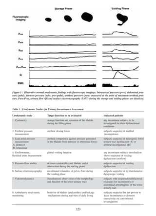
pressure. For a stress profile, the pressure transmission
ratio (PTR), defined as the ratio of increase in urethral
pressure to the increase in intravesical pressure (see
figure 6a), the maximum PTR, and the pressure transmission profile are important factors. Pressure transmission ratios can be recorded at any position along the
urethra; the position of the pressure sensor should be
stated, e.g. in terms of quartiles of the functional urethra length or in mm from the bladder neck. For continuous measurement of urethral pressure the intrinsic
variation in maximum urethral pressure should be
recorded.
119
mum urethral pressure[119]. Several authors have suggested that variations in excess of 30-33% of the
149 151 152
MUCP might be looked on as abnormal[149, 151, 152].
Although such variations have been reported to be associated with an increased likelihood of lower urinary
tract symptoms, their clinical significance remains in
150 153
some doubt[150, 153].
ÔUnstable urethraÕ is an involuntary fall in urethral pres154
sure in the absence of detrusor activity[154]. If sufficiently pronounced it may lead to zero maximum urethral closure pressure and allow leakage of urine. It is
thus distinguished from detrusor overactivity and from
incompetence of the urethral sphincter against increased
abdominal pressure. In the collated and revised ICS
report [1] it is suggested that Òterms such as Ôthe unstable
1
urethraÕ await further data and precise definitionÓ. The
authors of this chapter suggest that the term Ôunstable
urethraÕ be used only where a zero or negative closure
pressure results from involuntary urethral relaxation. The
term Ôurethral pressure variationÕ should be used for lesser degrees of pressure variation, where a positive urethral closure pressure is maintained. The former would
seem to be a well documented but uncommon cause of
incontinence [136, 155]; the latter is undoubtedly much
136 155
more common, but of doubtful significance.
3. INTERPRETATION OF ABNORMALITIES
a) Urethral incompetence
Studies have consistently shown that the resting maximum urethral closure pressure falls with increasing age
138
in both men [138] and women, and is lower in groups
of stress incontinent women than in continent women;
the severity of symptoms is inversely correlated with
urethral pressure[113, 121, 130, 139-141]. However,
113 121 130 139 - 141
significant overlap between the values found in continent and stress-incontinent women limits the discriminatory power of resting profile variables.
4. INDICATIONS IN INCONTINENT PATIENTS
Urethral pressure measurement has been advocated in
several conditions in the investigation and management
of incontinent patients, especially those suspected of
urethral dysfunction, although in few of these situations
has its role been unequivocally established.
Most agree that the variables of the stress urethral pressure profile are of greater diagnostic value than those of
the resting profile. Pressure transmission ratio [138,
138
142], maximum urethral closure pressure on stress, and
142
143 144
profile area on stress [143, 144] have each been found to
be the most reliable single variable by different authors,
although the sensitivity even with these remains poor.
Even on the basis of a discriminant analysis using a combined function of 30 resting and stress profile variables,
a correct classification was possible in only 78% of
patients, suggesting that the urethral pressure profile at
rest and on stress is not an accurate test for the diagnosis
of urodynamic stress incontinence [145].
145
5. GOOD URODYNAMIC PRACTICE
Good urodynamic practice must be based on an
understanding of the limitations of whatever method is
chosen, and the precautions required to overcome them.
If a microtransducer is used to record urethral pressure,
care should be taken to use the most flexible catheter
available. There should be a means of checking the
orientation when the catheter is in the patientÕs urethra.
b) Urethral pressure variations and urethral
instability
The orientational dependence (e.g., the differences between maximum urethral ÒpressuresÓ measured with the
transducer facing anteriorly, posteriorly and laterally)
should be measured in typical patients, so as to establish how carefully the catheter has to be oriented in routine practice.
During continuous urethrocystometry most patients
show fluctuations in maximum urethral closure pressure of variable degree. Numerous authors have suggested that variations in excess of 10[25], 15[93, 146, 147],
25
93 146 147
148
20[148], or 25cmH2O [149] might be looked on as
149
indicating abnormality. Such pressure variations are
however commonly seen in asymptomatic women
150
[150], and it has therefore been suggested that variation in urethral pressure might more appropriately be
looked on in relative terms, as a proportion of the maxi-
If a perfusion method is used, the rate of rise of pressure when the sidehole is completely blocked should be
determined, and reported. This is the maximum rate of
increase of urethral pressure (in cm H2O/s) that can be
measured, and it determines the response to rising pressures. A cough can result in a rate of increase of 250-
336](https://image.slidesharecdn.com/urodin-131127082012-phpapp01/85/Urodinamia-20-320.jpg)
![500 cm H2O/s. To measure a Òstress profileÓ the maximum measurable rate of pressure increase should be at
least as large as this (a criterion that may be difficult to
satisfy). Division of the maximum measurable rate of
pressure increase by the speed of catheter withdrawal
(in mm/s) yields the maximum urethral pressure gradient (in cm H2O/mm) that can be measured. In a normal urethra the urethral pressure gradient may be as
large as 5-10 cm H2O/mm. If the system cannot measure such a gradient, the urethral pressure profile is
truncated and the peak pressure is not recorded. The
remedy is to increase the rate of perfusion, decrease the
speed of withdrawal, or redesign the measuring system
to reduce its compliance.
156 - 162
(FUL) than those treated successfully [156-162]. In
patients treated by colposuspension, those with a preoperative resting MUCP less than 20 cmH2O were 3 to
4 times more likely to have an unsuccessful outcome
than those with higher values [163, 164]. Two recent
163 164
comparative studies however, one retrospective case
series[165], and one prospective randomised
165
study[166], have shown no difference in outcome bet166
ween colposuspension and sling in patients with low
pre-operative maximum urethral closure pressure.
Thus, whether a low maximum urethral closure pressure, suggesting intrinsic sphincter deficiency, is in fact
predictive of a poorer outcome of surgery awaits further
clarification.
With a perfusion method, frequently a single channel is
used both for perfusion and for measuring urethral pressure. In this case, an extra pressure, needed to drive the
perfusate through the channel, is added to the measured
pressure, which therefore exceeds the urethral pressure.
The extra pressure may be allowed for by first measuring the apparent bladder pressure with the perfusion
running, and then comparing urethral pressures to this
artificially elevated baseline value. This method works
reasonably well provided that the rate of perfusion
remains constant. To minimize the problem, the perfusion channel through the catheter may be made wider
(but this makes the catheter stiffer and may produce an
orientation artifact); or the rate of perfusion may be
decreased (but this will increase the response time and
reduce the maximum measurable pressure gradient).
Another solution is to use separate channels for perfusion and pressure measurement, which only come together at the sidehole; however, this may increase the
stiffness and the expense of the catheter.
7. SUMMARY
¥ The resting urethral pressure can be recorded by a
number of standard techniques. All involve some
degree of approximation.
¥ The results of both resting and stress urethral pressure measurements are highly influenced by methodological and biological factors.
¥ Because of the significant overlap of measured
variables between different groups of patients, the
diagnostic value of these variables is limited.
¥ Measurements of urethral pressure may have a role
in the identification of intrinsic sphincter deficiency
(see Section II.9).
8. FUTURE STUDY AREAS
¥ Development of new or improved methods of assessing urethral closure
¥ Developments in transducer design; especially the
incorporation of sensors into finer, more flexible
catheters
¥ Clearer definition of profile variables, especially
with respect to pressure variation and correlation
between these variables and other outcome measures.
¥ Relationship between urethral pressure variation /
urethral instability and detrusor overactivity.
If a balloon is used, it should be only slightly greater in
diameter than the catheter itself, and not more than 1-2
mm long. It must not be fully inflated, since inflation of
the balloon requires an extra pressure over and above
the urethral pressure: the balloon should appear half
empty. The optimum amount of fluid (gas or air) in the
balloon Ñ sufficient to make measurements, but not
enough to overinflate it Ñ has to be determined beforehand, and meticulously adhered to.
It is often recommended that intravesical pressure
should be recorded continuously throughout measurement of the urethral pressure profile. Unfortunately this
requires another measuring channel; the increased
catheter stiffness may cause an orientation artifact.
III. LEAK POINT PRESSURE
MEASUREMENT
The bladder pressure (pdet or pves) at which involuntary expulsion of urine from the urethral meatus is observed is the leak point pressure (LPP). The rise in bladder
pressure causing leakage may originate either from the
detrusor (caused e.g. by filling a low compliant bladder
or by detrusor contractions) or from an increase in
abdominal pressure. Thus there are two different LPPÕs
6. PREDICTIVE VALUE
Several studies have shown that patients undergoing
unsuccessful surgery for urodynamic stress incontinence by several different procedures have lower pre-operative resting MUCP and functional urethral length
337](https://image.slidesharecdn.com/urodin-131127082012-phpapp01/85/Urodinamia-21-320.jpg)
![Ñ the detrusor leak point pressure and the abdominal
leak point pressure Ñ and each is a direct measure of
the closure function of the entire bladder outlet under
different circumstances.
activity and low compliance during the test has to be
documented in a different way [46, 168], and that the
46 168
quality of the rectal or abdominal pressure measurement tends to be uncertain because it cannot be checked
by a cough test.
The detrusor leak point pressure (DLPP) is the value of
the detrusor pressure at which leakage occurs in the
absence of an abdominal pressure rise. Detrusor leak
point pressure measurement was introduced in myelodysplastic children as an indicator of the risk of upper
102
urinary tract deterioration [102]. In these patients and
others with neurogenic lower urinary tract dysfunction
15 102 167 168
the detrusor leak point pressure [15, 102, 167, 168]is
important [15, 102, 167, 169 - 171 [168, 172, 173]
15 102 167 169-171] 168 172 173
because a high value is correlated with a higher risk of
102 167, 170 171 168
upper urinary tract pathology [102, 167 170, 171] [168,
172 173
172, 173].
Besides the influence of the catheter, VLPP is also
15 183 - 186 18 168
dependent on bladder volume [15, 183-186] [18, 168,
188 190 201],
188, 190, 201 and may be influenced by pelvic muscle
contraction or urethral distortion during straining.
During the test the bladder should be adequately filled:
about half the cystometric capacity (200~300 ml in
adults) is suggested [183, 185, 188, 194, 202] [168,
183 185 188 194 202 168
190 191, 197
190, 191 197]. With proper precautions the VLPP is a
reasonably reproducible measurement [15, 168, 194,
15 168 194
197, 203]. The position of the patient at the time of
197 203
measurement should be noted.
In contrast, the intravesical pressure at leakage during
abdominal stress in the absence of a detrusor contraction is called the abdominal leak point pressure
(ALPP). The abdominal pressure increase during the
test is obtained voluntarily by coughing (CLPP) or Valsalva (VLPP) [174-178] [46, 179-182] [183-186] [18,
174 - 178 46 179 - 182 183 - 186 18
168 187 - 189 48 190 - 192
168, 187-189] [48, 190-192]. Although higher abdomi15 170 171,
nal pressures are reached on coughing [15, 170, 171
174 - 177 [18, 168 176 179, 191 193], the Valsalva
174-177] 18 168, 176, 179 191, 193
leak point pressure (VLPP) is better controlled and less
15 139 178 179 182 193 46
variable over time [15, 139, 178, 179, 182, 193] [46,
183 - 187 18 168 188-190, 192].
183-187] [18, 168, 188 - 190 192 CLPP then is used
for assessing leakage in patients who fail to leak on
VLPP. The abdominal leak point pressure is a diagnostic tool in patients with stress incontinence (Figure 7).
The contribution of the resting intravesical pressure to
the abdominal leak point pressure is controversial. The
increase in intravesical pressure from its resting level in
the empty bladder was proposed originally [178]; the
178
value of the intravesical pressure measured from its true
zero is sometimes used [102, 167, 174, 177, 193] [179,
102 167 174 177 193 179
184 186 187 198 200
184, 186, 187, 198, 200]; the increase in intravesical
pressure from its resting level immediately before straining or coughing has also been used [15, 139, 175, 176,
15 139 175 176
188, 193 194 (Figure 8). Particularly in the sitting and
188 193, 194]
standing positions, when the resting intravesical pressure may be high, there may be large differences (up to
about 50 cm H2O) between abdominal leak point pressures estimated in these different ways. CLPP is generally higher than VLPP in the same patient (Figure 8)
15 18, 168 189 191
[15, 18 168, 189, 191].
1. TECHNICAL ASPECTS
2. DESCRIPTION OF STUDY RESULTS
The leak point pressure is measured by marking on the
pressure recording the moment at which urine is expelled from the urethral meatus. This observation can be
48 102 167, 170 190
made by fluoroscopy [48, 102, 167 170, 190], direct
15 139 174-176, 193 180 181, 184visualisation [15, 139, 174 - 176 193] [180, 181 184 187 18 188, 189 191
187] [18, 188 189, 191] or electric conductance mea194 - 197
surements [194-197].
The leak point pressure depends on the calibre of the
15 168 194 198-200]
transurethral catheter [15, 168, 194, 198 - 200 and possibly on the method and rate of infusion [168, 198].
168 198
DLPP measurements must be made intravesically, but
for ALPP measurements rectal or vaginal pressure measurement appears feasible [15, 18 19 194]. However,
15 18, 19, 194
rectal or vaginal pressure measurements are inherently
less reliable than intravesical measurements, and one
group reported vaginal Valsalva leak point pressures to
be significantly lower than those measured concurrent15
ly in the bladder [15]. Most authors now accept that the
presence of a urethral catheter produces artificially high
abdominal leak point pressures, and thus favour vaginal
or, mostly, rectal abdominal pressure recording. This of
course has the consequence that the absence of detrusor
The following study conditions should be specified: the
position of patient, the type of leak point pressure
assessment, the bladder volume (and its relation to the
maximum bladder capacity), the location and type of
the pressure sensor, the presence and size of a urethral
catheter, and the method for detecting leakage. For the
abdominal leak point pressure the method of applying
abdominal stress and the baseline from which the pressure is measured (resting with empty bladder, resting
immediately before straining or coughing, or true zero
of intravesical pressure) should be specified. The
results are expressed as the lowest pressure at which
leakage is detected. Associated pathology, if present
(e.g. cystocele) should be included in the results. It
must be kept in mind that the quality of instructions
given to the patient may affect the results.
3. INTERPRETATION OF ABNORMALITIES
a) Detrusor leak point pressure
The primary aim of measuring the detrusor leak point
338](https://image.slidesharecdn.com/urodin-131127082012-phpapp01/85/Urodinamia-22-320.jpg)
![Leak point pressure
Detrusor leak point pressure
Abdominal leak point pressure
Valsalva leak point pressure
Cough leak point pressure
Figure 7 : Nomenclature for leak point pressures
cm H2O
cm H2O
Stop filling
P
P
VLPP
DLPP
a
b
Volume (ml)
Volume (ml)
Figure 8 : Measurement of detrusor leak point pressure (DLPP: left) and Valsalva leak point pressure (VLPP: right). The rise
in pressure from the base line (a) or from the zero level (b) represents VLPP depending on the investigators. Arrows indicate leakage.
pressure (DLPP) is to assess the risk to the upper urina168 182 183
ry tract [168, 182, 183]. The critical cut-off value between high and low leak point pressure is approximately 40 cm H2O. Patients with neurogenic bladder dys-
185, 188, 189, 202]. Low ALPP is likely to be associa185 188 189 202
ted with increased leakage on pad tests 191 197, 208
[191, 197 208,
209] and increased symptomatic severity of stress
209
46 48, 167 168, 184 202 210 211],
incontinence [46, 48 167, 168 184, 202, 210, 211 but
some reports contradict this association [59, 63 212
59 63, 212,
213]. There is a low concordance between low maxi213
15
mum urethral (closure) pressure and low ALPP [15,
167 176 178 180 181 193 48 168 183 185 197,
167, 176, 178, 180, 181, 193] [48, 168, 183, 185, 197
211, 214-216].
211 214 - 216 Thus, the gold standard method for the
diagnosis of intrinsic sphincter deficiency is controversial. The value of the ALPP is related to pelvic floor
48 208
muscle properties [48, 208]. A recent report has suggested that iatrogenic low ALPP might be caused by
simple hysterectomy [217].
217
function and a DLPP above this level are at risk for
102 168, 172, 173
upper urinary tract deterioration [102, 168 172 173,
183, 199] [177, 204 - 207
183 199 177 204-207].
b) Abdominal leak point pressure
The abdominal leak point pressure (ALPP) assesses the
abdominal pressure (or pressure increase) that causes
stress incontinence and thus offers a direct measure of
168 182 183
the urethral contribution to continence [168, 182, 183].
High values (greater than 90-100 cm H2O) suggest that
stress incontinence is associated with descent of the
168
bladder base descent or so called ÒhypermobilityÓ [168,
185, 188],
185 188 reflecting pelvic floor weakness. A co-existing cystocele may cause artifactually high leak point
pressures and should probably be repositioned during
the test [193]. Low values, less than about 60 cm H2O,
193
reflect more severe stress incontinence and suggest that
168 178 183,
there is intrinsic sphincter deficiency [168, 178, 183
4. INDICATION
FOR
LPP
MEASUREMENT IN
INCONTINENT PATIENTS
The detrusor leak point pressure should be assessed in
patients with overflow incontinence, particularly of
neurogenic origin. In those with neurogenic detrusor
overactivity detrusor pressure should be monitored
339](https://image.slidesharecdn.com/urodin-131127082012-phpapp01/85/Urodinamia-23-320.jpg)
![whether they are incontinent or not, particularly if
coexistent detrusor-sphincter dyssynergia allows high
detrusor pressures to be sustained. It is usually of no
interest in those with urge incontinence without serious
167
neuropathy[167] .
tions to the filling have subsided. The volume at which
the leakage occurs should also be compared to the functional bladder volume: if the leakage volume is much
higher than the regular bladder volume in everyday life,
then the measured leak point pressure has little patho177 225
logical impact [177, 225]. Typically the ALPP is measured at a bladder volume of 200 ml.
Abdominal leak point pressure measurements are used
frequently to evaluate urethral function in patients with
stress incontinence. A tentative diagnosis of intrinsic
urethral sphincter deficiency may be made from a
218
variety of tests [218], including X-ray classification and
168
low resting urethral pressure profile values [168], see
section II.9. At present however a low abdominal leak
point pressure (< 65 cm H2O) appears to be the most
139 167 178
widely accepted method of diagnosis [139, 167, 178,
180 181 193] [183-189] [203, 214, 218-223] [48, 168,
193 183 - 189 203 214 218 - 223 48 168
180, 181,
191 192 210 224 - 226
191, 192, 210, 224-226].
- Patient position
For detrusor leak point pressure measurement the position of the patient is not expected to have a significant
influence. This test is mostly performed in the supine
position.
5. GOOD URODYNAMIC PRACTICE
For abdominal leak point pressure measurement, the
patient position is of marked influence. The test should
be performed in the standing position, if possible for
the patient, but in any case in the position that will lead
to incontinence as judged from the patient history.
a) Transurethral catheter
- Marking of leakage
The catheter size must be considered in the assessment
of either type of leak point pressure, since a transurethral catheter might significantly increase the pressure
reading at leakage, and some patients may not leak at
168 169 191 194 197 - 200
all with such a catheter [168, 169, 191, 194, 197-200,
224].
224
Marking the exact moment of leakage is pivotal for this
assessment. As leakage is usually detected by visual or
X-ray observation, the fast pressure changes that occur
during coughing may easily lead to misinterpretation of
the actual value of the leak point pressure. Electrical
conductance recording appears to be the best method of
detecting the moment of leakage, but its complexity is
a drawback.
In the case of the detrusor leak point pressure, the effect
of the catheter may be increased by the presence of urethral obstruction (for example detrusor-sphincter dyssynergia, non-relaxing urethra, urethral stricture, or prostatic obstruction). If such a catheter artefact is suspected in DLPP measurements (for which intravesical
pressure measurement is essential), the ÒrealÓ DLPP
may be approximated by removing the urethral catheter
when the leakage occurs, and replacing it immediately
199
when this leakage stops [199]. The pressure value at
this last moment may be a better estimate for the DLPP.
An alternative would be to use suprapubic intravesical
pressure measurement.
- Patient instructions
The instructions to the patient for ALPP testing (for
example straining or coughing with increasing intensity) should be clear, concise, and unequivocal.
- ALPP: Absolute pressure value or increase from baseline?
Many investigators use the increase from the resting
pressure (measured either at the beginning of filling or
just before straining) as the value of the ALPP. This is
contrary to the idea that the ALPP represents the total
bladder outlet resistance. In a sitting or standing position, a resting intravesical or abdominal pressure of 20184
50 cm H2O is not uncommon [184], depending on body
228
mass [228]. The ICS recommended use of the absolute
intravesical pressure [227]. The consequence is that all
227
reports using the pressure increase should be re-evaluated by adding the resting pressure to the ALPP values
found (about 20 cm H2O for sitting and 40 cm H2O for
standing patients, if the actual values are unknown).
This of course raises the question of the reliability of
the ALPP cut-off values, as the actual data for these
values may be derived either from the absolute pressure or from the pressure increase. Fortunately, in clinical
practice this difference does not seem of great importance.
b) Bladder volume and filling rate
¥ DETRUSOR LEAK POINT PRESSURE
The bladder should usually be filled at a rate that will
not cause an artifactually rapid increase of detrusor
pressure, since this will reduce the bladder volume at
leakage. For patients with neurological pathology this
is of particular importance as they may experience
decreased compliance or detrusor contractions caused
by too fast filling even at filling rates as low as 10-20
227
ml/min [227].
¥ ABDOMINAL LEAK POINT PRESSURE
When the bladder is filled for ALPP measurements, the
test should be undertaken after possible detrusor reac-
340](https://image.slidesharecdn.com/urodin-131127082012-phpapp01/85/Urodinamia-24-320.jpg)
![6. PREDICTIVE VALUE
ding in a simple, non-invasive, and relatively inexpensive way. Residual volume is the volume in the bladder
immediately after voiding is completed.
a) Detrusor leak point pressure
A detrusor leak point pressure > 40 cm H2O is a defi29 30,
nite risk factor for upper tract deterioration [29, 30
168 172, 173 199 177 204-207]
168, 172 173, 199] [177, 204 - 207 especially if incon177
tinence occurs in everyday life [177]. Early intervention to reduce the pressure in such patients will protect
the upper urinary tract [30, 168, 172, 173 199] [177,
30 168 172 173, 199 177
204 - 207
204-207]
1. TECHNICAL ASPECTS
A closed room where the patient can void in privacy
and without interference is preferred whenever possible. Although one study in women was able to
demonstrate no difference between pre- and post-ins247
trumentation flow rates [247], uroflowmetry without
previous catheterization is desirable. It is important to
ensure that the micturition is representative and that an
adequate volume is voided. If possible, a voided volume of at least 150 ml, and preferably 200 ml (correspondingly less in children: 50 ml or more) should be
obtained. At least two uroflowmetries should be performed when obstruction is suspected, since a single flow
can be misleading [248]. Home uroflowmetry by means
248
of different portable devices has been accepted as a
more accurate technique because it facilitates multiple
measurements [249, 250].
249 250
b) Abdominal leak point pressure
When the diagnosis of intrinsic sphincter deficiency is
based on a low ALPP the therapy of choice is a sling
168 202].
procedure, or the injection of bulking agents [168, 202
The clinical outcome of these therapies for patients
46 216 223 229with low ALPP is not consistent [46, 216, 223, 229 231 232 - 236 219 220 237 - 239
231] [232-236] [219, 220, 237-239]. In addition, many
of these authors demonstrate a post-operative increase
in ALPP but not in urethral closure pressure.
7. SUMMARY
For residual urine measurement in-and-out transurethral catheterization or lower abdominal ultrasonography is usually performed. The investigation must insure that the residual urine is measured accurately and
completely, taking into account any urine retained due
to vesicoureteral reflux or bladder diverticula. It is also
important to make the measurement immediately after
voiding. If catheterization is used the catheter should be
withdrawn slowly to ensure complete emptying [251].
251
Transabdominal ultrasound is non-invasive but it
requires expensive equipment. Portable ultrasound
equipment is cheaper and provides a less accurate
assessment, but is sufficient in most cases.
¥ Detrusor leak point pressure measurements are indicated for patients with neuropathic conditions and
those who are unable to empty their bladder, because levels above 40 cm H2O place the individuals at
risk for upper urinary tract deterioration.
¥ Abdominal leak point pressure measurement is a
reasonably reliable method to quantify stress incontinence, and a low value is one way of identifying
intrinsic sphincter deficiency.
8. FUTURE STUDY AREAS
¥ Standardization and improvement of study methods
and conditions [240-246], including patient position,
240 - 246
transurethral catheter, bladder volume, and detection
of leakage.
¥ For detrusor leak point pressure: prospective studies
on the development of upper urinary tract changes
(not only in neurogenic cases) with elevated DLPP.
As it may be unethical to perform these studies on
patients, animal models should be used.
¥ For abdominal leak point pressure: Further standardization of methods and conditions, sharper definition of cut-off values, more evidence for significance of cut-off values for diagnosis and for prediction
of outcome.
2. DESCRIPTION OF STUDY RESULTS
The traces and relevant variables should be included in
the report of uroflowmetry. Uroflow traces should
always be examined to rule out the errors that can occur
with automatic reading [252]. The normal flow curve
252
usually has a smooth, near-Gaussian shape, and any
spikes of short duration (less than about 2 s) probably
represent artifacts, due either to sphincter contraction,
abdominal straining or movement of the urine stream in
the funnel.
The variables include voided volume, maximum flow
rate (Qmax), average flow rate (Qave), voiding time
and flow time, which are currently displayed by most
uroflowmeters. Qmax is the maximum measured value
of the flow rate, and Qave is voided volume divided by
voiding time. Flow time is the time over which measurable flow actually occurs.
Since there exists a non-linear relationship between
flow rates (Qmax and Qave) and the voided volume,
the voided volume and residual urine volume should be
IV. UROFLOWMETRY AND
RESIDUAL URINE MEASUREMENT
Uroflowmetry is a measurement of the rate of flow of
urine expelled via the urethra (the external urinary
stream) during voiding. It gives an assessment of voi-
341](https://image.slidesharecdn.com/urodin-131127082012-phpapp01/85/Urodinamia-25-320.jpg)
![stated along with the values of these flow rates. The
International Continence Society recommends presenting the results in a simple layout: voided volume/
Qmax/ residual volume [1]. Flow time and voiding time
1
can be of interest when interpreting some patterns
[253].
253
Incontinence can also be a presenting symptom in an
obstructed male, due either to overflow incontinence or
to detrusor instability secondary to obstruction.
A poor flow rate and elevated residual urine are more
commonly due to an inadequate detrusor contraction.
Repeated measurements of residual urine are needed to
confirm the results [261]. They may be associated with
261
a pronounced cystocele [262]. Residual urine may be
262
overestimated in patients with vesicoureteral reflux,
hydroureteral nephrosis, or bladder diverticula. In these
conditions, X-ray or ultrasound imaging of the bladder
at the completion of voiding will clarify the true postvoid residual volume.
3. INTERPRETATION OF ABNORMALITIES
Normally, the volume of residual urine should be very
small. In adults, values up to about 25 ml (measured
within a few minutes of voiding) are usually considered
within normal limits. Elevated residual urine (about
100 ml or more) warrants careful surveillance and/or
treatment, in relation to the other parameters of measurement and the clinical situation. In children, a residual
urine volume greater than about 10% of the maximum
cystometric bladder capacity may be considered elevated. In the frail elderly, residual urine volumes of about
100 ml are common and may be acceptable, although
they may affect choice of treatment for incontinence.
4. INDICATION
FOR
UROFLOWMETRY AND
RESIDUAL URINE DETERMINATION IN INCONTINENT PATIENTS
Residual urine should be determined in all patients.
Although the probability of an abnormal uroflowmetry
in an incontinent patient is low, several conditions that
alter the voiding phase can coexist with incontinence.
To exclude them, uroflowmetry is advisable for any
incontinent patient. Residual urine can be conveniently
determined immediately afterward.
When a normal Qmax and a normal voided volume
without residual urine are present, in either sex, infravesical obstruction or reduced detrusor contractility is
unlikely. However, there is a considerable overlap in
flow rates between obstructed and unobstructed
patients, in males as well as females [254, 255]. Thus
254 255
measurement of the flow rate alone has limited value
when determining if obstruction is present in a particular patient.
5. GOOD URODYNAMIC PRACTICE
¥ Flowmeter calibration should be checked regularly
by following the manufacturerÕs instructions or, at a
minimum, by pouring a known volume of water into
the flowmeter at a physiological rate and checking
the recorded volume.
Several different nomograms have been used to interpret the measured flow rates. SirokyÕs nomogram is
used for males and the Liverpool nomograms developed using a large number of subjects have scales for
254 256
males and females [254, 256]. A peak flow over the
90th percentile on the Liverpool nomogram may be
suspicious for detrusor instability, but this finding is not
uniformly accepted by all investigators. Flow acceleration (defined as the maximum flow divided by time to
maximum flow) has also been proposed as a sign of
detrusor instability [257]. However, many patients with
257
instability have uroflow values within the normal
range. Some initial work has been done on flow rates in
school children [258, 259], and there is some discre258 259
pancy from adult nomograms that awaits further evaluation.
¥ Patients should void with a Òcomfortably full bladderÓ.
Since this may be difficult to arrange, the concept of a
flow clinic, where patients may drink fluids and void a
number of times, may be considered.
¥ After voiding, patients should be asked whether the
void was representative of their usual pattern
¥ Flow traces and the computer output derived from
them should be carefully scrutinized for artifacts and
corrected if necessary.
6. PREDICTIVE VALUE
There is some evidence that poor bladder emptying, as
shown by an abnormally low flow rate [263] and/or an
263
108
elevated residual urine [108] may predict voiding difficulty after surgery for stress incontinence. These measurements may be useful for pre-operative counseling
of patients.
Low Qmax with or without residual urine is a clue to
obstruction and/or to reduced detrusor contractility,
260
although flow rate decreases with age [260]. Bladder
outlet obstruction in females is rare. However, up to
30% of females with obstruction will have some degree
of incontinence, although this will rarely be the only
symptom. A plateau-shaped flow curve with a low
Qmax is also suggestive of obstruction, but only pressure-flow studies can provide an accurate diagnosis.
7. SUMMARY
¥ Uroflowmetry in privacy with the residual urine mea-
342](https://image.slidesharecdn.com/urodin-131127082012-phpapp01/85/Urodinamia-26-320.jpg)
![surement should be included in the initial work-up of
any incontinent subject.
depends on pattern recognition, a large number of different variables have been defined to help describe
these patterns. Among the important variables are the
maximum flow rate Qmax and the detrusor pressure at
¥ Ultrasonography is a simple noninvasive way to
assess residual urine.
maximum flow (pdet.Qmax). The voided volume, the
volume of post-void residual urine, the presence or
absence of abdominal straining, and the flow pattern
also provide useful information.
¥ Uroflowmetry should be performed prior to any urethral instrumentation.
8. FUTURE STUDY AREAS
¥ Development of nomograms that are easy to use and
can be adjusted to a given population. (Nomograms
are also required for children of various age groups,
related to their sex and body size.)
3. INTERPRETATION OF ABNORMALITIES
¥ Definition of standardized flow patterns that may be
associated with certain conditions related to incontinence.
a simple way to classify the degree of obstruction in
males. The ICS obstruction nomogram is one example
264
[264]. A similar nomogram allows the detrusor contraction to be classified in 6 grades as very weak, weak-,
265
weak+, normal-, normal+, or strong [265]. Alternatively, similar classifications can be performed by simple
calculations based on the values of Qmax and
266
pdet.Qmax [266]. However, these nomograms or calculations have limitations. They were developed primarily for analysis of voiding in males. They do not take
into account any contribution from abdominal straining, nor do they indicate the etiology of any abnormality. The pressure-flow study is not a satisfactory way of
estimating the maximum flow rate or the residual urine
volume; free uroflowmetry is preferable.
As long as technical artifacts have been eliminated,
plotting Qmax and pdet.Qmax on a nomogram provides
V. PRESSURE-FLOW STUDIES OF
VOIDING
Pressure-flow studies are concerned with voiding and
therefore do not usually address incontinence directly.
In a given patient, however, a pressure-flow study may
shed light on the etiology of incontinence; or voiding
dysfunction may be combined with incontinence; or
voiding may be involuntary, so that voiding and incontinence are indistinguishable.
Urethral obstruction may be due to a structural abnormality or to overactivity of the urethral closure mechanism. Structural (anatomic) abnormalities include urethral stricture, posterior urethral valve, meatal stenosis,
prostatic enlargement or bladder neck contracture. Overactivity of the urethral closure mechanism may be a
voluntary act (e.g., an attempt to inhibit involuntary
voiding), or a physiological artifact of the examination,
or a learned behavior (dysfunctional voiding) or it may
be due to a neurological lesion. These possibilities can
be distinguished only with the help of auxiliary information. For example, in a patient with spinal cord injury, obstruction is likely to have a neurological basis and
to be due to overactivity of the striated urethral sphincter during voiding (detrusor-sphincter dyssynergia).
1. TECHNICAL ASPECTS
The standardization report of the International Continence Society should be studied for nomenclature but it
gives limited guidance about technical aspects of the
test [264]. Pressure-flow studies are subject to technical
264
as well as physiological artifacts caused by circumstances of the test. The following points should be
emphasized.
The urethral catheters for the study should be 8 French
gauge or smaller in size. To ensure accuracy, recording
a cough immediately before and after micturition is
mandatory, and more importantly, direct inspection of
the raw pressure and flow data is essential to enable
artifacts and unreliable data to be recognized and eliminated. If the flow pattern in the pressure-flow study is
not representative of free-flow studies in the same
patient, then the reliability of the interpretation may be
questionable.
The stream of voided liquid should flow directly into
the flowmeter (not via tubing) and the distance between
the external meatus and the flowmeter should be as
short as practicable.
¥ Obstruction in women
Recently, a number of authors have attempted to provide a urodynamic definition of obstruction applicable to
women. Such a definition would be useful both pretreatment (as a possible explanation of symptoms), and
post-surgery (to establish or rule out iatrogenic obstruction). Groutz et al [267] defined urethral obstruction as
267
a persistently low free flow rate of less than 12 ml/s
combined with a detrusor pressure at maximum flow
greater than 20 cm H2O during pressure-flow studies.
The prevalence was 6.5% among consecutive women
2. DESCRIPTION OF STUDY RESULTS
Since the interpretation of the pressure-flow study
343](https://image.slidesharecdn.com/urodin-131127082012-phpapp01/85/Urodinamia-27-320.jpg)
![falling in the green region of Figure 9 are unobstructed,
and that those falling in the amber or red regions are
indeed obstructed. In the pale yellow region there is
some disagreement, and this region therefore plays a
role similar to that of the equivocal region in the ICS
nomogram for male obstruction, outlined in blue in the
figure.
patients with various symptoms and etiologies. Lemack
268
& Zimmern [268] examined a population of women
with voiding symptoms but not necessarily with obstruction. They suggested that women with voiding
detrusor pressure of 21 cm H2O or more, together with
a flow rate 11 ml/s of lower, were obstructed. The pre269
valence was 20%. Kuo [269] defined bladder outlet
obstruction as a voiding detrusor pressure of 50 cm
H2O or greater together with a narrow urethra on voiding cystourethrography. 9.6% of their selected population of women with lower urinary tract symptoms were
diagnosed with bladder outlet obstruction. Salvatore et
270
al. [270] defined obstruction as a peak flow rate less
than 15 ml/s together with a maximum voiding detrusor
pressure greater than 60 cm H2O. The prevalence in a
large group of women with symptoms of urinary tract
271
dysfunction was 1.5%. Blaivas & Groutz [271] developed an obstruction nomogram based on statistical analysis of the maximum detrusor pressure (pdet.max)
during pressure-flow study of voiding, together with
the maximum flow rate Qmax in repeated free uroflow
studies. Patient with pdet.max greater than 57 cm H2O
were classified as either moderately or severely obstructed. Those with pdet below 57 cm H2O were classified as either mildly obstructed or unobstructed
depending on the value of free Qmax (Figure 9).
Among a group of 600 consecutive women, 6% were
mildly, 2% were moderately, and fewer than 1% were
severely obstructed.
There is evidence that in some types of urge incontinence voiding pressures are higher, perhaps even higher than
normal [273]. Women with stress incontinence, on the
273
contrary, tend to void at lower detrusor pressures than
normal subjects. Karram et al [274] showed that 30
274
continent women voided at a detrusor pressure of 20 ± 14
cm H2O (mean ± sd), while 70 women with urodynamic
stress incontinence voided at a detrusor pressure of 12 ±
11 cm H2O. Although the corresponding flow rates were
not specified, and although the authors interpreted the
detrusor pressure as a measure of contraction strength
rather than obstruction, these figures suggest that, as one
would expect, at least half of the continent women and
most of those with stress incontinence were unobstructed
according to Figure 9. Some of the continent women probably had pressure-flow studies in the equivocal region.
Methods of assessing detrusor contractility in women
have not yet been well defined.
4. INDICATIONS
FOR PRESSURE-FLOW STUDIES
IN INCONTINENT PATIENTS
If the free uroflow is normal with no significant residual urine there is usually no indication for a pressureflow study. If the free uroflow is abnormal and/or there
is elevated residual urine, a pressure-flow study may be
performed to distinguish between urethral obstruction
and detrusor underactivity (poor detrusor contractility),
or to help distinguish between neuropathy and a structural urethral obstruction as the cause of detrusor overactivity and related symptoms. If incontinence surgery
is being considered, a pressure-flow study may confirm
suspected detrusor underactivity and thus help to warn
patients who potentially may encounter post-operative
voiding difficulty. However, there is no evidence that
pressure-flow studies are more reliable predictors of
voiding difficulty than uroflowmetry and residual urine
determination [275].
275
These definitions use different pressures and different
flow rate measurements, but Figure 9 allows an
approximate comparison. It shows that there are 2 main
schools of thought: some define obstruction by detrusor
pressures above about 50-60 cm H2O; others take obstruction to begin at much lower pressures of about 20
cm H2O, provided that the flow rate is also low. The
Blaivas-Groutz nomogram suggests that the lower pressure cut-off may define mild obstruction, and the higher
cut-off may define moderate or severe obstruction. As
one would expect, the prevalence tends to be higher in
studies defining obstruction by lower cut-off pressures,
although of course it depends on the patient population
being studied.
272
Nitti et al [272] defined obstruction qualitatively as
radiographic evidence of narrowing in the presence of a
sustained detrusor contraction. For obstructed women
the mean values of pdet.qmax and Qmax were 43 cm
In a study of women with lower urinary tract symptoms, but without a primary complaint of simple stress
incontinence or overt neurological disease, pressureflow studies demonstrated significant abnormalities in
276
33% [276]. The abnormalities included obstruction
from various causes in 15%, dysfunctional voiding in
12%, and detrusor-sphincter dyssynergia as the initial
presentation of neurological disease in 4%. Thus pressure-flow studies can provide valuable information in
such cases.
H2O and 9 ml/s respectively, corresponding approximately to a point near the upper boundary of the mildly obstructed region. For unobstructed women their
values were 20 ml/s and 22 cm H2O, approximately on
the boundary of the unobstructed and mildly obstructed
regions in the figure.
To summarize, it appears generally agreed that studies
344](https://image.slidesharecdn.com/urodin-131127082012-phpapp01/85/Urodinamia-28-320.jpg)
![Figure 9 : Figure comparing various urodynamic definitions of obstruction in women. The ICS nomogram for the definition of obstruction (developed for males) is shown for comparison. Note that the pressure and flow rate variables plotted are
not identical for all definitions.
5. GOOD URODYNAMIC PRACTICE
a) In women
¥ Measurement of pressures requires the same standards
and precautions as for filling cystometry.
There is little evidence that pressure-flow studies can
predict success of surgery for stress incontinence. In
one retrospective review of 50 patients who underwent
a rectus fascia suburethral sling procedure, there was a
markedly higher risk of objective failure among those
who voided pre-operatively with the Valsalva maneuver
(54% versus 17%) [277]. There is tentative evidence
277
(requiring confirmation) that women who fail surgery
for stress incontinence have lower opening and closing
detrusor pressures in pre-operative pressure-flow stu278
dies than those in whom the surgery is successful [278].
The role of pre-operative voiding pressure in predicting
outcome of surgery requires further clarification.
¥ Secure taping of catheters is even more important
during pressure-flow studies, because the flow tends
to expel the urethral catheter.
¥ Catheter size is especially important because too large
a catheter can obstruct the urethra. 8 French gauge is
the recommended maximum.
6. PREDICTIVE VALUE
The evidence that pressure-flow studies can improve
the outcome of incontinence treatment is very limited.
Their main function is usually to help recognize or delineate possible neurogenic dysfunction, which would
make it unlikely that standard incontinence treatment
would be successful, and might rule it out entirely.
There is no evidence that pressure-flow studies (as opposed to uroflowmetry) can help to identify subjects with a
higher risk of post-operative retention [263, 275, 279].
263 275 279
The evidence that urodynamics can improve outcome
345](https://image.slidesharecdn.com/urodin-131127082012-phpapp01/85/Urodinamia-29-320.jpg)
![therefore is less subject to fluctuations caused by the normal physiological cycling of motor units. Surface EMG
recording of this type, as an indicator of appropriate pelvic floor function or of absence of abdominal straining
during voiding, has found a place in rehabilitation or biofeedback training [290-297] (e.g. flow-EMG recor290 - 297
dings).
for women with possible iatrogenic urethral obstruction
after incontinence surgery is unclear. In a retrospective
analysis of 51 women who underwent urethrolysis for
voiding dysfunction post cystourethropexy, urodynamic variables were not able to predict which of them
would benefit [280].
280
b) In men:
There is limited evidence that frail elderly men with
urge incontinence do better after TURP (in particular,
that their incontinence improves) if pre-operative pressure-flow study demonstrates clear obstruction [281]
281
1. TECHNICAL ASPECTS
The International Continence Society has published
technical advice on the use of urodynamic equipment,
with suggestions for EMG recording [298]. These sug298
gestions describe methods for recording EMG activity
and define the amplifier and signal-processing characteristics.
7. SUMMARY
A free uroflowmetry and a determination of post-void
residual urine should always precede pressure-flow studies.
Pressure-flow studies are not required in the diagnosis
of all types of urinary incontinence.
They may provide an answer to a specific question in
situations where neuropathy and/or urethral obstruction
are suspected.
Patch or surface electrodes are often placed so as to
record EMG activity in the anal sphincter because of its
relatively easy access. However, this activity is not
always correlated with the activity in the striated urethral sphincter [289, 299-304]. This is particularly true
289 299 - 304
in patients with neurogenic lower urinary tract dysfunction [301, 302], and in patients who strain to void but
301 302
relax the urethral sphincter simultaneously. In female
patients, vaginal surface electromyography may offer a
better approximation for the activity of the striated urethral sphincter [289].
289
8. FUTURE STUDY AREAS
Further refinement of nomograms for pressure-flow
studies in both normal and incontinent women.
Further study of the predictive value of pressure-flow
studies in incontinence treatment.
Surface EMG recordings are highly subject to artifacts
that cannot be recognised from the EMG trace provided
by standard urodynamic equipment. Therefore, the
EMG signal should always be monitored throughout
the test via an audio or video (oscilloscope) output, to
check that it has physiologically plausible characteristics. Patch electrodes in particular are subject to electrical artifacts if liquid runs over them. If this happens
during voiding, it can misleadingly suggest detrusorsphincter dyssynergia.
VI. SURFACE
ELECTROMYOGRAPHY
Electromyography (EMG) of the urethral sphincter, the
anal sphincter, or the pelvic floor is an established
method for the diagnosis of lower urinary tract dysfunc282 - 284
tion [282-284]. Two types of information can be obtained
from the EMG: (a) a simple indication of muscle behaviour (the kinesiological EMG); (b) an electrical correlate of muscle pathology. If one is looking for specific neuropathological information, a neurophysiological
approach, based on an EMG of type (b), as described in
chapter 4, is necessary. During urodynamic investigation
an EMG of type (a) is usually obtained. The interpretation is mainly restricted to recording of progressively
increasing EMG activity during filling of the bladder
(guarding reflex), and of timely relaxation of the pelvic
floor during voiding [285, 286]. While an EMG of type
285 286
(b) requires needle or wire electrodes, a kinesiological
EMG of type (a) can be obtained by needle electrodes or
surface (patch) electrodes [286-289]. Although needle
286 - 289
electrodes are somewhat less subject to artifacts, surface
electrodes are superior for this purpose, because the
signal is obtained from a larger volume of muscle and
2. DESCRIPTION OF STUDY RESULTS
The surface EMG is usually obtained simultaneously
with cystometry. Additional descriptive material that
should be provided includes the location or type of electrode, the characteristics of the recorder, and the method
of monitoring for artifacts. The amplitude of the EMG
signal is measured in mV (microvolts), and the scale
should be indicated on the tracing. However, the amplitude may be difficult to interpret because it depends on
the distance between the electrodes and the muscle.
3. INTERPRETATION OF ABNORMALITIES
If reduced EMG activity, or none, is found in the
sphincter during bladder filling, this may indicate an
incompetent urethral closure mechanism [47, 294].
47 294
Normally, the EMG should become almost silent
346](https://image.slidesharecdn.com/urodin-131127082012-phpapp01/85/Urodinamia-30-320.jpg)
![(sphincter relaxation) during voluntary voiding. If this
is not the case, urethral function may be overactive.
Waxing and waning of the sphincter EMG during voiding suggest a functional obstruction (detrusor-sphincter dyssynergia in neurological conditions, dysfunctional voiding in non-neuropathic patients). An increase of
EMG during voiding may indicate that there is abdominal straining.
4. INDICATION
FOR
EMG
¥ Therapeutic use of surface EMG may be advised in
rehabilitation or biofeedback procedures to improve
pelvic floor function.
8. FUTURE STUDY AREA
Definition of the most appropriate role for surface electromyography (see also chapter 4).
MEASUREMENT IN
VII. VIDEOURODYNAMICS
INCONTINENT PATIENTS
When urodynamic studies are performed in an office
setting, surface EMG recording can be used to test the
ability of the patient to control the pelvic floor area
voluntarily and to give a gross indication of possible
lack of co-ordination between the detrusor and the pelvic floor (during voiding). As a diagnostic tool, EMG in
a normal urodynamic setting is probably most useful
for detecting detrusor-sphincter dyssynergia [282-284,
282 - 284
286 287 290 305-309],
286, 287] [290, 305 - 309 although this can also be
inferred from fluoroscopic observations [305].
305
Surface EMG technique is also valuable in judging the
effect of pelvic floor training therapy and can be used
for biofeedback treatment.
Videourodynamics traditionally combines a routine
urodynamic study with X-ray or ultrasound imaging
[306, 310-314], although new imaging techniques such
306 310 - 314
as MRI and nuclear cystometrography are coming into
use. Videourodynamics is used for patients with complicated lower urinary tract dysfunction, for example
lower urinary tract dysfunction due to a neurological
311 313 - 320
condition [311, 313-320]. It also can offer a more accurate diagnosis in other patients [306, 312 314, 321306 312, 314 321
323 324 - 329
323] [324-329]. Overall, the invasiveness of the study
may adversely affect the functional results obtained by
this method of testing; therefore, it should not be considered a first line evaluation.
Most of the advantages of videourodynamics stem from
simultaneous measurement of pressure and visualization
of the anatomy. Incompetent bladder neck or inadequate
urethral closure during filling, or the location of urethral
obstruction during voiding, can be documented directly.
Descent of the bladder base, bladder base hypermobility
and intrinsic sphincter deficiency can be readily distinguished. Incontinence (leakage of contrast medium) can
be demonstrated fluoroscopically.
Dyssynergia between detrusor and external sphincter or
bladder neck can be documented. Moreover, videourodynamics has the advantage that any sign of reflux
during the filling or voiding phases can be detected
immediately, and that bladder diverticula or other anatomical malformations can be documented. The disadvantages of videourodynamics are radiation exposure, high
initial and running costs and less comfortable conditions
for the patient.
Ultrasound videourodynamics, although it has the advantage of no exposure to radiation, requires direct contact
of the ultrasound transducer with the patientÕs body in
the area of the lower urinary tract. Large probes placed
intravaginally or intrarectally may change the anatomical
conditions in this area. Other disadvantages include
inability to observe the upper urinary tract synchronously or to visualise the whole urethra, including the region
of the striated urethral sphincter, especially in men.
5. GOOD URODYNAMIC PRACTICE
For surface electromyography, the two signal electrodes should be placed as close as possible to the
muscle involved, usually the anal sphincter.
A third (ground) electrode, attached if possible over a
bony prominence, should be used.
Good mechanical adherence, resulting in good electrical contact with the skin, is vital. Preliminary skin preparation may be necessary.
It is advisable to test different types of electrode until
one is found that consistently provides good electrical
contact.
It is inadequate to rely only on the EMG trace provided
by standard urodynamic equipment. The EMG signal
should also be monitored throughout the test via an
audio or video (oscilloscope) output.
6. PREDICTIVE VALUE
There is no convincing evidence that surface EMG
measurement improves the outcome of incontinence
treatment.
7. SUMMARY
¥ Surface electromyography has limited value in the
routine urodynamic diagnostic work-up.
¥ A more accurate neurophysiological approach to
measurement and interpretation of the sphincter
EMG may be warranted in neuropathic conditions
(see chapter 4).
1. TECHNICAL ASPECTS
Videourodynamic investigation takes place under fluoroscopy if X-rays are used (Figure 10). It should always
347](https://image.slidesharecdn.com/urodin-131127082012-phpapp01/85/Urodinamia-31-320.jpg)
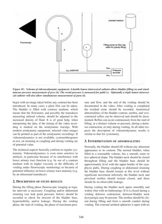
![intermittent or sustained narrowing during voiding
without proximal ballooning. If this is not the case,
detrusor-sphincter dyssynergia is possible.
4. INDICATION
sure is not recorded during AUM, the diagnostic sensitivity for stress incontinence may be low [336] and the
336
diagnosis of an unstable urethra or that of low urethral
closure pressure [335] is less feasible. Recording of
335
flow during micturition enables pressure to be related to
377
flow rate [337].
FOR VIDEOURODYNAMICS IN
INCONTINENT PATIENTS.
In patients with neurogenic lower urinary tract dysfunction and incontinence, videourodynamics may be
useful to define the cause of the incontinence. For
incontinent patients with other pathology, videourodynamics is considered when the history and simpler urodynamic tests do not lead to a definite diagnosis or after
failure of initial therapy based on less complicated
methods of diagnosis.
1. TECHNICAL ASPECTS
To record bladder pressure a catheter may be introduced
into the bladder transurethrally or suprapubically. Catheter-mounted microtransducers or fluid-filled catheters
are commercially available. It is recommended that the
urethral catheter should be 6 French gauge or less, and as
soft and flexible as cooked spaghetti to avoid pressure
artifacts due to bending. The catheter should be inserted
only a short distance into the bladder to avoid any contact
between the transducer and the bladder wall that might
cause pressure artifacts. Vaginal catheters used for recording abdominal pressure should be soft and the transducer should remain in the middle of the vagina without
coming in contact with the cervix. Rectal catheters are
usually wrapped in a balloon. Stiff catheters in contact
with the cervix and over-inflated rectal catheter balloons
may cause pressure artifacts as well.
If urethral pressure is recorded, the transducer should
be maintained at the maximum urethral pressure point.
This may be achieved by a single stitch to the female
urethral opening [335] or taping of the catheter to the
335
vulva, penis or thigh; However, movement artifacts and
catheter displacement as well as damage to the catheter,
remain a problem [333]. Conduction rings on the ure333
thral catheter outside the maximum urethral pressure
point can act as a simple, cheap and sensitive leak
335
338 339
detector [335]. The Urilos electronic nappy [338, 339]
340 341] are other
or a temperature-sensitive device [340, 341
methods of detecting leakage.
Recording times from 45 minutes up to 24 hours have
been described [335, 342]. Starting with a partially
335 342
filled bladder may reduce the overall recording time
343
[343]. Transducers should be zeroed to atmospheric
pressure and calibrated at a specified positive pressure
to ensure valid recordings. Empirically, a sample frequency of around 10 Hz gives clinically adequate pressure recordings of a cough.
5. GOOD URODYNAMIC PRACTICE
Videourodynamic investigation requires the same standards and precautions as filling cystometry and pressure-flow studies. Standard radiological precautions and
procedures should be followed to optimize image quality and minimize the exposure of patient and staff to
radiation.
6. PREDICTIVE VALUE
There is some evidence for clinical utility in myelodysplasia [330, 331
330 331].
7. SUMMARY
¥ Videourodynamics with fluoroscopy is indicated
when the diagnosis remains unclear after simpler
tests are performed orwhen complicated pathology is
expected from the history and symptoms.
¥ It has certain disadvantages; the patient is exposed to
extra radiation and the technique may influence the
lower urinary tract function and/or anatomy.
8. FUTURE STUDY AREA
Refinements in ultrasound imaging, and other techniques such as MRI and nuclear cystometrography, to
enhance their application in videourodynamics.
VIII. AMBULATORY URODYNAMIC
MONITORING
2. DESCRIPTION OF STUDY RESULTS
A portable solid state memory unit should record 2
pressures (pves and pabd) and optionally the urethral
pressure, together with flow and leak signals. A window
should display all recorded pressures to permit monitoring of the position of the transducer. Push buttons on
the solid state memory unit permit event markings
when the patient perceives urgency, leakage and micturition. Drinking and voiding should also be recorded
344
simultaneously in a diary [344].
Ambulatory urodynamic monitoring (AUM) is the
monitoring of leakage, flow recordings and pressure in
the bladder and abdomen (rectum or vagina) with or
without pressure in the urethra in an ambulatory setting
332 - 335
[332-335]. It permits monitoring in situations where the
patient usually leaks, unattended by medical staff and
laboratory equipment. Detection of leakage during the
recording is necessary and helps to separate clinically
relevant events from artifacts [335]. If the urethral pres335
349](https://image.slidesharecdn.com/urodin-131127082012-phpapp01/85/Urodinamia-33-320.jpg)
![urethral transducer out of the maximum urethral pressure zone. When no leakage is seen simultaneously
with a zero closure pressure, displacement of the transducer must be presumed. A decrease in the vaginal
pressure recording will cause a corresponding increase
in detrusor pressure even though the bladder pressure
remains stable. This may be wrongly interpreted as a
detrusor contraction.
Signals from the portable solid state memory unit are
downloaded to a computer after the recording is complete and urethral closure pressure and detrusor pressure are then calculated. The time scale and signal ranges
may be changed while the tracings are evaluated on the
display or print-out.
3. INTERPRETATION OF ABNORMALITIES
Conventional cystometry and AUM often give different
study results. In asymptomatic volunteers [345-347] as
345 - 347
well as in symptomatic patients [339, 348 - 354 an
339 348-354],
increase in the number of detrusor contractions on
AUM has been found, compared to conventional cystometry [355, 356]. With AUM, voiding volumes have
355 356
been found to be lower [346, 347 349, 353, 354], voi346 347, 349 353 354
ding pressures significantly greater [345, 346 354
345 346, 354,
357]
358
357 and the micturition flow rate higher [358], than
with conventional urodynamics.
4. INDICATIONS
FOR AMBULATORY MONITO-
RING IN INCONTINENT PATIENTS
350
Neurogenic lower urinary tract dysfunction [350], enu359 in older children, evaluation of the effect
resis [359]
360
pharmacotherapy [360] and electrotherapy [361], or
361
failure of previous treatment are other possible indications for AUM.
5. GOOD URODYNAMIC PRACTICE
During the filling phase, detrusor contractions without
subsequent leakage have been found in 38 to 69% of
normal volunteers undergoing AUM [345-347]. The
345 - 347
significance of detrusor contractions not associated
with leakage in asymptomatic patients is unknown. It is
probably more valuable to concentrate on the detrusor
contractions associated with clinical symptoms. The
increased number of contractions observed during
AUM may be caused by catheter irritation of the bladder wall. Microtip transducer catheters introduced deeply into the bladder cavity may record artifacts due to
contact between the transducer and the bladder wall.
Signals should be recorded as recommended in the ICS
recommendations on ambulatory monitoring [362].
362
There are a number of cautions that are especially
important in AUM. Stringent checks on signal quality
should be incorporated in the measurement protocol. At
the start of monitoring, these should include testing of
recorded pressures on-line by coughing and abdominal
straining in the supine, sitting and erect positions. The
investigator must be convinced that signal quality is
adequate before proceeding with the ambulatory phase
of the investigation. Before termination of the investigation and at regular intervals during monitoring, similar checks of signal quality such as cough tests should
be carried out. Such tests will serve as a useful retrospective quality check during the interpretation of traces.
Patients with neuropathic conditions and low compliance during conventional cystometry were not found to
have a high end-filling pressure during AUM. Instead a
significantly increased frequency of phasic detrusor
contractions was observed [350]. Low compliance may
350
thus be an artifact of artificially rapid filling during
conventional cystometry.
The following considerations must be taken into
account when using microtip transducers. They will
record direct contact with solid material (the wall of a
viscus or faecal material) as a change in pressure. The
use of multiple transducers may eliminate this source of
artefact. The transducers should be calibrated before
every investigation. The zeroing to atmosphere, the difficulties with the pressure reference level, and the possibility of a negative detrusor pressure are all just as
relevant as in regular cystometry (see section II.2.e,
Pressure measurement).
An index to classify detrusor activity observed during the
filling phase of ambulatory monitoring may be useful in
344
patients complaining of mixed incontinence [344].
The detrusor pressure recorded during the voiding
phase of ambulatory monitoring is often found to
consist of 2 waves, where the second is larger than the
first one. The second wave or Òafter-contractionÓ may
be caused by contact between the collapsed bladder
wall and the microtip transducer and thus be an artifact,
but it may alternatively be a physiological phenomenon
345
[345]. The vaginal or rectal catheters used for recording
the abdominal pressure may record artifacts due to kinking of the catheter in the vagina or to rectal contractions, respectively.
Because of these difficulties, the recording of urethral
pressure is usually a qualitative measurement with
emphasis on changes in pressure rather than absolute
values. The use of urethral electrical conductance to
identify leakage in association with pressure monitoring facilitates interpretation of urethral pressure traces.
Precise positioning and secure fixation of the catheters
are essential to maintain signal quality. The orientation
of the transducer should be documented.
During recording of the urethral pressure, the closure
pressure may drop to zero due to displacement of the
350](https://image.slidesharecdn.com/urodin-131127082012-phpapp01/85/Urodinamia-34-320.jpg)
![6. PREDICTIVE VALUE
A diagnosis of intrinsic sphincter deficiency may be
based on measurements of the resting urethral pressure
profile. A maximum urethral closure pressure of less
than 20 - 30 cm H2O [363] suggests intrinsic sphincter
363
deficiency. On the other hand, a low value of the abdominal leak point pressure (below about 60 cm H2O) is
also accepted as evidence of intrinsic sphincter deficien178
cy (ISD) [178]. Additionally, videourodynamics offers a
classification of stress incontinence that covers similar
ground [364]. Type I stress incontinence is leakage that
364
occurs during stress with only slight (< 2 cm) descent of
the bladder base below the upper border of the symphysis. Type II stress incontinence is leakage on stress
accompanied by marked bladder base descent (> 2 cm
below the same reference level); the descent may occur
during the stress (type IIA) or be permanently present
(type IIB). In type III stress incontinence the bladder
neck and proximal urethra are already open at rest (with
full bladder), with or without descent. Type II clearly
corresponds to hypermobility. Types I and III represent
different grades of intrinsic sphincter deficiency.
The predictive value of ambulatory urodynamic studies
has not been well documented.
7. SUMMARY
¥ Ambulatory urodynamic monitoring is most commonly used as a second line test, especially when a
conventional urodynamic investigation has failed to
produce or explain the symptoms.
¥ It is a sensitive (but not very specific) way of detecting urine leakage, and may be indicated for patients
with mixed incontinence symptoms, or those complaining of incontinence without objective evidence
of leakage.
¥ Artifacts associated with catheter displacement may
make interpretation difficult.
¥ A means of detecting leakage is advisable..
8. FUTURE STUDY AREAS
¥ Further study of the place and advantages of ambulatory monitoring
¥ Reproducibility of results in normal and abnormal
subjects
¥ Clinical significance of non-symptomatic detrusor
contractions and variables such as contraction number, frequency and amplitude
¥ Development of a mechanism for catheter fixation
¥ Improved quality control of recordings
¥ Development of automatic trace analysis related to
leakage episodes
2. CONCORDANCE
The videourodynamic classification is based on direct
observation of hypermobility. The urethral pressure
profile provides a direct measurement of possible ISD
at rest but no information about hypermobility. The
interpretation of the abdominal leak point pressure in
terms of ISD and/or hypermobility seems to be empirical, as there is no obvious reason why a low LPP should
correspond to intrinsic sphincter deficiency or a high
LPP to hypermobility. Abdominal leak point pressure
measurement and urethral pressure measurement both
raise difficult issues of standardization, which up to
now have not been resolved (see sections II.2 and II.3).
Not surprisingly, these 3 different methods have some
correlation but do not always give concordant results
(see section I.2.1). ISD and hypermobility may coexist
[365]; cough-induced abdominal LPP is not always
365
consistent with urethral closure pressure measurement
197
[197]. Therefore, at present, ISD is not a well-defined
concept.
IX. INTRINSIC SPHINCTER
DEFICIENCY
1. BACKGROUND
Stress incontinence is believed to result from two different anomalies: (a) hypermobility of the bladder base
and pelvic floor; (b) intrinsic deficiency of the urethral
sphincter itself (intrinsic sphincter deficiency, ISD).
The urethral pressure profile, the abdominal leak point
pressure, and videourodynamics may all be used to
determine the competence of the urethral closure
mechanism and, if it is incompetent, to distinguish between hypermobility and intrinsic sphincter deficiency.
These tests are discussed more fully in sections I.2.1,
Urethral competence and persistent stress incontinence; II.2, Urethral pressure measurement; II.3, Leak
point pressure measurement; and II.7, Videourodynamics.
3. PREDICTIVE VALUE
In spite of its limitations the concept of intrinsic sphincter deficiency has been used as a guide to treatment.
Urethral bulking (e.g. collagen implantation) and suburethral sling operations have been recommended for use
in ISD as alternatives to suspension operations that are
218 366
clearly aimed at correction of hypermobility [218, 366].
However, there is little evidence that the outcome of
surgery is in fact improved by patient selection based
on assessment of ISD or hypermobility. Urethral bulking seems to work just as well (or as poorly) with
351](https://image.slidesharecdn.com/urodin-131127082012-phpapp01/85/Urodinamia-35-320.jpg)
![367 368
hypermobility as without [367, 368]. Burch colposuspension and sling surgery may have similar success
366
rates whether or not there is evidence of ISD [366].
Most clinicians agree that urodynamic studies should be
performed in patients with complicated conditions
and/or neurogenic disorders, with upper urinary tract
dysfunction, or after treatment failures, especially failure
of surgery. This argument is not evidence-based. Whenever correct diagnosis of the underlying type of incontinence is important, invasive urodynamic studies are
essential. This is especially true in clinical research units.
4. CONCLUSION
Intrinsic sphincter deficiency (ISD) is not a clearly defined concept; different methods of assessing it give different results; it is not clear which method should be
preferred. Whenever this term is used it is mandatory to
specify the method used to define it, and the standards
followed in applying that method.
In contrast, the necessity of urodynamic evaluation in
non-neurogenic and uncomplicated incontinence is
369
debatable [369]. In fact randomized trials using patients
to be treated with physiotherapy demonstrated no difference in cure or improvement rate whether they had
been examined with urodynamics (50 Ð 59%) or not
370
371
(57%) [370, 371]. Surgical outcome in incontinent
women aged 50 or less was not affected by prior urodynamic evaluation, if they were free of certain adver108
se symptoms and signs [108]. However, this is a retrospective analysis, and in reality extreme care should be
taken to minimize untoward and sometimes irreversible
consequences of surgery, as far as they are avoidable by
pre-surgical urodynamic assessment.
C. CLINICAL APPLICATION
OF URODYNAMIC STUDIES
I. GENERAL
Other factors which determine strategy include the probability of failure of diagnosis, based solely on signs
and symptoms, the cost and invasiveness of the planned
treatment, and the reversibility of the planned treatment, in particular the possible consequences of treatment failure. In the following sections, strategies for
urodynamic investigation in various populations of
incontinent patients will be discussed.
As pointed out in the introduction, urodynamic studies
may be performed:
¥ to determine the mechanism of incontinence and the
relative importance of different contributory factors
in complex or mixed cases.
¥ to obtain information about other aspects of upper
and lower urinary tract function.
¥ to predict the outcome of treatment or no treatment,
in terms of both success and side-effects, thus allowing appropriate counselling
II. ADULTS (MALE)
Males complaining of incontinence will often have
additional concerns, especially associated with the presence of benign prostatic enlargement in older men.
Thus, joint evaluation of the filling and voiding phases
is required for a precise diagnosis. In addition, the length of the urethra, the possibility of obstruction, and the
likelihood that incontinence is of the urge type, all
make it more difficult to reproduce leakage in males
than in females [372]. In the case of previous surgery,
372
evaluation of the urethra by whatever means the physician feels most comfortable (urethrography or urethroscopy) is often done, and a full urodynamic study is
mandatory. Urethral or anastomotic stricture and bladder neck contracture are important concerns. Should
there be a suspicion of a stricture, a uroflowmetry will
provide an accurate assessment of the voiding phase.
Preferably this should be followed by an ultrasound
determination of the post-void residual urine volume.
Strictures can cause great difficulty in passing the urodynamic catheter. In those cases, a urodynamic study
using a suprapubic catheter should be considered.
¥ to determine the reasons for failure of treatment or to
confirm or refute the effects of treatment
The approach to investigation depends on which of
these issues is most significant in a specific patient. The
approach the clinician adopts for an individual patient
will vary depending on:
¥ the clinical presentation (i.e. type of incontinence)
¥ the extent of the problem (i.e. the quantity of leakage and its impact on the patientÕs quality of life)
¥ the availability of diagnostic equipment and trained
personnel to interpret the findings.
¥ the likelihood that urodynamic investigation will
influence decisions regarding treatment
¥ the predictive value of particular investigations in
the specific circumstances presented
¥ the available treatment options
¥ the acceptability of those options to the patient
¥ the risk and cost associated with these investigations
352](https://image.slidesharecdn.com/urodin-131127082012-phpapp01/85/Urodinamia-36-320.jpg)
![1. INCONTINENCE RELATED TO OBSTRUCTION
cm H2O
A significant percentage of men with obstruction will
present with incontinence, which may be related to
detrusor overactivity or overflow incontinence. The
presence of incontinence by no means rules out obs- Pves
Stop filling
truction in the male.
50
There is evidence that men with infravesical obstruction (due either to prostatic enlargement or to urethral
stricture) may develop detrusor overactivity and incontinence if no treatment for their obstruction is given
Volume (ml)
0
100
200
373
[373]. In most men with obstruction, overactivity rather
than overflow incontinence will be the mechanism
Figure 11 : A 63-year-old man with stress incontinence after
involved in their leakage. In most cases, overactivity
a radical prostatectomy. He had a stable bladder, but leaked
urine by Valsalva maneuver at pves of 30 cm H2O or higher.
will resolve after relief of the obstruction, although this
Arrows indicate leakage.
281
has been reported to be rarer in elderly patients [281].
A substantial number of obstructed patients will have
incontinence, and the incontinence has been found to be
ding on the type of surgery (either transurethral resection of the prostate, open prostatectomy or radical prosassociated with obstruction but not the residual urine
tatectomy) [138, 379, 380]. Two factors may play a
volume in a large survey examining symptoms and uro138 379 380
374
role in bladder dysfunction: 1) asymptomatic detrusor
dynamic findings [374]. There is substantial evidence
138
overactivity before surgery [138], and 2) an acute
that if only overactivity without obstruction is present,
decrease in bladder blood flow after prostatic surgery
the patientÕs incontinence can worsen after prostatic
[381]. However, sphincter incompetence seems to be
381
surgery [281]. A discussion of the issue may be found
281
the most common cause [210]. The mechanism contri210
in the Proceedings of the 4th International Consultation
buting most to incontinence should be identified and
on BPH. A neurogenic cause should always be sought,
managed properly.
since there is a higher rate of incontinence after transurethral resection of the prostate in patients with ParThe proliferation of techniques for orthotopic neobladkinsonÕs disease who have poor voluntary sphincter
der diversion has inevitably resulted in incontinence in
control [375] and in those with clinical features of Par375
the long term survivors, both females and males. Nockinsonism who in fact have multiple system atrophy
turnal incontinence is more frequent, and, as in the pre[376]. The latter findings may be suspected if urinary
376
vious paragraph, a full urodynamic assessment is mansymptoms or erectile failure precede or present with
datory, sometimes with the aid of endoscopy, since all
Parkinsonism, and if there is incontinence and a high
kind of alterations can be found. Storage problems are
376
residual urine [376].
common and the can be managed by intermittent self
382 383
catheterization [382, 383].
2. INCONTINENCE AFTER SURGERY
Post-traumatic incontinence should be examined similarly to incontinence in patients who present with it after
lower urinary tract surgery. Detrusor overactivity or low
compliance may be present in the filling phase, whereas
obstruction or an underactive detrusor can occur in the
voiding phase [384]; all of these, alone or in combina384
tion, can cause incontinence. In these patients, multiple
anatomic and neurologic lesions may be present, and a
complete evaluation of the bladder and urethra is necessary. Adult enuresis is also frequently associated with
Òdetrusor instabilityÓ [385, 386].
385 386
Urinary incontinence may develop in men who have
undergone surgery for lower urinary tract problems
other than incontinence. A thorough evaluation includes
a urodynamic study, and urethrography or urethroscopy. If urodynamic evaluation reveals that there is obstruction as well as incontinence, a combined surgical
treatment program can be planned [377].
377
The term Òpost-prostatectomy incontinenceÓ is currently used in the literature to describe a wide variety of
conditions (Figure 11). There are discrepancies in the
reported prevalence of this type of incontinence, since
the continence rate is lower when the patients are questioned by an independent observer [378]. Spontaneous
378
recovery within the first year has been reported in a
significant percentage of patients [378-380], while
378 - 380
others advocate prompt evaluation and management
[378]. Urodynamic findings are also different depen378
III. ADULT (FEMALE)
This section summarises the role of urodynamic studies
in commonly encountered clinical problems in the nonneuropathic adult female with urinary incontinence.
353](https://image.slidesharecdn.com/urodin-131127082012-phpapp01/85/Urodinamia-37-320.jpg)
![1. STRESS INCONTINENCE (AS THE ONLY
SYMPTOM)
drug therapy or behaviour modification techniques.
Filling cystometry should allow confirmation of the
diagnosis in most cases. When this proves to be inconclusive either because it fails to reproduce the patientÕs
complaints, or fails to provide a pathophysiological
explanation for the complaints, ambulatory urodynamics or urethral pressure measurement to search for
urethral instability should be considered.
In women whose sole or main symptom is stress incontinence, the likelihood of a diagnosis of urodynamic
387 388
stress incontinence approaches 100% [387, 388], particularly when the physical sign of stress incontinence is
also present. Such patients are uncommon in most specialist centers; if a detailed history is taken, they represent only 2 to 10% of referrals. [389] When other
389
symptoms are present, the likelihood of a pure diagnosis of urodynamic stress incontinence may fall to
32 35
approximately 60% [32, 35].
3. MIXED STRESS AND URGE INCONTINENCE
In patients with mixed symptoms of stress and urge
incontinence the likelihood of a diagnosis of urodynamic stress incontinence varies between 30 and 60%,
depending on what additional symptoms are present.
Initially treatment should be conservative, either directed at the clinical Ôbest guessÕ or using combination therapy.
When non-surgical treatment is being considered the
most appropriate strategy would be to rely on clinical
assessment. Where surgery is being considered, the
role of urodynamic investigation seems to be expanding. The study is aimed not only at the confirmation
of urodynamic stress incontinence but also the exclusion of detrusor overactivity and underactivity. It is
geared to assessing the voiding pattern, and hence the
counselling of patients about the likelihood of post-operative voiding difficulty, and the assessment of sphincter function, which may influence the choice of surgical
390
procedure to be undertaken [390].
Where conservative treatment fails, urodynamic investigation is appropriate to direct future management
(Figure 12). It is important to establish whether stress
or urge incontinence is the more frequent complaint,
which is the more troublesome to the patient, whether
both sphincter weakness and detrusor overactivity are
seen on urodynamic investigation, and whether the
symptoms and the urodynamic findings correlate with
one another. Filling cystometry is the investigation of
choice. It should be remembered that in the presence of
low urethral resistance, especially in the face of urine
leakage during an investigation, detrusor pressures will
give an inaccurately low value for detrusor contractility. Repeat cystometry with the bladder neck occluded,
e.g. by a Foley balloon catheter, may be a useful
adjunct in this situation.
A stress test would be the Ôgold standardÕ test for the
diagnosis of stress incontinence. The diagnosis of urodynamic stress incontinence should be confirmed urodynamically at cystometry by the absence of involuntary contractions during leakage caused by a stress
maneuver. The absence of detrusor overactivity during
filling cystometry, however, may not be definitive. It
would be important to perform a provocative cystometry; in ambiguous cases, videourodynamics or ambulatory urodynamics should be considered. In patients
with previous unsuccessful surgery, or where marked
stress incontinence is demonstrated despite limited urethral mobility, the measurement of resting urethral pressure and abdominal leak point pressure may help to
163 164 390
identify intrinsic sphincter deficiency [163, 164, 390].
4. INCONTINENCE WITH SYMPTOMS OR SIGNS
OF VOIDING DIFFICULTY
Fewer than 10% of patients presenting with incontinence will complain of symptoms of voiding difficulty,
although 25-90% will report such symptoms on direct
cm H2O
2. URGE INCONTINENCE
In general, detrusor overactivity is identified during
filling cystometry in about 70% of women with com391
plaints suggesting urge incontinence [391], although
among those complaining of urge incontinence without
stress, the likelihood of observing detrusor overactivity
388
approaches 100% [388].
Stop filling
c
a
Pves
b
50
100
Therefore, if non-surgical treatment is being considered, the most appropriate strategy is to rely on clinical
assessment provided other conditions have been ruled
out.. Urodynamic investigation is only appropriate in
urge incontinence in the research setting, or when invasive treatments are being considered after failure of
150
Volume (ml)
Figure 12 : Motor urge incontinence in a 69-year-old
woman combined with stress incontinence. At a bladder
volume of 110ml, urinary leakage was caused by sequential
coughs (a) but not by Valsalva (b). Involuntary contraction
at 160ml resulted in leakage (c). Arrows indicate leakage.
354](https://image.slidesharecdn.com/urodin-131127082012-phpapp01/85/Urodinamia-38-320.jpg)
![392 393
questioning [392, 393]. When there are significant
symptoms of voiding difficulty or previous histories of
394
anti-incontinence surgery [394], filling cystometry, uroflowmetry and pressure-flow studies would be appropriate. Since, in women, values of pressure and flow
rate indicating obstruction are not clearly defined,
videourodynamics may have an additional role in this
field (see section II.5.c Pressure flow studies). Videourodynamics has no proven advantage in the absence of
neurological disease. When there is evidence of increased detrusor pressure, either in the form of impaired
bladder compliance during filling, or an obstructive
voiding pattern, assessment of the upper urinary tract
should also be undertaken.
IV. NEUROGENIC LOWER URINARY
TRACT DYSFUNCTION
Neurogenic incontinence may express itself as urge
incontinence, reflex incontinence, overflow incontinence or stress incontinence. Not all patients with neurogenic conditions develop typical urinary symptoms or
urodynamic findings. For example, urinary symptoms
associated with stroke cannot always be assumed to be
due to detrusor overactivity, and the site of the lesion
does not always provide convincing evidence for the
expected urodynamic features. In patients with neurogenic lower urinary tract dysfunction, a specific individual diagnosis of the dysfunction is an absolute prere313 395 396
quisite for a correct choice of therapy [313, 395, 396].
cm H2O
Pves
50
a
b
a
b
Pabd
50
Pdet
50
Pura
50
c
d
EMG
The diagnosis is aimed at describing the (dys)function
of the bladder, the urethra and the pelvic floor, their coordination during filling and voiding, and their influence on other pathologic conditions (e.g. autonomic dysreflexia); thus it always requires urodynamic investigation (Figure 13). Because many patients with this
condition also show anatomical abnormalities involving the lower urinary tract and dyssynergia that can be
demonstrated most easily by imaging, videourodynamics is the urodynamic test of choice [305, 313, 315,
305 313 315
395, 396]. Detrusor-sphincter dyssynergia or detrusor395 396
bladder neck dyssynergia are the main neurogenic dysfunctions in the urethra. The first condition may be diagnosed by sphincter EMG recording, but it appears that
videourodynamics is an equally accurate a diagnostic
305
tool for this condition [305]. Detrusor-bladder neck
dyssynergia can be diagnosed only by videourodyna397
mics [397]. Ambulatory urodynamics might offer a better approach to recording the actual lower urinary tract
dysfunction under normal circumstances. The ice water
100
200
Volume
(ml)
Figure 13 : Urge incontinence and interrupted voiding in a
50-year-old man with multiple sclerosis. High pressure
involuntary contractions (a, b) and detrusor sphincter dyssynergia with increased surface EMG activity (c, d) were
recorded. Anatomical deformity of the bladder might be present.
A comprehensive classification of neurogenic lower
urinary tract dysfunction was adapted by H. Burgdšrfer
from the literature[402]. The neurological lesion has an
402
immediate impact on the type of dysfunction and predicts incontinence, detrusor function, sphincteric func402
tion, and residual urine [402].
398 - 400
test [398-400] is believed to be specific for neurogenic
detrusor dysfunction, but it must be used judiciously in
401
young children [401].
Therapy for neurogenic lower urinary tract dysfunction
aims to achieve the most nearly physiological filling
355](https://image.slidesharecdn.com/urodin-131127082012-phpapp01/85/Urodinamia-39-320.jpg)
![regimen should be assessed by repeating cystometric,
urethral pressure profilometric and EMG studies, if
408
available [408]. A considerable number of children
(40% or more) have progressive neurologic deficits as
they grow up and reach puberty [409, 410]. Monitoring
409 410
their external urethral sphincter EMG periodically
during the first 3 to 6 years of life and then periodically with (1) any change in leg function, (2) development
of incontinence despite strict adherence to current bladder and bowel continence programs, or (3) back pain or
increasing scoliosis, is the most accurate way to detect
a change in function. A CMG/EMG should be performed 3 months following any neurosurgical intervention
to correct a tethered cord, or if scoliosis surgery has
been performed that leads to changes in leg function or
increased incontinence.
and voiding condition [313, 395, 396] as well as social
313 395 396
acceptability. Timely and adequate diagnosis is of paramount importance for the patientÕs quality of life [395,
395
396, 403, 404]. Urodynamic investigation is also essen396 403 404
tial in following up the natural history of the disease or
for checking the efficacy of treatment. Elevated detrusor pressure during filling or voiding puts the upper urinary tract at risk. The primary aim of therapy in patients
with this condition is conversion to a low pressure bladder during filling [313, 396], even if this causes incom313 396
plete emptying. Adequate therapy depends on whether
the detrusor is hyperreflexic or has reduced compliance, and only urodynamics can answer those questions
unequivocally.
Since the neuropathic bladder is sensitive to the filling
rate, filling may start at 10ml/min and be slowly increased slowly up to 20 or 30 ml/min. If the detrusor pressure begins to rise, the filling should be temporarily
halted until the pressure falls or equilibrates. During the
filling phase, patients with a neuropathy, especially
those with a high spinal cord injury, may develop autonomic dysreflexia. This is a dangerous syndrome
necessitating continuous blood pressure monitoring. If
the blood pressure rises, the test should be terminated
and the bladder should be emptied immediately.
For children with an occult spinal dysraphism, cystometric and EMG studies are necessary preoperatively,
and 3 months postoperatively following correction of
411
412
the spinal defect [411, 412]. This helps the neurosurgeon to clarify the status of the sacral spinal cord preand postoperatively, and to use this information to
determine if progressive changes occur over time, postoperatively. Twenty to 35% of infants and children
under 2 have no apparent neurologic deficits grossly
but suffer from upper and/or lower motor neuron injury
to the bladder and urethral sphincter, preoperatively
411 413 414
[411, 413, 414]. Less than 10% have a change in function from their surgery directly, while a variable number improve their sacral cord function, depending on
the age of intervention [411, 415, 416]. With axial
415 416
411
growth, 25 to 35% develop progressive denervation
414 417 418
[414, 417, 418]; documenting this change and treating
the incontinence problems require a detailed urodynamic study.
V . CHILDREN
The indications for urodynamic evaluation in children
can be divided along neurologic, anatomic and functional lines. The types of studies to be performed are based
on the underlying pathologic condition rather than the
presenting symptom.
1. NEUROGENIC BLADDER DYSFUNCTION
Sacral agenesis is a lesion that is often missed in infancy because of its subtle manifestations, with generally
no loss of lower extremity motor and sensory function
[419]. Urinary and/or fecal incontinence at an older age
419
are usually the issues that lead to the diagnosis. Because both upper and/or lower motor neuron lesions of the
bladder and external urethral sphincter are possible
regardless of the level of absent vertebrae, a detailed
420 - 422
urodynamic study is indicated [420-422]. The lesion
tends to be non-progressive with this disorder, so characterizing its extent and treating it appropriately at the
time of diagnosis is sufficient.
For children with myelodysplasia, urodynamic studies
need to be performed soon after the back has been closed and the child is stable neurosurgically, generally
266
within the first 2 to 3 weeks of life [266]. Cystometry
looking for hyperreflexia and/or low compliance, detrusor leak point pressure, electromyography (EMG) of
the external urethral sphincter and residual urine measurement are the key elements of a detailed urodynamic
study. This provides not only a baseline for future comparison but also a means of determining those children
102
at risk for deterioration of their upper urinary tract [102,
405]. Cystometry is repeated or indicated for the first
405
time in older infants and children if they develop new
406
onset hydronephrosis, reflux or incontinence [406,
407
407]. In older children on a continence program already (i.e. clean intermittent catheterization and/or drug
therapy) any new incontinence not related to urinary
infection nor easily treated by increasing their current
Spinal cord injuries in children are fortunately quite
rare [423]. For those patients who regain the ability to
423
void spontaneously, it is imperative to know if they
have low filling and emptying pressures. Thus, after the
initial spinal shock from the injury wears off cystometry, sphincter EMG and residual urine measurements
are necessary [424-426]. Cauda equina injuries may
424 - 426
356](https://image.slidesharecdn.com/urodin-131127082012-phpapp01/85/Urodinamia-40-320.jpg)
![require assessment of urethral pressure as well, due to
the neurologic lesion affecting the external sphincter
muscle. For those children who are continent on intermittent catheterization cystometry with sphincter EMG
is needed regardless, in order to insure low bladder
filling pressures and absence of hyperreflexia during
filling. Balanced voiding with pressures below 40 cm
H2O will insure a stable upper urinary tract [427].
427
When filling and voiding pressures are elevated there is
a 30% incidence of upper urinary tract deterioration
207
[207].
The VATER or VACTERL association is a group of
diverse abnormalities that include Vertebral bony, Anal
atresia, Cardiac, Tracheo-Esophageal fistula, Renal and
435
Limb anomalies [435]. Imperforate anus may occur as
an isolated lesion or in conjunction with this association. Urinary incontinence is not common unless the
spinal cord is involved (20%). Various defects of the
spinal cord can be noted producing a picture of either
upper and/or lower motor neuron type lesions to the
lower urinary tract [420, 434, 436]. Videourodynamic
420 434 436
studies are indicated soon after the diagnosis is made,
especially if radiologic imaging demonstrates a spinal
cord abnormality. These studies serve as both a reason
to explore and treat any intraspinal abnormality to
improve the childÕs chances of becoming continent of
both urine and feces, and as a basis for comparison,
especially if incontinence should become a problem in
the future.
Cerebral palsy is a non-progressive disease of the cere428
bral cortex or brainstem [428]. Affected children tend
to toilet train completely, albeit at an age later than normal children [424]. Most of the time their incontinence
424
is secondary to urgency and an inability to be toiletted
on time. Dribbling in between voidings is almost never
seen. Studies have shown that the children have either
a normal urodynamic examination or exhibit signs of
upper motor neuron dysfunction with hyperreflexia;
very rarely is dyssynergia between the bladder and urethral sphincter noted during voiding [429, 430]. As a
429 430
result, cystometry and sphincter EMG are needed when
anticholinergic therapy fails to control the incontinence, the children are unable to empty their bladder completely when they void or an ultrasound shows hydronephrosis and a thick walled bladder.
3. ANATOMIC
Anatomic abnormalities constitute a whole different set
of indications for urodynamic evaluation. Boys with
posterior urethral valves may present with hydronephrosis in utero, urinary infection in infancy and incontinence in childhood. Ablation of the valves often leads
to improvement of upper urinary tract dilation and
symptoms of incontinence, but in a substantial number
(20 to 25%), there is no appreciable change in bladder
function [437, 438]. Characterizing the bladder func437 438
tion during its filling and emptying phases, pre- and
post resection of the valves provides the necessary
parameters for improving the functional state of the
lower urinary tract. [439, 440] Medical treatment may
439 440
be instituted based on persistent abnormal findings of
detrusor muscle function [441]. Exstrophy of the blad441
der can be a daunting problem to manage. Most children have their exstrophied bladder closed in the neonatal period with subsequent staged reconstruction of
the bladder neck and epispadiac urethra performed at
variable times in infancy and early childhood. There is
a tendency now to close the bladder and complete the
urethroplasty simultaneously in the newborn period.
Although 15 to 20% achieve continence following the
initial closure only [442], cystometry, urethral pressure
442
profilometry and residual urine measurements are needed to characterize the status of the lower urinary tract
at various stages of the reconstruction and to determine
what surgical procedure or medical regimen may be
needed next, in order to achieve continence [443]. Uri443
nary incontinence following surgical excision of a ureterocele that extends down past the bladder neck to the
external urethra (caecosphincteric ureterocele) may be
due to an injury to the bladder neck and/or the external
urethral sphincter mechanism. There is sparse evidence that this form of bladder outlet obstruction affects
2. IMPERFORATE ANUS
Imperforate anus, as the name implies, occurs when the
anal canal fails to cannulize to the surface ectoderm in
the area of the future rectum. A classification system
has been devised in which lesions are divided into high,
intermediate or low depending on whether or not the
rectum ends above, at or below the levator ani muscle.
In the past imperforate anus repair for high lesions
often resulted in urinary incontinence due to pudendal
nerve injury that occurred from the perineal approach to
431
bringing the rectum down to the anal verge [431]. With
the advent of the posterior sagittal anoplasty this complication was eliminated as a cause for subsequent urinary incontinence, although bladder neck incompetence may be a consequence of extensive mobilization of
the sigmoid colon which helps transfer the rectum to its
final location. In addition, it has been noted recently,
that distal spinal cord abnormalities are present on
magnetic resonance imaging even in the absence of
overlying skin manifestations, in 35% of children with
imperforate anus [420, 432, 433]. Therefore, videouro420 432 433
dynamics and EMG of the sphincter are indicated in
any of these children who have a bony abnormality of
the spine, an abnormal renal ECHO or VCUG suggestive of neurogenic dysfunction or are incontinent
beyond the normally accepted toilet trained age [434].
434
357](https://image.slidesharecdn.com/urodin-131127082012-phpapp01/85/Urodinamia-41-320.jpg)
![is present on radiologic studies it is unlikely there will be
any aberration on urodynamic testing. Girls with day
wetting only may have vaginal trapping of urine with
subsequent postmicturition leakage. A thorough physical
examination and careful questioning of when the incontinence occurs should help to define and treat the problem. Urgency or sudden incontinence throughout the
day in the absence of infection should be treated with
anticholinergic drugs initially; cystometry is indicated if
incontinence persists despite medical therapy.
detrusor muscle function. Static and dynamic urethral
pressure profilometry and videourodynamic studies of
the bladder neck are indicated when incontinence follows treatment of this condition.
The presence of vesicoureteral reflux after apparently
successful antireflux surgery warrants cystometry and a
voiding pressure study. Hyperreflexia, elevated voiding pressure, significant residual urine and poor elimination habits should be determined and managed in
order to help resolve the persistent postoperative reflux
in a non-invasive manner. Urethral stricture disease in
boys is a rare event, arising most likely from an unsuspected straddle injury in the past. A urinary flow rate
pre- and postoperatively after either endoscopic or open
surgery for this condition can demonstrate to the boy,
the marked change in his ability to void normally.
Because recurrence of a stricture may be insidious, periodic monitoring of the urinary flow rate for peak and
mean values, and evaluating the characteristics of the
urine stream may alert the clinician to early signs of
renarrowing.
Fecal incontinence in the absence of any anatomic
and/or neurologic deficit often affects lower urinary
tract function and contributes to urinary incontinence in
a number of ways. Constipation and fecal impaction
have been shown to cause detrusor overactivity and
reduced functional bladder capacity [450]. Monitoring
450
the effectiveness of colonic emptying and assessing
bowel contents during videourodynamics may provide
an explanation for the lower urinary tract dysfunction
noted during these studies and pose an easy, effective
answer for treatment. Alternatively, fecal soiling from
constipation or poor hygiene may lead to urinary infection which can then alter both detrusor and urethral
function. Understanding and eliminating this etiology
may normalize lower urinary tract function without the
451
need for urodynamic studies [451].
4. FUNCTIONAL
When assessing functional disorders involving the
lower urinary tract in children, one must take into
account the dynamics of the maturing nervous system,
learned habits of elimination for bladder and bowel
function and social influences that might modulate the
444 445
childÕs behavior [444, 445].
Urinary infection is a major cause of incontinence in
girls, because it can lead to low compliance, and detrusor and/or urethral instability. It can arise from a multitude of causes. As noted, bowel dysfunction may lead
452
to its occurrence [452]. In fact, dysfunctional elimination ÒsyndromesÓ are now considered the primary and
not a secondary cause of recurrent urinary infection and
in some cases the etiology for vesicoureteral reflux
451
[451]. Treating these elimination disorders is para451
mount to surgery for reflux in many instances [451,
453
453]. In addition to systemic and local host factors,
abnormal voiding dynamics from learned behavior patterns, aberrant voiding habits and poor hygiene are likely causes of recurrent UTI in girls [454].
454
Day and nighttime incontinence is not considered a
worrisome issue until age 5 or 6. Then, most children
without an unsuspected anatomic or neurologic lesion
should be dry [446]. Persistent day incontinence in the
446
absence of urinary infection and with a normal bladder
and bowel emptying regimen warrants cystometry,
pressure flow studies and a urinary flow rate. Stress
incontinence in girls and adolescent females is rare but
should be delineated with cystometry, pressure flow
studies and video urodynamics or ambulatory urodynamics, if simple measures to insure regular emptying do
not work.
Urodynamic studies in infants < 6 months of age with
reflux reveal an overactive bladder in 50% and high
455
voiding pressure in 95%[455]. With increasing age,
voiding pressures diminish to normal values and overactivity resolves in most children, suggesting the presence of a transient form of bladder outflow obstruction
456
in infants with severe reflux [456]. Similar findings
were noted by several investigators assessing newborns
with severe reflux [457] [458]. These studies in infants
457 458
and the association of dysfunctional elimination syndromes with reflux and infection in older children
strongly suggest that vesicoureteral reflux is a secondary disorder related more to abnormal bladder function
than to a primary anatomic defect at the ureterovesical
Nighttime wetting (enuresis) is a condition that often
improves over time [445-448]. Although there may be
445 - 448
social and familial pressures to resolve the condition in
various cultures before puberty, in western cultures it is
generally not necessary to conduct urodynamic studies
until adolescence, to determine why it has not resolved.
Urodynamic testing in a home setting, such as provided
by ambulatory monitoring may be an ideal method to
characterize why these children have continued enuresis.
Daytime wetting as an isolated symptom in boys without
nocturnal enuresis nocturna rarely warrants lower urina449
ry tract investigation [449]. If no anatomic abnormality
358](https://image.slidesharecdn.com/urodin-131127082012-phpapp01/85/Urodinamia-42-320.jpg)
![munity survey, only 12% of incontinent women and
almost 5% of continent women were found to have
involuntary detrusor contractions [467]. Meanwhile,
467
other studies showed that 11% to 42% [464, 468] of
464 468
continent elderly women demonstrated involuntary
contractions on conventional cystometry. These observations indicate that more detailed examinations are
needed to disclose the underlying pathophysiology in
the incontinent elderly [461-463]. In fact diverse types
461 - 463
of lower urinary tract dysfunction are shown to be
involved in incontinence in the elderly by multichannel
videourodynamics [465].
465
junction. Urodynamic studies are indicated (1) in the
absence of reflux, but when voiding cystography
reveals trabeculation and/or diverticulum formation,
with abnormalities in the appearance of the bladder
neck and urethra; (2) when there is persistent incontinence and/or recurrent infection despite long term antibiotics and anticholinergics, respectively; and (3) with
an interrupted urine stream and an elevated residual
urine [459]. A urinary flow rate prior to any instrumen459
tation in a relaxed setting in order to recreate a representative flow rate is probably the single most important test to obtain [460]. An intermittent flow pattern
460
secondary to incomplete relaxation of the sphincter
throughout voiding and incomplete emptying are a likely consequence of, as well as etiology for, recurrent
UTI. Videourodynamics which include bladder and
urethral pressure monitoring during the filling and emptying phases of the micturition cycle can provide numerous clues, when the actual flow rate curves are normal.
Sphincter EMG may be necessary as well when dyssynergia between the detrusor and sphincter is suspected.
On the other hand, frail elderly patients may require
special consideration. Most of them will not undergo
surgical or invasive treatments for their incontinence.
In this setting the Minimum Data Set and Resident
Assessment Protocol [469], when administered by trai469
ned staff, provided a stepwise and non-urodynamic diagnosis of the type of incontinence without serious misclassification [470].
470
Among urodynamic investigations, measurement of
post-void residual urine is important. If it is small,
significant infravesical obstruction or detrusor underactivity or acontractility is unlikely, and a small dose of
anticholinergic medication may be tried. If a large
amount of residual urine is found, overflow incontinence or incontinence associated with infection is suspected, and intermittent catheterization may be indicated.
The measurement is included in the Resident Assessment Protocol along with the stress test [470].
470
5. TECHNICAL CONCERNS
A reduced rate of filling, i.e. 10% of the expected bladder volume per minute, is recommended. Expected
bladder volumes are noted in section 2.1.d. The smallest dual-channel urethral catheter available should be
used in children for the same reasons as specified for
adults. Most children can undergo studies without premedication; only the most agitated may require some
degree of sedation. A suprapubic catheter placed under
anesthesia the day before may make the subsequent
investigations more accurate in cases of a very small
caliber urethra or when a precise assessment of urethral
competence is needed. It has been employed to monitor
bladder pressure during natural fill cystometry.
If cystometry is indicated and no equipment or referral
is available, it can be modified to Òsimple cystometryÓ
[87] The procedure needs only an open syringe attached
to a single lumen catheter, sterile water or saline and a
tape measure. Fluid is infused by gravity at a pressure
head of 15-20 cm H2O. Bladder capacity, sensation of
filling, and presence of a detrusor contraction or overactivity can be semiquantified. Pressure is measured
by observing the height of the column of water. These
simple measures can be carried out at the bedside and
may be useful for disabled patients [89-91, 471 472].
89 - 91 471, 472
Simple cystometry, as compared with multichannel
cystometry, has a specificity of 79% or 75% and a sensitivity of 75% or 88% for the observation of detrusor
overactivity [89, 90]. The accuracy can be improved by
89 90
combining it with simpler tests [90, 91 or with a stress
90 91]
21
test to exclude stress incontinence [21]. Thus, simple
cystometry can be helpful to assess bladder function
among geriatric patients in whom formal urodynamics
are either unavailable or impractical.
VI. FRAIL ELDERLY
Elderly patients should not be considered differently
from younger subjects simply because of their chronological age. That symptoms poorly predict urodynamic
diagnosis is also true in the elderly [461, 462]. Ques461 462
tionnaire surveys, which have been frequently used in
epidemiological studies, are known to correlate poorly
with extended urodynamic testing [463]. Urodynamic
463
findings in the elderly tend to demonstrate an overacti464 465
ve detrusor [464, 465], and a reduction in bladder capacity, urinary flowrate and detrusor contractility [466].
466
Symptoms such as urgency, frequency and incontinence are more frequently detected in aged men and
women [466]. However, these changes in urodynamic
466
and symptomatic findings associated with aging are not
necessarily interrelated. For example, in a large com-
However, the clinical significance of these findings is
limited. Detrusor overactivity is found in up to 50% of
symptom-free elderly, while detrusor overactivity
incontinence is the most likely finding in incontinent
465 473
frail elderly in any case [465, 473]. Furthermore most
359](https://image.slidesharecdn.com/urodin-131127082012-phpapp01/85/Urodinamia-43-320.jpg)
![of the studies recommending simple cystometry were
conducted before a high prevalence of detrusor hyperactivity with impaired contractility (DHIC) was reco465
gnised [465]. This dysfunction is the most common
abnormality in incontinence observed in frail elderly
populations [465, 473], and is easily misdiagnosed as a
465 473
stable detrusor by simple cystometry [21], because
21
single channel cystometry is less sensitive for detecting
low-pressure detrusor contractions. If a contraction
coincides with a cough, the leakage may be regarded as
the sign of a positive stress test. Further research is needed to estimate predictive values of non-urodynamic
assessment, simple cystometry with or without simpler
tests, or further referral investigation in the frail elderly.
¥ Investigation should only be performed in women if
voiding difficulty or neuropathy is suspected, if previous surgical or non-surgical treatments have failed,
or if invasive or surgical treatments are considered
(Recommendation grade C).
¥ In men and children detailed urodynamic investigation should be undertaken (Recommendation grade
D).
¥ Uroflowmetry with residual urine measurement and
filling cystometry with and without provocation (tailored to the patientÕs condition) should be employed
as a first-line investigation (Recommendation grade
C).
¥ Gas cystometry is not recommended (Recommendation grade D).
¥ If it is necessary to estimate urethral competence
during the filling phase, urethral pressure profiles
and/or abdominal leak point pressure measurements
are indicated (Recommendation grade C).
¥ Urethral function during the voiding phase should be
evaluated by pressure-flow studies or videourodynamics, ± electromyography especially if neuropathy is
evident (Recommendation grade D).
¥ Videourodynamics may be used to detect leakage
and morphological abnormality simultaneously with
pressure monitoring (Recommendation grade D).
¥ Ambulatory urodynamic monitoring might be considered for those who fail to demonstrate leakage or
detrusor overactivity in other investigations (Recommendation grade D).
E. CONCLUSIONS
I. GENERAL CONCLUSIONS
¥ Urodynamic investigation is the only means of
objectively assessing lower urinary tract function or
dysfunction in incontinent patients. Therefore it
should be performed in clinical research and is
required in clinical practice whenever detailed
knowledge of lower urinary tract function is needed
to decide on treatment, to help predict the outcome
of treatment, or to understand the reasons for failure
of treatment.
¥ Adherence to standardized well-documented
methods is critically important to ensure the clinical
and scientific reliability and reproducibility of study
results. Clearly defined and widely accepted standard terminology, e.g. the terminology developed by
the ICS, for describing urodynamic results facilitates
straightforward and unambiguous communication.
¥ Urodynamic investigation by itself is inadequate to
define the cause of lower urinary tract dysfunction
and its consequences. Integration of symptoms, voiding diary, history, physical findings, radiologic imaging and all the results of urodynamic study are
essential for the correct assessment.
¥ The indication for each urodynamic examination
should be tailored to the individual patient in the
light of the diagnostic strategy and the potential predictive value of the results it will provide.
III. RECOMMENDATIONS FOR
FUTURE RESEARCH
1. RESEARCH ON CLINICAL RELEVANCE
¥ To remedy the paucity of high-quality evidence for
or against the utility of urodynamics in the investigation of incontinence, properly conducted, largescale, randomized controlled trials are urgently needed to determine the role, predictive value, and
effect on treatment outcomes of urodynamics,
videourodynamics and ambulatory monitoring. Such
trials should be conducted for various patient groups
in various clinical settings
¥ Investigation of the predictive value of urodynamic
findings for treatment outcomes
II. RECOMMENDATIONS FOR
CLINICAL PRACTICE
2. RESEARCH ON METHODOLOGICAL ASPECTS
The following recommendations have been graded as
either grade C (based on level 4 evidence from case
series) or grade D (based on level 5 evidence: i.e.,
expert opinion). All have unanimous committee agreement.
¥ Standardization of:
methods for quality control of urodynamic equipment;
techniques and instrumentation;
360](https://image.slidesharecdn.com/urodin-131127082012-phpapp01/85/Urodinamia-44-320.jpg)
![11. Groutz, A., Blaivas, J.G. and Sassone, A.M.: Detrusor pressure
uroflowmetry studies in women: effect of a 7Fr transurethral
catheter. J Urol, 164: 109,2000
training and qualifications of personnel;
documentation of results.
¥ Development of improved techniques for assessing
bladder sensation.
¥ Minimization of the invasiveness of urodynamic
techniques (e.g., refinement of ultrasonic imaging
techniques) so that they are more widely applicable
and can be carried out in more natural settings, with
a minimum of personnel and expense.
¥ Development of methods of quantification of urodynamic observations, to improve reliability, interchangeability, and clinical relevance.
12. Klingler, H.C., Madersbacher, S. and Schmidbauer, C.P.: Impact
of different sized catheters on pressure-flow studies in patients
with benign prostatic hyperplasia. Neurourol Urodyn, 15:
473,1996
13. Reynard, J.M., Lim, C., Swami, S. et al.: The obstructive effect
of a urethral catheter. J Urol, 155: 901,1996
14. Walker, R.M., Di Pasquale, B., Hubregtse, M. et al.: Pressureflow studies in the diagnosis of bladder outlet obstruction: a
study comparing suprapubic and transurethral techniques. Br J
Urol, 79: 693,1997
15. Bump, R.C., Elser, D.M., Theofrastous, J.P. et al.: Valsalva leak
point pressures in women with genuine stress incontinence:
reproducibility, effect of catheter caliber, and correlations with
other measures of urethral resistance. Continence Program for
Women Research Group. Am J Obstet Gynecol, 173: 551,1995
3. RESEARCH ON INTERPRETATIONAL ASPECTS
¥ Precise determination of physiological, technical
and interpretation variability of urodynamic results.
¥ Revised terminology to describe urodynamic findings more precisely and clearly, especially in the
field of urge incontinence, detrusor overactivity, and
bladder sensation.
¥ Development of an improved urodynamic definition
for intrinsic sphincter deficiency based on the
understanding and characterization of the underlying
pathophysiology.
16. Ask, P. and Hok, B.: Pressure measurement thechniques in urodynamic investigations. Neurourol Urodyn, 9: 1,1990
17. Sullivan, M.P. and Yalla, S.V.: Penile urethral compressionrelease maneuver as a non-invasive screening test for diagnosing
prostatic obstruction. Neurourol Urodyn, 19: 657,2000
18. Miklos, J.R., Sze, E.H. and Karram, M.M.: A critical appraisal
of the methods of measuring leak-point pressures in women with
stress incontinence. Obstet Gynecol, 86: 349,1995
19. Theofrastous, J.P., Wyman, J.F., Bump, R.C. et al.: Relationship
between urethral and vaginal pressures during pelvic muscle
contraction. The Continence Program for Women Research
Group. Neurourol Urodyn, 16: 553,1997
20. Wall, L.L., Hewitt, J.K. and Helms, M.J.: Are vaginal and rectal
pressures equivalent approximations of one another for the purpose of performing subtracted cystometry? Obstet Gynecol, 85:
488,1995
21. Resnick, N.M., Brandeis, G.H., Baumann, M.M. et al.: Misdiagnosis of urinary incontinence in nursing home women: prevalence and a proposed solution. Neurourol Urodyn, 15: 599,1996
22. Griffiths, D.: The pressure within a collapsed tube, with special
reference to urethral pressure. Phys Med Biol, 30: 951,1985
23. Brown, M. and Wickham, J.E.A.: The urethral pressure profile.
Br J Urol, 41: 211,1969
24. Abrams, P.H., Martin, S. and Griffiths, D.J.: The measurement
and interpretation of urethral pressures obtained by the method
of Brown and Wickham. Br J Urol, 50: 33,1978
25. Plevnik, S., Vrtacnik, P. and Janez, J.: Detection of fluid entry
into the urethra by electric impedance measurement: electric
fluid bridge test. Clin Phys Physiol Meas, 4: 309,1983
26. Miller, J.M., Ashton-Miller, J.A. and Delancey, J.O.: Quantification of cough-related urine loss using the paper towel test. Obstet Gynecol, 91: 705,1998
27. Thind, P., Bagi, P., Lose, G. et al.: Characterization of pressure
changes in the lower urinary tract during coughing with special
reference to the demands on the pressure recording equipment.
Neurourol Urodyn, 13: 219,1994
28. Ask, P.: Measurement techniques for urodynamic investigations.
Crit Rev Biomed Eng, 17: 413,1989
29. Madersbacher, H. and Dietl, P.: Urodynamic practice in neurourological patients: techniques and clinical value. Paraplegia,
22: 145,1984
30. Kulseng-Hanssen, S.: Reliability and validity of stationary cystometry, stationary cysto- urethrometry and ambulatory cystourethro-vaginometry. Acta Obstet Gynecol Scand Suppl, 166:
33,1997
31. Barnes, D.G., Ralph, D., Hill, P.D. et al.: A consumerÕs guide to
commercially available urodynamic equipment. Br J Urol, 68:
138,1991
REFERENCES
1.
Abrams, P., Blaivas, J.G., Stanton, S.L. et al.: Standardisation of
terminology of lower urinary tract function. Neurourol Urodyn,
7: 403,1988
2.
Steers, W.D., Barrett, D.M. and Wein, A.J.: Voiding dysfunction,
diagnosis, classification and management. In: Adult and Pediatric Urology, Edited by J.Y. Gillenwater, J.T. Grayhack, S.S.
Howards, et al. Chicago: Year Book Medical Publishers, pp.
1220, 1996
3.
Blaivas, J.G., Romanzi, L.J. and Heritz, D.M.: Urinary incontinence: pathophysiology, evaluation, treatmentoverview and
nonsurgical management. In: CampbellÕs Urology, Edited by
P.C. Walsh, A.B. Retik, E.D. Vaughan, et al. Philadelphia: Saunders, pp. 1007-1043, 1997
Abrams, P.: Urodynamics. London: Springer.1997
Blaivas, J.G. and Chancellor, M.B.: Atlas of Urodynamics. Baltimore: Williams and Wilkins.1996
Mundy, A.R., Stephenson, T.P. and Wein, A.J.: Urodynamics:
principles, practice and application. Edinburgh: Churchill
Livingstone.1994
Sand, P.K. and Ostergard, D.R.: Urodynamics and the evaluation
of female incontinence: a practical guide. London: Springer-Verlag.1995
Ostergard, D.R.: Urogynecology and urodynamics: theory and
practice, 4th edn. Baltimore: Williams and Wilkins.1996
Blaivas, J.G.: Techniques of evaluation. In: Neurourology and
urodynamics: principles and practice, Edited by S.V. Yalla, E.J.
McGuire, A. Elbadawi, et al. New York: Macmillan, pp. 155198, 1988
4.
5.
6.
7.
8.
9.
10. Hermieu, J.F., Ravery, V., Le Coent, R. et al.: [Effects of a 6F
urethral catheter on uroflowmetry in men with benign prostatic
hypertrophy]. Prog Urol, 8: 1035,1998
361](https://image.slidesharecdn.com/urodin-131127082012-phpapp01/85/Urodinamia-45-320.jpg)
![74.
75.
76.
77.
95. Geirsson, G., Fall, M. and Lindstrom, S.: Subtypes of overactive bladder in old age. Age Ageing, 22: 125,1993
96. Wyndaele, J.J.: The normal pattern of perception of bladder
filling during cystometry studied in 38 young healthy volunteers. J Urol, 160: 479,1998
97. Hjalmas, K.: Urodynamics in normal infants and children. Scand
J Urol Nephrol Suppl, 114: 20,1988
98. Kaefer, M., Zurakowski, D., Bauer, S.B. et al.: Estimating normal bladder capacity in children. J Urol, 158: 2261,1997
Comparative studies in normal and pathologic dogs. Invest Urol,
11: 459,1974
Bates, C.P., Whiteside, C.G. and Turner-Warwick, R.: Synchronous cine-pressure-flow-cysto-urethrography with special reference to stress and urge incontinence. Br J Urol, 42: 714,1970
Hellstrom, P.A., Tammela, T.L., Kontturi, M.J. et al.: The bladder cooling test for urodynamic assessment: analysis of 400 examinations. Br J Urol, 67: 275,1991
Geirsson, G., Fall, M. and Lindstrom, S.: The ice-water testÑa
simple and valuable supplement to routine cystometry. Br J
Urol, 71: 681,1993
Fall, M. and Geirsson, G.: Positive ice-water test: a predictor of
neurological disease? World J Urol, 14: S51,1996
99. Toppercer, A. and Tetreault, J.P.: Compliance of the bladder: an
attempt to establish normal values. Urology, 14: 204,1979
100. Wyndaele, J.J.: Normality in urodynamics studied in healthy
adults. J Urol, 161: 899,1999
78. Ronzoni, G., Menchinelli, P., Manca, A. et al.: The ice-water test
in the diagnosis and treatment of the neurogenic bladder. Br J
Urol, 79: 698,1997
101. Gilmour, R.F., Churchill, B.M., Steckler, R.E. et al.: A new technique for dynamic analysis of bladder compliance. J Urol, 150:
1200,1993
79. Ismael, S.S., Epstein, T., Bayle, B. et al.: Bladder cooling reflex
in patients with multiple sclerosis. J Urol, 164: 1280,2000
102. McGuire, E.J., Woodside, J.R., Borden, T.A. et al.: Prognostic
value of urodynamic testing in myelodysplastic patients. J Urol,
126: 205,1981
80. Blaivas, J.G., Groutz, A. and Verhaaren, M.: Does the method of
cystometry affect the incidence of involuntary detrusor contractions? A prospective randomized urodynamic study. Neurourol
Urodyn, 20: 141,2001
103. McGuire, E.J., Woodside, J.R. and Borden, T.A.: Upper urinary
tract deterioration in patients with myelodysplasia and detrusor
hypertonia: a followup study. J Urol, 129: 823,1983
81. Arnold, E.P., Webster, J.R., Loose, H. et al.: Urodynamics of
female incontinence: factors influencing the results of surgery.
Am J Obstet Gynecol, 117: 805,1973
104. McLorie, G.A., Perez-Marero, R., Csima, A. et al.: Determinants
of hydronephrosis and renal injury in patients with myelomeningocele. J Urol, 140: 1289,1988
82. Mayer, R., Wells, T.J., Brink, C.A. et al.: Handwashing in the
cystometric evaluation of detrusor instability. Neurourol Urodyn: 563,1991
105. Wiskind, A.K., Miller, K.F. and Wall, L.L.: One hundred unstable bladders. Obstet Gynecol, 83: 108,1994
106. Holley, R.L., Richer, H.E., Goode, P.S. et al.: Cost- effectiveness
of the cough stress test with simple cystometrogram versus urodynamics in the diagnosis of genuine stress urinary incontinence. J Gynecol Techniques, 5: 135,1999
83. Gallo, M.L., Choe, J.M., Breslin, D. et al.: Provocative testing
during cystometry to elicit detrusor instability: Measured instability at maximum infusion.,1998
84. Kerr, L.A., Bauer, S.B. and Staskin, D.R.: Abnormal detrusor
function precipitating hydronephrosis identified by extended
voiding cystometry. J Urol, 152: 89,1994
107. Griffiths, D.: Clinical aspects of detrusor instability and the
value of urodynamics: a review of the evidence. Eur Urol, 34:
13,1998
85. Ness, T.J., Richter, H.E., Varner, R.E. et al.: A psychophysical
study of discomfort produced by repeated filling of the urinary
bladder. Pain, 76: 61,1998
108. Thompson, P.K., Duff, D.S. and Thayer, P.S.: Stress incontinence in women under 50: does urodynamics improve surgical outcome? Int Urogynecol J Pelvic Floor Dysfunct, 11: 285,2000
86. Oliver, S., Susser, J., Fowler, C. et al.: Sensations during urodynamics and sensations scored in voiding diaries: are they comparable? Neurourol Urodyn, 19: 479,2000
109. Schrepferman, C.G., Griebling, T.L., Nygaard, I.E. et al.: Resolution of urge symptoms following sling cystourethropexy. J
Urol, 164: 1628,2000
87. Wall, L.L., Wiskind, A.K. and Taylor, P.A.: Simple bladder
filling with a cough stress test compared with subtracted cystometry for the diagnosis of urinary incontinence. Am J Obstet
Gynecol, 171: 1472,1994
110. del Campo-Rodriguez, M., Batista-Miranda, J.E., Errando-Smet,
C. et al.: Outcome of colposuspension in patients with stress
urinary incontinence and abnormal cystometry. Arch Esp Urol,
52: 810,1999
88. Swift, S.E.: The reliability of performing a screening cystometrogram using a fetal monitoring device for the detection of
detrusor instability. Obstet Gynecol, 89: 708,1997
111. Golomb, J., Dotan, Z., Leibovitch, I. et al.: [Can preoperative
urodynamic examination allow us to predict the risk of incontinence after radical prostatectomy?]. Prog Urol, 9: 288,1999
89. Fonda, D., Brimage, P.J. and DÕastoli, M.: Simple screening for
urinary incontinence in the elderly: comparison of simple and
multichannel cystometry. Urology, 42: 536,1993
112. Barnes, A.: The method of evaluating the stress of urinary incontinence. Obstet Gynecol, 83: 108,1961
113. Enhorning, G.: Simultaneous recording of intravesical and
intraurethral pressure. Acta Obstet Gynecol Scand, 176: 1,1961
90. Ouslander, J., Leach, G., Abelson, S. et al.: Simple versus multichannel cystometry in the evaluation of bladder function in an
incontinent geriatric population. J Urol, 140: 1482,1988
114. Zinner, N., Ritter, R. and Sterling, A.M.: The mechanism of micturition. In: Scientific foundation of urology, Edited by D.
Williams and G. Chisholm. London: William Heinemann, pp.
39-51, 1976
91. Sutherst, J.R. and Brown, M.C.: Comparison of single and multichannel cystometry in diagnosing bladder instability. Br Med J
(Clin Res Ed), 288: 1720,1984
115. Griffiths, D.: Urodynamics: medical physics handbook. Bristol:
Adams Hilger.1980
92. Bombieri, L., Dance, D.A., Rienhardt, G.W. et al.: Urinary tract
infection after urodynamic studies in women: incidence and
natural history. BJU Int, 83: 392,1999
93. Vereecken, R.L. and Das, J.: Urethral instability: related to stress
and/or urge incontinence? J Urol, 134: 698,1985
94. Fall, M., Ohlsson, B.L. and Carlsson, C.A.: The neurogenic overactive bladder. Classification based on urodynamics. Br J Urol,
64: 368,1989
116. Harrison, N.W.: The urethral pressure profile. Urol Res, 4:
95,1976
117. Lose, G.: Simultaneous recording of pressure and cross-sectional area in the female urethra: a study of urethral closure function in healthy and stress incontinent women. Neurourol Urodyn, 11: 55,1992
363](https://image.slidesharecdn.com/urodin-131127082012-phpapp01/85/Urodinamia-47-320.jpg)
![118. Hilton, P.: Urethral pressure measurement at rest: an analysis of
variance. Neurourol Urodyn, 1: 303,1982
139. Theofrastous, J.P., Bump, R.C., Elser, D.M. et al.: Correlation of
urodynamic measures of urethral resistance with clinical measures of incontinence severity in women with pure genuine
stress incontinence. The Continence Program for Women
Research Group. Am J Obstet Gynecol, 173: 407,1995
140. Toews, H.A.: Intraurethral and intravesical pressures in normal
and stress- incontinent women. Obstet Gynecol, 29: 613,1967
141. Low, J.A. and Kao, M.S.: Patterns of urethral resistance in deficient urethral sphincter function. Obstet Gynecol, 40: 634,1972
142. Bump, R.C., Copeland, W.E., Jr., Hurt, W.G. et al.: Dynamic
urethral pressure/profilometry pressure transmission ratio determinations in stress-incontinent and stress-continent subjects.
Am J Obstet Gynecol, 159: 749,1988
119. Hilton, P.: Urethral pressure measurement by microtransducer:
observations on methodology, the pathophysiology ofgenuine
stress incontinence and the effects of treatment in the female
[MD]. Newcastle upon Tyne.1981
120. Amundsen, M.: Urethrocysometry in women [MD].
Sweden: Lund.1975
Lund,
121. Hilton, P. and Stanton, S.L.: Urethral pressure measurement by
microtransducer: the results in symptom-free women and in
those with genuine stress incontinence. Br J Obstet Gynaecol,
90: 919,1983
122. Haeusler, G., Tempfer, C., Heinzl, H. et al.: Value of urethral
pressure profilometry in the female incontinent patient: a prospective trial with an 8-channel urethral catheter. Urology, 52:
1113,1998
143. Versi, E., Cardozo, L.D., Studd, J.W. et al.: Evalution of urethral
pressure profilometry for the diagnosis of genuine stress incontinence. World J Urol, 4: 6,1986
144. Meyer, S., De Grandi, P., Schmidt, N. et al.: Urodynamic parameters in patients with slight and severe genuine stress incontinence: is the stress profile useful? Neurourol Urodyn, 13:
21,1994
123. Van Geelen, J.M., Doesburg, W.H. and Martin, C.B., Jr.: Female urethral pressure profile; reproducibility, axial variation and
effects of low dose oral contraceptives. J Urol, 131: 394,1984
124. Teague, C.T. and Merrill, D.C.: Laboratory comparison of urethral profilometry techniques. Urology, 13: 221,1979
125. Hilton, P.: Urethral pressure measurement: a comparison of profiles obtained by conventional and fibre-optic transducers. Neurourol Urodyn, 8: 481,1989
126. Elser, D.M., London, W., Fantl, J.A. et al.: A comparison of urethral profilometry using microtip and fiberoptic catheters. Int
Urogynecol J Pelvic Floor Dysfunct, 10: 371,1999
127. Awad, S.A., Bryniak, S.R., Lowe, P.J. et al.: Urethral pressure
profile in female stress incontinence. J Urol, 120: 475,1978
128. Glen, E.S. and Rowan, D.: Continuous flow cystometry and urethral pressure profile measurement with monitored intravesical
pressure: a diagnostic and prognostic investigation. Urol Res, 1:
97,1973
129. Hendriksson, L., Ulmsten, U. and Andersson, K.E.: The effects
of changes in posture on the urethral closure pressure in stress
incontinent women. Scand J Urol Nephrol, 11: 207,1977
130. Hendriksson, L., Andersson, K.E. and Ulmsten, U.: The urethral
pressure profiles in continent and stress-incontinent women.
Scand J Urol Nephrol, 13: 5,1979
131. Ghoniem, M., Rottenberg, J., Fretin, J. et al.: Urethral pressure
profile: standardisation of technique and study of reproducibility. Urology, 5: 632,1973
132. Van Geelen, J.M., Doesburg, W.H., Thomas, C.M.G. et al.: Urodynamic studies in the normal menstrual cycle: The relationship
between hormonal changes during the menstrual cycle and the
urethral pressure profile. Am J Obstet Gynecol, 141: 384,1981
145. Versi, E.: Discriminant analysis of urethral pressure profilometry data for the diagnosis of genuine stress incontinence. Br J
Obstet Gynaecol, 97: 251,1990
146. Ulmsten, U., Henriksson, L. and Iosif, S.: The unstable female
urethra. Am J Obstet Gynecol, 144: 93,1982
147. Weil, A., Miege, B., Rottenberg, R. et al.: Clinical significance
of urethral instability. Obstet Gynecol, 68: 106,1986
148. Kulseng Hanssen, S.: Prevalence and pattern of unstable urethral
pressure in one hundred seventy-four gynecologic patients
referred for urodynamic investigation. Am J Obstet Gynecol,
146: 875,1983
149. Versi, E. and Cardozo, L.: Urethral instability: Diagnosis based
on variations of the maximum urethral pressure in normal climacteric women. Neurourol Urodyn, 5: 535,1986
150. Sorensen, S.: Urethral pressure variations in healthy and incontinent women. Neurourol Urodyn, 11: 549,1992
151. Vereecken, R.L.: Physiological and pathological urethral pressure variations. Urol Int, 57: 145,1996
152. Hilton, P.: Unstable urethral pressure-toward a more relevant
definition. Neurourol Urodyn, 6: 411,1988
153. Tapp, A.J., Cardozo, L.D., Versi, E. et al.: The prevalence of
variation of resting urethral pressure in women and its association with lower urinary tract function. Br J Urol, 61: 314,1988
154. Bates, C., Bradley, W., Glen, E.S. et al.: Fourth report on the
standardisation of terminology of lower urinary tract function.
Terminology related to neuromuscular dysfunction of the lower
urinary tract. Br J Urol, 53: 333,1981
133. James, E.D.: The design and use of a system to investigate normal behaviour and diagnose disorders ofthe loer urinary tract
[PhD]. Exter.1980
155. Sand, P.K., Bowen, L.W. and Ostergard, D.R.: Uninhibited urethral relaxation: an unusual cause of incontinence. Obstet Gynecol, 68: 645,1986
134. Schick, E.: Objective assessment of resistance of female urethra
to stress. A scale to establish degree of urethral incompetence.
Urology, 26: 518,1985
156. Behr, J., Winkler, M. and Schwiersch, U.: [Urodynamic observations on the Marshall-Marchetti-Krantz operation]. Geburtshilfe Frauenheilkd, 46: 649,1986
135. Cundiff, G.W., Harris, R.L., Theofrastous, J.P. et al.: Pressure
transmission ratio reproducibility in stress continent and stress
incontinent women. Neurourol Urodyn, 16: 161,1997
157. Hilton, P. and Stanton, S.L.: A clinical and urodynamic evaluation of the polypropylene (Marlex) sling for genuine stress
incontinence. Neurourol Urodyn, 2: 145,1983
136. Hilton, P.: Urethral pressure measurement on stress: an anylysis
of profiles abtained on coughing and straining. Neurourol Urodyn, 2: 55,1983
137. Swift, S.E., Rust, P.F. and Ostergard, D.R.: Intrasubject variability of the pressure-transmission ratio in patients with genuine
stress incontinence. Int Urogynecol J Pelvic Floor Dysfunct, 7:
312,1996
138. Hammerer, P. and Huland, H.: Urodynamic evaluation of
changes in urinary control after radical prostatectomy. J Urol,
157: 233,1995
158. Hilton, P. and Stanton, S.L.: A clinical and urodynamic assessment of the Burch colposuspension for genuine stress incontinence. Br J Obstet Gynaecol, 90: 934,1983
159. Hilton, P.: A clinical and urodynamic study comparing the Stamey bladder neck suspension and suburethral sling procedures
in the treatment of genuine stress incontinence. Br J Obstet
Gynaecol, 96: 213,1989
160. Hilton, P. and Mayne, C.J.: The Stamey endoscopic bladder neck
364](https://image.slidesharecdn.com/urodin-131127082012-phpapp01/85/Urodinamia-48-320.jpg)
![suspension: a clinical and urodynamic investigation, including
actuarial follow-up over four years. Br J Obstet Gynaecol, 98:
1141,1991
181. Swift, S.E. and Ostergard, D.R.: A comparison of stress leakpoint pressure and maximal urethral closure pressure in patients
with genuine stress incontinence. Obstet Gynecol, 85: 704,1995
161. Weil, A., Reyes, H., Bischoff, P. et al.: Modifications of the urethral rest and stress profiles after different types of surgery for
urinary stress incontinence. Br J Obstet Gynaecol, 91: 46,1984
182. Griffiths, D.J. and Versi, E.: Urethral function. Curr Opin Obstet
Gynecol, 8: 372,1996
183. McGuire, E.J., Cespedes, R.D. and OÕConnell, H.E.: Leak-point
pressures. Urol Clin North Am, 23: 253,1996
162. Francis, L.N., Sand, P.K., Hamrang, K. et al.: A urodynamic
appraisal of success and failure after retropubic urethropexy. J
Reprod Med, 32: 693,1987
184. Nitti, V.W. and Combs, A.J.: Correlation of Valsalva leak point
pressure with subjective degree of stress urinary incontinence in
women. J Urol, 155: 281,1996
163. Sand, P.K., Bowen, L.W., Panganiban, R. et al.: The low pressure urethra as a factor in failed retropubic urethropexy. Obstet
Gynecol, 69: 399,1987
185. Bump, R.C., Coates, K.W., Cundiff, G.W. et al.: Diagnosing
intrinsic sphincteric deficiency: comparing urethral closure
pressure, urethral axis, and Valsalva leak point pressures. Am J
Obstet Gynecol, 177: 303,1997
164. Bowen, L.W., Sand, P.K., Ostergard, D.R. et al.: Unsuccessful
Burch retropubic urethropexy: a case-controlled urodynamic
study. Am J Obstet Gynecol, 160: 452,1989
186. Desautel, M.G., Kapoor, R. and Badlani, G.H.: Sphincteric
incontinence: the primary cause of post-prostatectomy incontinence in patients with prostate cancer. Neurourol Urodyn, 16:
153,1997
165. Maher, C.F., Dwyer, P.L., Carey, M.P. et al.: Colposuspension or
sling for low urethral pressure stress incontinence? Int Urogynecol J Pelvic Floor Dysfunct, 10: 384,1999
187. Petrou, S.P. and Kollmorgen, T.A.: Valsalva leak point pressure
and bladder volume. Neurourol Urodyn, 17: 3,1998
166. Sand, P.K., Winkler, H., Blackhurst, D.W. et al.: A prospective
randomized study comparing modified Burch retropubic urethropexy and suburethral sling for treatment of genuine stress
incontinence with low-pressure urethra. Am J Obstet Gynecol,
182: 30,2000
188. Theofrastous, J.P., Cundiff, G.W., Harris, R.L. et al.: The effect
of vesical volume on Valsalva leak-point pressures in women
with genuine stress urinary incontinence. Obstet Gynecol, 87:
711,1996
167. McGuire, E.J.: Urodynamic evaluation of stress incontinence.
Urol Clin North Am, 22: 551,1995
168. Lane, T.M. and Shah, P.J.: Leak-point pressures. BJU Int, 86:
942,2000
189. Nitti, V.W., Kim, Y. and Combs, A.J.: Voiding dysfunction following transurethral resection of the prostate: symptoms and
urodynamic findings. J Urol, 157: 600,1997
169. Wheeler, J.S., Jr. and Walter, J.W.: Acute urologic management
of the patient with spinal cord injury. Initial hospitalization.
Urol Clin North Am, 20: 403,1993
190. Faerber, G.J. and Vashi, A.R.: Variations in Valsalva leak point
pressure with increasing vesical volume. J Urol, 159:
1909,1998
170. Hernandez, R.D., Hurwitz, R.S., Foote, J.E. et al.: Nonsurgical
management of threatened upper urinary tracts and incontinence in children with myelomeningocele. J Urol, 152: 1582,1994
191. Peschers, U.M., Jundt, K. and Dimpfl, T.: Differences between
cough and Valsalva leak-point pressure in stress incontinent
women. Neurourol Urodyn, 19: 677,2000
171. Szollar, S.M. and Lee, S.M.: Intravesical oxybutynin for spinal
cord injury patients. Spinal Cord, 34: 284,1996
192. Petrou, S.P. and Wan, J.: VLPP in the evaluation of the female
with stress urinary incontinence. Int Urogynecol J Pelvic Floor
Dysfunct, 10: 254,1999
172. Bloom, D.A., Knechtel, J.M. and McGuire, E.J.: Urethral dilation improves bladder compliance in children with myelomeningocele and high leak point pressures. J Urol, 144: 430,1990
193. Sultana, C.J.: Urethral closure pressure and leak-point pressure
in incontinent women. Obstet Gynecol, 86: 839,1995
194. Siltberg, H., Larsson, G. and Victor, A.: Reproducibility of a new
method to determine cough-induced leak-point pressure in
women with stress urinary incontinence. Int Urogynecol J Pelvic Floor Dysfunct, 7: 13,1996
195. Plevnik, S., Brown, M., Sutherst, J.R. et al.: Tracking of fluid in
urethra by simultaneous electric impedance measurement at
three sites. Urol Int, 38: 29,1983
196. Sutherst, J. and Brown, M.: The fluid bridge test for urethral
incompetence. A comparison of results in women with incontinence and women with normal urinary control. Acta Obstet
Gynecol Scand, 62: 271,1983
197. Siltberg, H., Larsson, G., Hallen, B. et al.: Validation of coughinduced leak point pressure measurement in the evaluation of
pharmacological treatment of stress incontinence. Neurourol
Urodyn, 19: 591,1999
198. Decter, R.M. and Harpster, L.: Pitfalls in determination of leak
point pressure. J Urol, 148: 588,1992
199. Combs, A.J. and Horowitz, M.: A new technique for assessing
detrusor leak point pressure in patients with spina bifida. J Urol,
156: 757,1996
200. Belville, W.D., Swierzewski, S.J., 3rd, Wedemeyer, G. et al.:
Fiberoptic microtransducer pressure technology: urodynamic
implications. Neurourol Urodyn, 12: 171,1993
201. Haab, F., Dmochowski, R., Zimmern, P. et al.: [The variability
of the leakage pressure threshold due to exertion Òthe Valsalva
Leak Point PressureÓ as a function of the filling volume of the
bladder]. Prog Urol, 7: 422,1997
173. Juma, S., Mostafavi, M. and Joseph, A.: Sphincterotomy: longterm complications and warning signs. Neurourol Urodyn, 14:
33,1995
174. Blok, C., van Riel, M.P., van Venrooij, G.E. et al.: Continuous
quantification of urethral competence with a new tube- foil sleeve catheter. J Urol, 132: 1004,1984
175. Kujansuu, E., Wirta, P. and Yla-Outinen, A.: Quantification of
urethral closure function by SUI threshold after pubococcygeal
sling operation. Ann Chir Gynaecol Suppl, 197: 19,1985
176. van Venrooij, G.E., Blok, C., van Riel, M.P. et al.: Relative urethral leakage pressure versus maximum urethral closure pressure. The reliability of the measurement of urethral competence
with the new tube-foil sleeve catheter in patients. J Urol, 134:
592,1985
177. McCormack, M., Pike, J. and Kiruluta, G.: Leak point of incontinence: a measure of the interaction between outlet resistance
and bladder capacity. J Urol, 150: 162,1993
178. McGuire, E.J., Fitzpatrick, C.C., Wan, J. et al.: Clinical assessment of urethral sphincter function. J Urol, 150: 1452,1993
179. Wan, J., McGuire, E.J., Bloom, D.A. et al.: Stress leak point
pressure: a diagnostic tool for incontinent children. J Urol, 150:
700,1993
180. Swift, S.E. and Ostergard, D.R.: Evaluation of current urodynamic testing methods in the diagnosis of genuine stress incontinence. Obstet Gynecol, 86: 85,1995
365](https://image.slidesharecdn.com/urodin-131127082012-phpapp01/85/Urodinamia-49-320.jpg)
![223. Stricker, P.D.: Proper patient selection for Contigen Bard Collagen implant. Int J Urol, 2 Suppl 1: 2,1995
202. Cummings, J.M., Boullier, J.A., Parra, R.O. et al.: Leak point
pressures in women with urinary stress incontinence: correlation with patient history. J Urol, 157: 818,1997
224. Flood, H.D., Alevizatos, C. and Liu, J.L.: Sex differences in the
determination of abdominal leak point pressure in patients with
intrinsic sphincter deficiency. J Urol, 156: 1737,1996
203. Song, J.T., Rozanski, T.A. and Belville, W.D.: Stress leak point
pressure: a simple and reproducible method utilizing a fiberoptic microtransducer. Urology, 46: 81,1995
225. De Giovanni, L., Menchinelli, P., Rubino, F. et al.: Valsalva leak
point pressure: how to chose the best method. Arch Ital Urol
Androl, 72: 25,2000
204. Wang, S.C., McGuire, E.J. and Bloom, D.A.: A bladder pressure management system for myelodysplasiaÑclinical outcome. J
Urol, 140: 1499,1988
226. Hsu, T.H., Rackley, R.R. and Appell, R.A.: The supine stress
test: a simple method to detect intrinsic urethral sphincter dysfunction. J Urol, 162: 460,1999
205. Linsenmeyer, T.A., Bagaria, S.P. and Gendron, B.: The impact of
urodynamic parameters on the upper tracts of spinal cord injured men who void reflexly. J Spinal Cord Med, 21: 15,1998
227. Stohrer, M., Goepel, M., Kondo, A. et al.: The standardization of
terminology in neurogenic lower urinary tract dysfunction: with
suggestions for diagnostic procedures. International Continence
Society Standardization Committee. Neurourol Urodyn, 18:
139,1999
206. Kurzrock, E.A. and Polse, S.: Renal deterioration in myelodysplastic children: urodynamic evaluation and clinical correlates.
J Urol, 159: 1657,1998
207. Giannantoni, A., Scivoletto, G., Di Stasi, S.M. et al.: Clean intermittent catheterization and prevention of renal disease in spinal
cord injury patients. Spinal Cord, 36: 29,1998
228. Noblett, K.L., Jensen, J.K. and Ostergard, D.R.: The relationship
of body mass index to intra-abdominal pressure as measured by
multichannel cystometry. Int Urogynecol J Pelvic Floor Dysfunct, 8: 323,1997
208. Takahashi, S., Homma, Y., Fujishiro, T. et al.: Electromyographic study of the striated urethral sphincter in type 3 stress
incontinence: evidence of myogenic-dominant damages. Urology, 56: 946,2000
229. Mikhail, M.S. and Rosa, H.: The relationship between preoperative abdominal leak point pressure and surgical outcome following retropubic urethropexy for genuine stress incontinence.
Obstet Gynecol, 95: S25,2000
209. Pycha, A., Klingler, C.H., Haitel, A. et al.: Implantable microballoons: an attractive alternative in the management of intrinsic sphincter deficiency. Eur Urol, 33: 469,1998
210. Ficazzola, M.A. and Nitti, V.W.: The etiology of post-radical
prostatectomy incontinence and correlation of symptoms with
urodynamic findings. J Urol, 160: 1317,1998
211. Larosa, M., Simonazzi, M., Pozzoli, G.L. et al.: [Correlation between the leak point pressure and the clinical grade of incontinence]. Arch Ital Urol Androl, 70: 71,1998
212. Lemack, G.E. and Zimmern, P.E.: Predictability of urodynamic
findings based on the Urogenital Distress Inventory-6 questionnaire. Urology, 54: 461,1999
213. Winters, J.C., Appell, R.A. and Rackley, R.R.: Urodynamic findings in postprostatectomy incontinence. Neurourol Urodyn,
17: 493,1998
214. Gudziak, M.R., McGuire, E.J. and Gormley, E.A.: Urodynamic
assessment of urethral sphincter function in post- prostatectomy
incontinence. J Urol, 156: 1131,1996
215. McGuire, E.J.: Diagnosis and treatment of intrinsic sphincter
deficiency. Int J Urol, 2 Suppl 1: 7,1995
216. Clemens, J.Q., Bushman, W. and Schaeffer, A.J.: Urodynamic
analysis of the bulbourethral sling procedure. J Urol, 162:
1977,1999
217. Morgan, J.L., OÕConnell, H.E. and McGuire, E.J.: Is intrinsic
sphincter deficiency a complication of simple hysterectomy? J
Urol, 164: 767,2000
218. Fantl, J.A., Newman, D.K., Colling, J. et al.: Urinary incontinence in adults: acute and chronic management. Clinical practice guideline #2, 1996 Update, US Department of Health and
Human Services, Public Health Service, Agency for Health
Care Policy and Research,AHCPR publication # 96-0682.
Rockville,1996
219. Richardson, T.D., Kennelly, M.J. and Faerber, G.J.: Endoscopic
injection of glutaraldehyde cross-linked collagen for the treatment of intrinsic sphincter deficiency in women. Urology, 46:
378,1995
220. Milam, D.F. and Franke, J.J.: Prevention and treatment of incontinence after radical prostatectomy. Semin Urol Oncol, 13:
224,1995
221. McLennan, M.T. and Bent, A.E.: Supine empty stress test as a
predictor of low valsalva leak point pressure. Neurourol Urodyn, 17: 121,1998
230. Klutke, J.J., Subir, C., Andriole, G. et al.: Long-term results after
antegrade collagen injection for stress urinary incontinence following radical retropubic prostatectomy. Urology, 53: 974,1999
231. Kim, Y.H., Kattan, M.W. and Boone, T.B.: Correlation of urodynamic results and urethral coaptation with success after transurethral collagen injection. Urology, 50: 941,1997
232. McGuire, E.J. and English, S.F.: Periurethral collagen injection
for male and female sphincteric incontinence: indications, techniques, and result. World J Urol, 15: 306,1997
233. Sanchez-Ortiz, R.F., Broderick, G.A., Chaikin, D.C. et al.: Collagen injection therapy for post-radical retropubic prostatectomy incontinence: role of Valsalva leak point pressure. J Urol,
158: 2132,1997
234. Kremer, C.C. and Freeman, R.M.: Which patients are at risk of
voiding difficulty immediately after colposuspension? Int J
Gynaecol Obstet, 6: 257,1995
235. Litwiller, S.E., Nelson, R.S., Fone, P.D. et al.: Vaginal wall sling:
long-term outcome analysis of factors contributing to patients
satisfaction and surgical success. J Urol, 157: 1279,1997
236. Zaragoza, M.R.: Expanded indications for the pubovaginal
sling: treatment of type 2 or 3 stress incontinence. J Urol, 156:
1620,1996
237. Darson, M.F., Malizia, A.A. and Barrett, D.M.: Periurethral
injection of the genitourinary spheroidal membrane. J Endourol,
10: 283,1996
238. Faerber, G.J.: Endoscopic collagen injection therapy in elderly
women with type I stress urinary incontinence. J Urol, 155:
512,1996
239. OÕConnell, H.E., McGuire, E.J., Aboseif, S. et al.: Transurethral
collagen therapy in women. J Urol, 154: 1463,1995
240. Howard, D., Miller, J.M., Delancey, J.O. et al.: Differential
effects of cough, valsalva, and continence status on vesical neck
movement. Obstet Gynecol, 95: 535,2000
241. Handa, V.L., Jensen, J.K. and Ostergard, D.R.: The effect of
patient position on proximal urethral mobility. Obstet Gynecol,
86: 273,1995
242. Norton, P.A. and Baker, J.E.: Postural changes can reduce leakage in women with stress urinary incontinence. Obstet Gynecol, 84: 770,1994
243. Cruikshank, S.H. and Kovac, S.R.: The functional anatomy of
the urethra: role of the pubourethral ligaments. Am J Obstet
Gynecol, 176: 1200,1997
222. McGuire, E.J. and Cespedes, R.D.: Proper diagnosis: a must
before surgery for stress incontinence. J Endourol, 10: 201,1996
366](https://image.slidesharecdn.com/urodin-131127082012-phpapp01/85/Urodinamia-50-320.jpg)
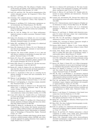
![urethrocystography and simultaneous pressure-flow measurements. Obstet Gynecol, 74: 417,1989
289. Lose, G., Tanko, A., Colstrup, H. et al.: Urethral sphincter electromyography with vaginal surface electrodes: a comparison
with sphincter electromyography recorded via periurethral
coaxial, anal sphincter needle and perianal surface electrodes. J
Urol, 133: 815,1985
311. Shabsigh, R., Fishman, I.J. and Krebs, M.: Combined transrectal
ultrasonography and urodynamics in the evaluation of detrusorsphincter dyssynergia. Br J Urol, 62: 326,1988
290. Barrett, D.M. and Wein, A.J.: Flow evaluation and simultaneous
external sphincter electromyography in clinical urodynamics. J
Urol, 125: 538,1981
312. Keane, D.P., Winder, A., Lewis, P. et al.: A combined urodynamic and continence unitÑa review of the first 19 years. Br J
Urol, 71: 161,1993
291. OÕDonnell, P.D. and Doyle, R.: Biofeedback therapy technique
for treatment of urinary incontinence. Urology, 37: 432,1991
313. Rivas, D.A. and Chancellor, M.B.: Neurogenic vesical dysfunction. Urol Clin North Am, 22: 579,1995
292. van Gool, J.D., Vijverberg, M.A., Messer, A.P. et al.: Functional
daytime incontinence: non-pharmacological treatment. Scand J
Urol Nephrol Suppl, 141: 93,1992
314. McGuire, E.J., Cespedes, R.D., Cross, C.A. et al.: Videourodynamic studies. Urol Clin North Am, 23: 309,1996
315. Rickwood, A.M. and Arnold, A.J.: Current management of
childhood neuropathic bladder: review of 156 cases. Z Kinderchir, 45: 238,1990
293. McIntosh, L.J., Frahm, J.D., Mallett, V.T. et al.: Pelvic floor
rehabilitation in the treatment of incontinence. J Reprod Med,
38: 662,1993
316. Perez, L.M., Khoury, J. and Webster, G.D.: The value of urodynamic studies in infants less than 1 year old with congenital spinal dysraphism. J Urol, 148: 584,1992
294. Gunnarsson, M. and Mattiasson, A.: Circumvaginal surface electromyography in women with urinary incontinence and in healthy volunteers. Scand J Urol Nephrol Suppl, 157: 89,1994
317. Ghoniem, G.M., Roach, M.B., Lewis, V.H. et al.: The value of
leak pressure and bladder compliance in the urodynamic evaluation of meningomyelocele patients. J Urol, 144: 1440,1990
295. Fried, G.W., Goetz, G., Potts-Nulty, S. et al.: A behavioral
approach to the treatment of urinary incontinence in a disabled
population. Arch Phys Med Rehabil, 76: 1120,1995
318. Kaplan, S.A., Chancellor, M.B. and Blaivas, J.G.: Bladder and
sphincter behavior in patients with spinal cord lesions. J Urol,
146: 113,1991
296. Noble, J.G., Dixon, P.J., Rickards, D. et al.: Urethral sphincter
volumes in women with obstructed voiding and abnormal
sphincter electromyographic activity. Br J Urol, 76: 741,1995
319. Kaplan, S.A., Te, A.E. and Blaivas, J.G.: Urodynamic findings
in patients with diabetic cystopathy. J Urol, 153: 342,1995
297. Thorp, J.M., Jones, L.H., Wells, E. et al.: Assessment of pelvic
floor function: a series of simple tests in nulliparous women. Int
Urogynecol J Pelvic Floor Dysfunct, 7: 94,1996
320. Chao, R. and Mayo, M.E.: Long-term urodynamic follow up in
pediatric spinal cord injury. Paraplegia, 32: 806,1994
321. Saxton, H.M.: Urodynamics in the investigation of women with
frequency, urgency, and incontinence, and voiding difficulties.
Urol Radiol, 13: 48,1991
322. Weerasinghe, N. and Malone, P.S.: The value of videourodynamics in the investigation of neurologically normal children who
wet. Br J Urol, 71: 539,1993
323. Griffiths, D.J., McCracken, P.N., Harrison, G.M. et al.: Characteristics of urinary incontinence in elderly patients studied by
24-hour monitoring and urodynamic testing. Age Ageing, 21:
195,1992
324. Passerini-Glazel, G., Cisternino, A., Camuffo, M.C. et al.:
Video-urodynamic studies of minor voiding dysfunctions in
children: an overview of 13 yearsÕ experience. Scand J Urol
Nephrol Suppl, 141: 70,1992
325. Goluboff, E.T., Chang, D.T., Olsson, C.A. et al.: Urodynamics
and the etiology of post-prostatectomy urinary incontinence: the
initial Columbia experience. J Urol, 153: 1034,1995
326. Trockman, B.A., Gerspach, J., Dmochowski, R. et al.: Primary
bladder neck obstruction: urodynamic findings and treatment
results in 36 men. J Urol, 156: 1418,1996
327. Javle, P., Jenkins, S.A., West, C. et al.: Quantification of voiding
dysfunction in patients awaiting transurethral prostatectomy. J
Urol, 156: 1014,1996
328. Porter, T., Weerasinghe, N. and Malone, P.S.: Modification of
therapy based on videourodynamics in neurologically normal
children: Southampton 1988-1993. Br J Urol, 76: 779,1995
329. Glazier, D.B., Murphy, D.P., Fleisher, M.H. et al.: Evaluation of
the utility of video-urodynamics in children with urinary tract
infection and voiding dysfunction. Br J Urol, 80: 806,1997
330. Ilker, Y., Tarcan, T., Yucel, S. et al.: Re- techered cord syndorome in myelodysplasia and the importance of urological early
follow- up in the early diagnosis. Neurourol Urodyn, 19:
517,2000
331. Lackner, J., Kiss, G. and Madersbacher, H.: Can early urological
management improve the outcome (upper urinary tract, continence) in patients with myelomeningoceles? -Long term results.
Neurourol Urodyn, 19: 532,2000
298. Rowan, D., James, E.D., Kramer, A.E. et al.: Urodynamic equipment: technical aspects. Produced by the International Continence Society Working Party on Urodynamic Equipment. J
Med Eng Technol, 11: 57,1987
299. Vereecken, R.L. and Grisar, P.: Perineal electromyographic patterns in urge incontinence. Arch Gynecol, 237: 235,1986
300. Siroky, M.B.: Electromyography of the perineal floor. Urol Clin
North Am, 23: 299,1996
301. Rossier, A.B., Fam, B.A., Dibenedetto, M. et al.: Urodynamics
in spinal shock patients. J Urol, 122: 783,1979
302. Perkash, I.: Urodynamic evaluation: periurethral striated EMG
versus perianal striated EMG. Paraplegia, 18: 275,1980
303. Kaneko, S., Watabe, Y., Mizunaga, M. et al.: Automatic analysis
of urethral electromyography for accurate diagnosis of voiding
dysfunction. Urol Int, 47: 55,1991
304. Jost, W.H., Derouet, H. and Kaiser, T.: [Electromyography of the
sphincter vesicae externus muscle. Technique, indications and
outcome]. Urologe A, 36: 356,1997
305. McGuire, E.J. and Woodside, J.R.: Diagnostic advantages of
fluoroscopic monitoring during urodynamic evaluation. J Urol,
125: 830,1981
306. Abdel-Rahman, M., Coulombe, A., Devroede, G. et al.: Urorectodynamic evaluation of healthy volunteers. Urology, 19:
559,1982
307. Lockhart, J.L., Shessel, F., Weinstein, D. et al.: Urodynamics in
women with stress and urge incontinence. Urology, 20:
333,1982
308. Rudy, D.C., Awad, S.A. and Downie, J.W.: External sphincter
dyssynergia: an abnormal continence reflex. J Urol, 140:
105,1988
309. Bo, K., Stien, R., Kulseng-Hanssen, S. et al.: Clinical and urodynamic assessment of nulliparous young women with and
without stress incontinence symptoms: a case-control study.
Obstet Gynecol, 84: 1028,1994
310. Koelbl, H. and Bernaschek, G.: A new method for sonographic
368](https://image.slidesharecdn.com/urodin-131127082012-phpapp01/85/Urodinamia-52-320.jpg)
![353. Heslington, K. and Hilton, P.: The incidence of detrusor instability by ambulatory monitoring and conventional cystometry pre
and post colposuspension. Neurourol Urodyn, 14: 416,1995
332. van Waalwijk van Doorn, E.S. and Gommer, E.D.: Ambulatory
urodynamics. Curr Opin Obstet Gynecol, 7: 378,1995
333. Heslington, K. and Hilton, P.: Ambulatory urodynamic monitoring. Br J Obstet Gynaecol, 103: 393,1996
354. Young, C.K., Godley, M.L., Duffy, P.G. et al.: Natural filling
cystometry in infants and children. Br J Urol, 67: 531,1995
334. Bristow, S.E. and Neal, D.E.: Ambulatory urodynamics. Br J
Urol, 77: 333,1996
355. Brown, K. and Hilton, P.: The incidence of detrusor instability
before and after colposuspension: a study using conventional
and ambulatory urodynamic monitoring. BJU Int, 84: 961,1999
356. Groen, J., van Mastrigt, R. and Bosch, R.: Factors causing differences in voiding parameters between conventional and ambulatory urodynamics. Urol Res, 28: 128,2000
357. Webb, R.J., Griffiths, C.J., Ramsden, P.D. et al.: Measurement of
voiding pressures on ambulatory monitoring: comparison with
conventional cystometry. Br J Urol, 65: 152,1990
358. Webb, R.J., Griffiths, C.J., Zachariah, K.K. et al.: Filling and
voiding pressures measured by ambulatory monitoring and
conventional studies during natural and artificial bladder filling.
J Urol, 146: 815,1991
359. McInerney, P.D., Harris, S.A., Pritchard, A. et al.: Night studies
for primary diurnal and nocturnal enuresis and preliminary
results of the ÒclamÓ ileocystoplasty. Br J Urol, 67: 42,1991
360. Rosario, D.J., Leaker, B.R., Smith, D.J. et al.: A pilot study of
the effects of multiple doses of the M3 muscarinic receptor
antagonist darifenacin on ambulatory parameters of detrusor
activity in patients with detrusor instability. Neurourol Urodyn,
14: 464,1995
361. Hasan, S.T., Robson, W.A., Pridie, A.K. et al.: Outcome of transcutaneous electrical stimulation in patients with detrusor instability. Neurourol Urodyn, 13: 349,1994
362. van Waalwijk van Doorn, E., Anders, K., Khullar, V. et al.: Standardisation of ambulatory urodynamic monitoring: Report of
the Standardisation Sub-Committee of the International Continence Society for Ambulatory Urodynamic Studies. Neurourol
Urodyn, 19: 113,2000
363. Lose, G.: Urethral pressure measurement. Acta Obstet Gynecol
Scand Suppl, 166: 39,1997
364. Blaivas, J.G. and Olsson, C.A.: Stress incontinence: classification and surgical approach. J Urol, 139: 727,1988
365. Kayigil, O., Iftekhar Ahmed, S. and Metin, A.: The coexistence
of intrinsic sphincter deficiency with type II stress incontinence. J Urol, 162: 1365,1999
366. Leach, G.E., Dmochowski, R.R., Appell, R.A. et al.: Female
Stress Urinary Incontinence Clinical Guidelines Panel summary report on surgical management of female stress urinary
incontinence. The American Urological Association. J Urol,
158: 875,1997
367. Herschorn, S., Steele, D.J. and Radomski, S.B.: Followup of
intraurethral collagen for female stress urinary incontinence. J
Urol, 156: 1305,1996
335. Kulseng-Hanssen, S. and Klevmark, B.: Ambulatory urodynamic monitoring of women. Scand J Urol Nephrol Suppl, 179:
27,1996
336. Anders, K., Khullar, V., Cardozo, S. et al.: Ambulatory urodynamic monitoring in clinical urogynaecological practice. Neurourol Urodyn, 16: 510,1997
337. Rosario, D.J., Potts, K.L., Woo, H.H. et al.: Ambulatory pressure-flow studies in young asymptomatic males. Neurourol Urodyn, 15: 278,1996
338. James, E.D., Flack, F.C., Caldwell, K.P. et al.: Continuous measurement of urine loss and frequency in incontinent patients.
Preliminary report. Br J Urol, 43: 233,1971
339. Webb, R.J., Ramsden, P.D. and Neal, D.E.: Ambulatory monitoring and electronic measurement of urinary leakage in the diagnosis of detrusor instability and incontinence. Br J Urol, 68:
148,1991
340. Wijkstra, H., van Kerrebroek, E.V.A., Koldewijn, E. et al.: The
use of heat sensors in detecting urine leakage during long term
urudynamic. Neurourol Urodyn, 10: 422,1991
341. Eckford, S.D., Finney, R., Jackson, S.R. et al.: Detection of urinary incontinence during ambulatory monitoring of bladder
function by a temperature-sensitive device. Br J Urol, 77:
194,1996
342. van Waalwijk van Doorn, E.S. and Zwiers, W.: Ambulant monitoring to assess the efficacy of oxybutynin chloride in patients
with mixed incontinence. Eur Urol, 18: 49,1990
343. Kulseng-Hanssen, S., Kristofferson, M. and Larsen, E.: Physioogical bladder filling with 2 and 4 hours recording versus 45
minutes recording with 300 ml. saline bladder? prefilling using
ambulatory urodynamic equipment. Neurourol Urodyn, 14:
151,1995
344. van Waalwijk van Doorn, E.S., Ambergen, A.W. and Janknegt,
R.A.: Detrusor activity index: quantification of detrusor overactivity by ambulatory monitoring. J Urol, 157: 596,1997
345. van Waalwijk van Doorn, E.S., Remmers, A. and Janknegt, R.A.:
Conventional and extramural ambulatory urodynamic testing of
the lower urinary tract in female volunteers. J Urol, 147:
1319,1992
346. Robertson, A.S., Griffiths, C.J., Ramsden, P.D. et al.: Bladder
function in healthy volunteers: ambulatory monitoring and
conventional urodynamic studies. Br J Urol, 73: 242,1994
347. Heslington, K. and Hilton, P.: A comparison of ambulatory
monitoring and conventional cystometry in asymptomatic
female volunteers. Neurourol Urodyn, 14: 534,1995
348. Kulseng-Hanssen, S. and Klevmark, B.: Ambulatory urethrocysto- rectometry: A new technique. Neurourol Urodyn, 7:
119,1988
349. Styles, R.A., Neal, D.E. and Ramsden, P.D.: Comparison of
long-term monitoring and standard cystometry in chronic retention of urine. Br J Urol, 58: 652,1986
350. Webb, R.J., Styles, R.A., Griffiths, C.J. et al.: Ambulatory monitoring of bladder pressures in patients with low compliance as a
result of neurogenic bladder dysfunction. Br J Urol, 64:
150,1989
368. Winters, J.C., Chiverton, A., Scarpero, H.M. et al.: Collagen
injection therapy in elderly women: long-term results and
patient satisfaction. Urology, 55: 856,2000
369. Vereecken, R.L.: A critical view on the value of urodynamics in
non-neurogenic incontinence in women. Int J Gynaecol Obstet,
11: 188,2000
370. Ramsay, I.N., Ali, H.M., Hunter, M. et al.: A randomized
controlled trial of urodynamic investigations prior to consevative treatment of urinary incontinence in the female. Int J Gynaecol Obstet, 6: 277,1995
371. Holtedahl, K., Verelst, M., Schiefloe, A. et al.: Usefulness of
urodynamic examination in female urinary incontinenceÑ lessons from a population-based, randomized, controlled study of
conservative treatment. Scand J Urol Nephrol, 34: 169,2000
351. van Waalwijk van Doorn, E.S., Remmers, A. and Janknegt, R.A.:
Extramural ambulatory urodynamic monitoring during natural
filling and normal daily activities: evaluation of 100 patients. J
Urol, 146: 124,1991
372. Haab, F., Ciofu, C., Pedron, P. et al.: [Feasibility of ÒValsalva
Leak Point PressureÓ. Prospective study]. Prog Urol, 7:
611,1997
352. McInerney, P.D., Vanner, T.F., Harris, S.A. et al.: Ambulatory
urodynamics. Br J Urol, 67: 272,1991
369](https://image.slidesharecdn.com/urodin-131127082012-phpapp01/85/Urodinamia-53-320.jpg)
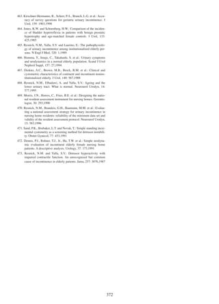

This document summarizes the key points regarding urodynamics studies for assessing urinary incontinence. It discusses the general aspects of urodynamics, including informing patients, test conditions, the role of the investigator, use of catheters and transducers, and definitions of various pressure measurements. The document emphasizes that urodynamics aims to objectively observe lower urinary tract function and dysfunction in order to appropriately manage incontinence and its underlying causes.


![Incontinence & Female Urology [Dr.Edmond Wong]](https://cdn.slidesharecdn.com/ss_thumbnails/incontinencefemaleedmond-140716213134-phpapp01-thumbnail.jpg?width=640&height=640&fit=bounds)


