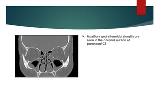
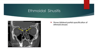
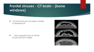
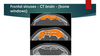
Frontal sinuses -CT brain - (bone
windows)
The frontal sinuses are highly variable
in appearance
Many people have no frontal
sinuses (bottom image)
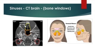
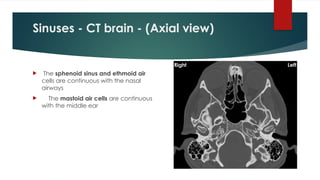
Sinuses - CTbrain - (Axial view)
The sphenoid sinus and ethmoid air
cells are continuous with the nasal
airways
The mastoid air cells are continuous
with the middle ear
16.
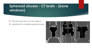
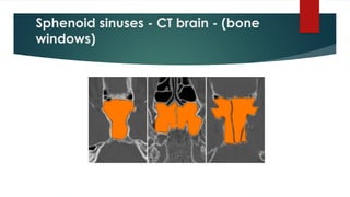
Sphenoid sinuses -CT brain - (bone
windows)
The sphenoid sinus can be single or
septated into multiple separate sinuses