Cerebral Small Vessel
Disease
Dr. Dwaipayan Sen
Consultant Stroke Physician
Salford Royal Hospital
Overview
• Definition
• Radiological Markers
• Pathophysiology
• Clinical syndromes
• Aetiology/Associations/Work up
• Evidence base for treatments
• Future directions/Candidate Drugs
Definitions
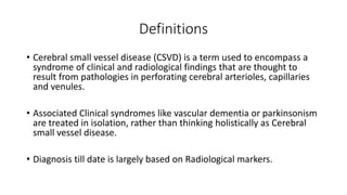
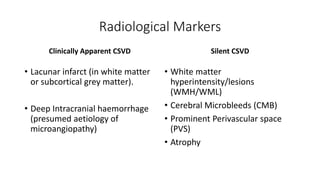
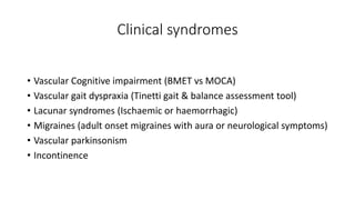
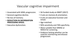
• Cerebral small vessel disease (CSVD) is a term used to encompass a
syndrome of clinical and radiological findings that are thought to
result from pathologies in perforating cerebral arterioles, capillaries
and venules.
• Associated Clinical syndromes like vascular dementia or parkinsonism
are treated in isolation, rather than thinking holistically as Cerebral
small vessel disease.
• Diagnosis till date is largely based on Radiological markers.
Radiological Markers
Clinically Apparent CSVD
• Lacunar infarct (in white matter
or subcortical grey matter).
• Deep Intracranial haemorrhage
(presumed aetiology of
microangiopathy)
Silent CSVD
• White matter
hyperintensity/lesions
(WMH/WML)
• Cerebral Microbleeds (CMB)
• Prominent Perivascular space
(PVS)
• Atrophy
Lacunar infarct (LI)
• A Lacunar infarct is defined as an
infarct < 1.5 cm in diameter in an
area of brain supplied by
perforator arteries (white matter,
basal ganglia and brainstem) and
is consistent with a clinical lacunar
syndrome
Deep Intracranial
Haemorrhage
• A deep Intracranial Haemorrhage
is described as a haemorrhage in
white matter, basal ganglia or
brainstem consistent with a
clinical lacunar syndrome
Lacunar syndromes
Histology
• Lacunes first described by Fisher in
1960’s.
• Ovoid subcortical fluid filled
cavities.
• Diameter 3 – 15 mm.
• Healed infarct or residual lesion of
Haemorrhage.
Clinical
• Acute presentation of hemiparesis,
hemisensory loss (Oxford
Classification – LACS).
• 8 – 28% of elderly population.
• Burden of disease over time leads
to:
1. Cognitive impairment
2. Dementia
3. Gait disturbance
4. Increased risk of stroke
White matter
Hyperintensities/Lesions
(WMH/WML
• Hyperintense lesions on T2 or
FLAIR imaging (patchy or confluent
depending on their stage in
development & severity) which
are symmetrically and bilaterally
distributed in white matter,
including pons and brain stem and
also occur in deep grey matter
WMH/WML
Histology
• Imprecise due to very few samples.
• Demyelination
• Loss of oligodendrocytes
• Axonal damage
• Oedema (suggestive of leakage of
fluid from impaired BBB)
Clinical
• Cognitive impairment
• Dementia
• Depression
• Migraine (Association)
• Epidemiological study in BMJ (2010):
1. Triples risk of lacunar stroke
2. Doubles risk of dementia
3. Increases risk of death
Prominent Perivascular Spaces
PVS
Histology
• Extension of Subarachnoid
spaces that surround cerebral
microvessels
• Fluid filled spaces that follow
the course of a small vessel
through the brain parenchyma
Clinical
• Few PVS may be normal – doubt
as to whether they should be
considered as a marker of CSVD.
• Some epidemiological studies
report that a higher number of
PVS is associated with:
1. Worsening cognitive function
2. Dementia
3. Hypertension
Cerebral Microbleed
(CMB)
• Small, round, homogenous foci of
hypointensity in SWI or GRE
sequence of MRI.
• Lipohyalinosis related to
Hypertension leads to Deep CMB.
• Amyloid deposition leads to
cortical distribution of CMB.
• In real life there is often a overlap.
CMB
Histology
• Very few studies.
• Perivascular haemosiderin laden
macrophages.
• Lipofibrohyalinosis
(Hypertension).
• Amyloid deposition (Cerebral
amyloid angiopathy).
Clinical
• 11 – 15% in normal population
(Rotterdam scan study & AGES –
Reykjavik studies).
• 33 – 67% in population with
Ischameic and haemorrhagic
stroke.
• Intracranial haemorrhages
• Ischaemic events
• Transient neurological episodes
(seizures/Amyloid spells)
Pathogenesis
Conventional Cardiovascular risk factors (Age,
HTN, Smoking)
Disruption of Cerebral autoregulation (Angiotensin
II implicated)
Hemodynamic alteration
Endothelial dysfunction
BBB disruption & permeability change
Seepage of fluid and plasma components
Perivascular inflammation
• Conventional Atherosclerosis (LI or
WMH> 7 mm)
1. Arteriosclerosis
2. Microatheroma in distal arterioles
• Non Atherosclerotic causes (Lacunes or
WMH < 7 mm, PVS, CMB)
1. Blood Brain Barrier (BBB) disruption
(Phosphodiesterase inhibitors, cilostazol)
2. Endothelial dysfunction (NO signaling)
3. Perivascular inflammation (IL6
implicated)
Clinical syndromes
• Vascular Cognitive impairment (BMET vs MOCA)
• Vascular gait dyspraxia (Tinetti gait & balance assessment tool)
• Lacunar syndromes (Ischaemic or haemorrhagic)
• Migraines (adult onset migraines with aura or neurological symptoms)
• Vascular parkinsonism
• Incontinence
Vascular cognitive impairment
• Associated with WML progression.
• General cognitive decline.
• No loss of memory.
• REDUCTION IN INFORMATION
PROCESSING SPEED.
• Executive dysfunction.
• SA funded study on BMET (2017)
• 4 sets on memory & orientation.
• 4 sets on executive function and
timed.
• Age matched.
• 92% sensitivity and 76% specificity.
• More suitable than MMSE or
MOCA for screening.
• Evidence lacking whether can be
used for monitoring and disease
progression.
Natural History (Rotterdam Scan Study)
Factors associated with progression
• Age
• Baseline WML severity
• Lacunar infarcts
• Female gender
• HTN
• Current smoking
Factors not associated
• Carotid stenosis
• AF
• Homocysteine levels
• Previous smoking
• At very old age and significant
WML burden – none of the
standard CVS risk reduction
worked.
Causes/Associations
Standard CVS risk factors
• Age
• HTN
• Smoking
• Dyslipidaemia
• DM
• Coronary artery disease
Others
• Hypothyroidism (subclinical)
• Homocysteinaemia
• Vitamin B12 & Folate deficiency
• OSA
• Acquired thrombophilia
• Small vessel vasculitis
• CTD
• Amyloid deposition (CAA)
• Genetic diseases
(CADASIL/CARASIL/COL4 A1/Fabry’s)
Treatment
• Thrombolysis
• Antiplatelets
• Statins
• Antihypertensive treatment
• Carotid stenosis
• Candidate drugs
Thrombolysis
Efficacy
• NINDS and subsequent trial data
shows equal efficacy of thrombolysis
in lacunar strokes.
• Patients with lacunar strokes did
better with thrombolysis than placebo
(Canadian Stroke registry, 2013).
• Lacunar strokes showed least
improvement in NIHSS on day 7
(Madrid Stroke registry, 2012).
• Current guidelines recommend
treatment.
Safety
sICH PH Reference
No CMB 1.2 5.7 Dannenberg S
et al; Stroke
2014
1 CMB 3.8 3.8
2 – 4 CMB 10.5 21.1
> 5 CMB 30 30
1 CMB
increases OR
for sICH by
1.19
Antiplatelets
• Various trials over years suggest they are effective in LI
(Aspirin/Clopidogrel/Dipyridamole/Ticlopidine).
• Dual antiplatelets not effective and cause more hemorrhagic complications
(Secondary prevention of small subcortical strokes; SPS3).
• Role in silent CSVD with WMH & CMB less clear.
• Antiplatelets in ICH – RESTART trial.
1. Reduced haemorrhage
2. Reduced non fatal MI and Stroke (major vascular occlusion)
3. Effect seen even in presence of CMBs (irrespective of location)
4. Hypothesis unclear.
Hypertension
• SPS3 study with intensive BP lowering arm (SBP < 130 mm hg)
1. Reduced recurrent Ischaemic strokes
2. Reduced ICH
• PROGRESS Trial (ACE inhibitors)
1. RR in composite outcome of stroke and dementia (34%)
2. Delay in WML progression in Stroke patients (MRI Sub study)
• Rotterdam Scan Study
1. In very old age group (>75 years) and established WML burden – no
effect.
Hyperlipidaemia/Statins
Hypotheses
• Some observational trials
suggest that hyperlipidaemia
has a protective effect in WML
progression.
• Statins have additional anti
inflammatory and antioxidant
properties that may negate the
effect of hyperlipidaemia
reduction
Clinical trial results
• SPARCL – Atorvastatin in Ischaemic Stroke patients
1. Reduced Stroke and IHD
2. Trend towards increase in number of haemorrhagic
strokes
3. 1,409 amongst 4,731 patients had lacunar strokes
4. Post hoc analysis shows total benefit similar to overall
study population
• ROCAS (Regression of cerebral artery stenosis, 2009)
1. Lower incidence of lacunes.
2. Reduction in WML progression.
• VITATOPS MRI Substudy (Pre stroke statin use)
• Reduced WML progression
• Reduced cognitive decline
• No effect on incident lacunes or CMBs
Carotid Artery Stenosis (CAS)
Hypothesis
• Primate models show only 6% of
injected carotid emboli enter
Lenticulostriate vessels.
• TCD trials show little or no
microembolic signals in small vessel
disease
• CAS is an incidental finding in LI
• Is Carotid endarterectomy useful in
lacunar infarct?
Clinical Trial results
• NASCET post hoc analysis (n < 500)
1. CAS less common in LI
2. Ipsilateral and contralateral CAS has equal
incidence
3. Absolute RR is far lower in LI (9% vs 15%)
compared to embolic strokes.
4. Beneficial effect on severe CAS (>70% stenosis); nil
on lesser degree on stenosis (50 – 69% stenosis)
• Honnenberg et al (Stroke, 2010)
1. Multidetector CT assessed for plaque ulceration
and rupture
2. Independent association with non LI.
3. No association of plaque ulceration or rupture with
LI
CAS & CEA in LI - Summary
• CAS can be causative in a small number of atheroembolic lacunar
strokes.
• In the vast majority it may be an incidental finding and classed as
asymptomatic.
• CEA has a small benefit in LI in severe stenosis (> 70%), but not in
moderate stenosis (50 – 69%); possibly a combination benefit derived
from small symptomatic and large asymptomatic cohort.
• Is this benefit > advancements in medical treatment?
• Is this benefit > perioperative risk?
Candidate drugs
Hypothesis
• Cilostazol
1. Weak antiplatelet action
2. BBB preservation
(Phosphodiestaerase 3’ inhibitor)
• NO donors
1. BBB preservation
(Very limited clinical data, some from
ENOS trial)
Clinical trial results for Cilostazol
• Pooled data in Asia Pacific population (5 trials, 4780
patients, Asia Pacific population)
1. Less risk of bleeding compared to Aspirin
2. Reduced Ischaemic Stroke (OR 0.64, 95%CI 0.51–
0.79)
• Pooled data in Asia pacific population
1. Population registry analysis
2. 9148 dementia-free subjects
3. Reduced incidence of dementia (adjusted HR 0.75,
95%CI 0.61–0.92, p for dose trend, 0.001)
• Studies in Progress
1. LACI – 2 (UK population)
2. Cilostazol or Isosorbide mononitrate or both
3. Used in addition to standard treatment
Candidate Drugs (Vitamins)
Hypothesis
• Homocysteinaemia associated with CSVD in
some population based studies.
• Vitamin B12 and Folic acid supplements will
reduce Homocysteine levels, therefore may
reduce CSVD
• Folic Acid has atherogenic potential, so can
worsen CSVD or vascular disease
Clinical Trial data
• VITATOPS (Cerebrovascular disease, 2002).
1. Vitamin B6, Vitamin B12 and Folic Acid
supplemented.
2. All Stroke patients included (Ischaemic and
Haemorrhagic)
3. No difference in vascular event outcome
(Stroke, MI or death due to vascular cause.
4. MRI Sub study – showed reduction in WML
progression in moderate to severe disease
5. Potential for testing in CSVD and LI patients
exclusively; and for outcomes in all clinical
syndromes like cognition, gait, recurrent
LI?
Concept of CSVD clinic in SRFT
Clinical Assessment
• Vascular cognitive impairment
(BMET/MOCA)
• Vascular gait dyspraxia (Tinetti gait and
balance tool)
• Lacunar syndromes
• Migraine (Complicated/with aura)
• Vascular Parkinsonism
• Continence
• Associated disease in other vascular bed
(Coronary vascular disease/Peripheral
vascular disease)
Investigations/work up
• Standard cardiovascular risk
(DM/Lipids/ECG/Smoking/HTN)
• TFT, acquired Thrombophilia, Vitamin B12
or Folate deficiencies, Coeliac.
• Neuroimaging
• CTD screen, ANCA, Vascular imaging, CSF
in suspected Small vessel vasculitis.
• Genetic tests (CADASIL including
extended testing for all
mutations/Fabry’s)
• Skin Biopsy
Thank You & Questions?

CSVD

  • 1.
    Cerebral Small Vessel Disease Dr.Dwaipayan Sen Consultant Stroke Physician Salford Royal Hospital
  • 2.
    Overview • Definition • RadiologicalMarkers • Pathophysiology • Clinical syndromes • Aetiology/Associations/Work up • Evidence base for treatments • Future directions/Candidate Drugs
  • 3.
    Definitions • Cerebral smallvessel disease (CSVD) is a term used to encompass a syndrome of clinical and radiological findings that are thought to result from pathologies in perforating cerebral arterioles, capillaries and venules. • Associated Clinical syndromes like vascular dementia or parkinsonism are treated in isolation, rather than thinking holistically as Cerebral small vessel disease. • Diagnosis till date is largely based on Radiological markers.
  • 4.
    Radiological Markers Clinically ApparentCSVD • Lacunar infarct (in white matter or subcortical grey matter). • Deep Intracranial haemorrhage (presumed aetiology of microangiopathy) Silent CSVD • White matter hyperintensity/lesions (WMH/WML) • Cerebral Microbleeds (CMB) • Prominent Perivascular space (PVS) • Atrophy
  • 5.
    Lacunar infarct (LI) •A Lacunar infarct is defined as an infarct < 1.5 cm in diameter in an area of brain supplied by perforator arteries (white matter, basal ganglia and brainstem) and is consistent with a clinical lacunar syndrome
  • 6.
    Deep Intracranial Haemorrhage • Adeep Intracranial Haemorrhage is described as a haemorrhage in white matter, basal ganglia or brainstem consistent with a clinical lacunar syndrome
  • 7.
    Lacunar syndromes Histology • Lacunesfirst described by Fisher in 1960’s. • Ovoid subcortical fluid filled cavities. • Diameter 3 – 15 mm. • Healed infarct or residual lesion of Haemorrhage. Clinical • Acute presentation of hemiparesis, hemisensory loss (Oxford Classification – LACS). • 8 – 28% of elderly population. • Burden of disease over time leads to: 1. Cognitive impairment 2. Dementia 3. Gait disturbance 4. Increased risk of stroke
  • 8.
    White matter Hyperintensities/Lesions (WMH/WML • Hyperintenselesions on T2 or FLAIR imaging (patchy or confluent depending on their stage in development & severity) which are symmetrically and bilaterally distributed in white matter, including pons and brain stem and also occur in deep grey matter
  • 9.
    WMH/WML Histology • Imprecise dueto very few samples. • Demyelination • Loss of oligodendrocytes • Axonal damage • Oedema (suggestive of leakage of fluid from impaired BBB) Clinical • Cognitive impairment • Dementia • Depression • Migraine (Association) • Epidemiological study in BMJ (2010): 1. Triples risk of lacunar stroke 2. Doubles risk of dementia 3. Increases risk of death
  • 10.
  • 11.
    PVS Histology • Extension ofSubarachnoid spaces that surround cerebral microvessels • Fluid filled spaces that follow the course of a small vessel through the brain parenchyma Clinical • Few PVS may be normal – doubt as to whether they should be considered as a marker of CSVD. • Some epidemiological studies report that a higher number of PVS is associated with: 1. Worsening cognitive function 2. Dementia 3. Hypertension
  • 12.
    Cerebral Microbleed (CMB) • Small,round, homogenous foci of hypointensity in SWI or GRE sequence of MRI. • Lipohyalinosis related to Hypertension leads to Deep CMB. • Amyloid deposition leads to cortical distribution of CMB. • In real life there is often a overlap.
  • 13.
    CMB Histology • Very fewstudies. • Perivascular haemosiderin laden macrophages. • Lipofibrohyalinosis (Hypertension). • Amyloid deposition (Cerebral amyloid angiopathy). Clinical • 11 – 15% in normal population (Rotterdam scan study & AGES – Reykjavik studies). • 33 – 67% in population with Ischameic and haemorrhagic stroke. • Intracranial haemorrhages • Ischaemic events • Transient neurological episodes (seizures/Amyloid spells)
  • 14.
    Pathogenesis Conventional Cardiovascular riskfactors (Age, HTN, Smoking) Disruption of Cerebral autoregulation (Angiotensin II implicated) Hemodynamic alteration Endothelial dysfunction BBB disruption & permeability change Seepage of fluid and plasma components Perivascular inflammation • Conventional Atherosclerosis (LI or WMH> 7 mm) 1. Arteriosclerosis 2. Microatheroma in distal arterioles • Non Atherosclerotic causes (Lacunes or WMH < 7 mm, PVS, CMB) 1. Blood Brain Barrier (BBB) disruption (Phosphodiesterase inhibitors, cilostazol) 2. Endothelial dysfunction (NO signaling) 3. Perivascular inflammation (IL6 implicated)
  • 15.
    Clinical syndromes • VascularCognitive impairment (BMET vs MOCA) • Vascular gait dyspraxia (Tinetti gait & balance assessment tool) • Lacunar syndromes (Ischaemic or haemorrhagic) • Migraines (adult onset migraines with aura or neurological symptoms) • Vascular parkinsonism • Incontinence
  • 16.
    Vascular cognitive impairment •Associated with WML progression. • General cognitive decline. • No loss of memory. • REDUCTION IN INFORMATION PROCESSING SPEED. • Executive dysfunction. • SA funded study on BMET (2017) • 4 sets on memory & orientation. • 4 sets on executive function and timed. • Age matched. • 92% sensitivity and 76% specificity. • More suitable than MMSE or MOCA for screening. • Evidence lacking whether can be used for monitoring and disease progression.
  • 17.
    Natural History (RotterdamScan Study) Factors associated with progression • Age • Baseline WML severity • Lacunar infarcts • Female gender • HTN • Current smoking Factors not associated • Carotid stenosis • AF • Homocysteine levels • Previous smoking • At very old age and significant WML burden – none of the standard CVS risk reduction worked.
  • 18.
    Causes/Associations Standard CVS riskfactors • Age • HTN • Smoking • Dyslipidaemia • DM • Coronary artery disease Others • Hypothyroidism (subclinical) • Homocysteinaemia • Vitamin B12 & Folate deficiency • OSA • Acquired thrombophilia • Small vessel vasculitis • CTD • Amyloid deposition (CAA) • Genetic diseases (CADASIL/CARASIL/COL4 A1/Fabry’s)
  • 19.
    Treatment • Thrombolysis • Antiplatelets •Statins • Antihypertensive treatment • Carotid stenosis • Candidate drugs
  • 20.
    Thrombolysis Efficacy • NINDS andsubsequent trial data shows equal efficacy of thrombolysis in lacunar strokes. • Patients with lacunar strokes did better with thrombolysis than placebo (Canadian Stroke registry, 2013). • Lacunar strokes showed least improvement in NIHSS on day 7 (Madrid Stroke registry, 2012). • Current guidelines recommend treatment. Safety sICH PH Reference No CMB 1.2 5.7 Dannenberg S et al; Stroke 2014 1 CMB 3.8 3.8 2 – 4 CMB 10.5 21.1 > 5 CMB 30 30 1 CMB increases OR for sICH by 1.19
  • 21.
    Antiplatelets • Various trialsover years suggest they are effective in LI (Aspirin/Clopidogrel/Dipyridamole/Ticlopidine). • Dual antiplatelets not effective and cause more hemorrhagic complications (Secondary prevention of small subcortical strokes; SPS3). • Role in silent CSVD with WMH & CMB less clear. • Antiplatelets in ICH – RESTART trial. 1. Reduced haemorrhage 2. Reduced non fatal MI and Stroke (major vascular occlusion) 3. Effect seen even in presence of CMBs (irrespective of location) 4. Hypothesis unclear.
  • 22.
    Hypertension • SPS3 studywith intensive BP lowering arm (SBP < 130 mm hg) 1. Reduced recurrent Ischaemic strokes 2. Reduced ICH • PROGRESS Trial (ACE inhibitors) 1. RR in composite outcome of stroke and dementia (34%) 2. Delay in WML progression in Stroke patients (MRI Sub study) • Rotterdam Scan Study 1. In very old age group (>75 years) and established WML burden – no effect.
  • 23.
    Hyperlipidaemia/Statins Hypotheses • Some observationaltrials suggest that hyperlipidaemia has a protective effect in WML progression. • Statins have additional anti inflammatory and antioxidant properties that may negate the effect of hyperlipidaemia reduction Clinical trial results • SPARCL – Atorvastatin in Ischaemic Stroke patients 1. Reduced Stroke and IHD 2. Trend towards increase in number of haemorrhagic strokes 3. 1,409 amongst 4,731 patients had lacunar strokes 4. Post hoc analysis shows total benefit similar to overall study population • ROCAS (Regression of cerebral artery stenosis, 2009) 1. Lower incidence of lacunes. 2. Reduction in WML progression. • VITATOPS MRI Substudy (Pre stroke statin use) • Reduced WML progression • Reduced cognitive decline • No effect on incident lacunes or CMBs
  • 24.
    Carotid Artery Stenosis(CAS) Hypothesis • Primate models show only 6% of injected carotid emboli enter Lenticulostriate vessels. • TCD trials show little or no microembolic signals in small vessel disease • CAS is an incidental finding in LI • Is Carotid endarterectomy useful in lacunar infarct? Clinical Trial results • NASCET post hoc analysis (n < 500) 1. CAS less common in LI 2. Ipsilateral and contralateral CAS has equal incidence 3. Absolute RR is far lower in LI (9% vs 15%) compared to embolic strokes. 4. Beneficial effect on severe CAS (>70% stenosis); nil on lesser degree on stenosis (50 – 69% stenosis) • Honnenberg et al (Stroke, 2010) 1. Multidetector CT assessed for plaque ulceration and rupture 2. Independent association with non LI. 3. No association of plaque ulceration or rupture with LI
  • 25.
    CAS & CEAin LI - Summary • CAS can be causative in a small number of atheroembolic lacunar strokes. • In the vast majority it may be an incidental finding and classed as asymptomatic. • CEA has a small benefit in LI in severe stenosis (> 70%), but not in moderate stenosis (50 – 69%); possibly a combination benefit derived from small symptomatic and large asymptomatic cohort. • Is this benefit > advancements in medical treatment? • Is this benefit > perioperative risk?
  • 26.
    Candidate drugs Hypothesis • Cilostazol 1.Weak antiplatelet action 2. BBB preservation (Phosphodiestaerase 3’ inhibitor) • NO donors 1. BBB preservation (Very limited clinical data, some from ENOS trial) Clinical trial results for Cilostazol • Pooled data in Asia Pacific population (5 trials, 4780 patients, Asia Pacific population) 1. Less risk of bleeding compared to Aspirin 2. Reduced Ischaemic Stroke (OR 0.64, 95%CI 0.51– 0.79) • Pooled data in Asia pacific population 1. Population registry analysis 2. 9148 dementia-free subjects 3. Reduced incidence of dementia (adjusted HR 0.75, 95%CI 0.61–0.92, p for dose trend, 0.001) • Studies in Progress 1. LACI – 2 (UK population) 2. Cilostazol or Isosorbide mononitrate or both 3. Used in addition to standard treatment
  • 27.
    Candidate Drugs (Vitamins) Hypothesis •Homocysteinaemia associated with CSVD in some population based studies. • Vitamin B12 and Folic acid supplements will reduce Homocysteine levels, therefore may reduce CSVD • Folic Acid has atherogenic potential, so can worsen CSVD or vascular disease Clinical Trial data • VITATOPS (Cerebrovascular disease, 2002). 1. Vitamin B6, Vitamin B12 and Folic Acid supplemented. 2. All Stroke patients included (Ischaemic and Haemorrhagic) 3. No difference in vascular event outcome (Stroke, MI or death due to vascular cause. 4. MRI Sub study – showed reduction in WML progression in moderate to severe disease 5. Potential for testing in CSVD and LI patients exclusively; and for outcomes in all clinical syndromes like cognition, gait, recurrent LI?
  • 28.
    Concept of CSVDclinic in SRFT Clinical Assessment • Vascular cognitive impairment (BMET/MOCA) • Vascular gait dyspraxia (Tinetti gait and balance tool) • Lacunar syndromes • Migraine (Complicated/with aura) • Vascular Parkinsonism • Continence • Associated disease in other vascular bed (Coronary vascular disease/Peripheral vascular disease) Investigations/work up • Standard cardiovascular risk (DM/Lipids/ECG/Smoking/HTN) • TFT, acquired Thrombophilia, Vitamin B12 or Folate deficiencies, Coeliac. • Neuroimaging • CTD screen, ANCA, Vascular imaging, CSF in suspected Small vessel vasculitis. • Genetic tests (CADASIL including extended testing for all mutations/Fabry’s) • Skin Biopsy
  • 29.
    Thank You &Questions?