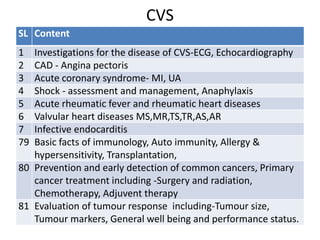
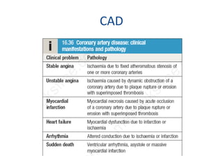
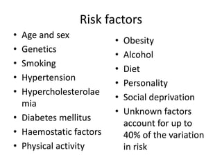
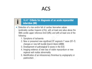
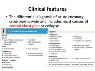
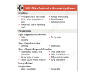
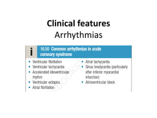
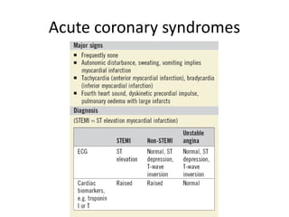
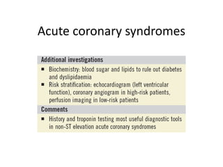
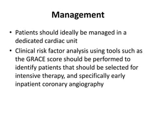
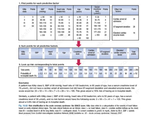
CAD
Dr Md Main Uddin
Assistant Professor
(Medicine)
Cox’s Bazar Medical
College
CVS
SL Content
1 Investigations for the disease of CVS-ECG, Echocardiography
2 CAD - Angina pectoris
3 Acute coronary syndrome- MI, UA
4 Shock - assessment and management, Anaphylaxis
5 Acute rheumatic fever and rheumatic heart diseases
6 Valvular heart diseases MS,MR,TS,TR,AS,AR
7 Infective endocarditis
79 Basic facts of immunology, Auto immunity, Allergy &
hypersensitivity, Transplantation,
80 Prevention and early detection of common cancers, Primary
cancer treatment including -Surgery and radiation,
Chemotherapy, Adjuvent therapy
81 Evaluation of tumour response including-Tumour size,
Tumour markers, General well being and performance status.
CAD
Risk factors
• Age and sex
• Genetics
• Smoking
• Hypertension
• Hypercholesterolae
mia
• Diabetes mellitus
• Haemostatic factors
• Physical activity
• Obesity
• Alcohol
• Diet
• Personality
• Social deprivation
• Unknown factors
account for up to
40% of the variation
in risk
Angina pectoris
• Angina pectoris is a symptom complex caused
by transient myocardial ischaemia, which
occurs whenever there is an imbalance
between myocardial oxygen supply and
demand.
Pathogenesis
• Atherosclerosis, aortic valve disease,
hypertrophic cardiomyopathy, vasculitis or
aortitis.
Clinical features
• Stable angina is
characterised by central
chest pain, discomfort
or breathlessness that is
predictably precipitated
by exertion or other
forms of stress and is
promptly relieved by
rest
Clinical features
• Physical examination is frequently
unremarkable but should include a careful
search for evidence of valve disease
(particularly aortic), important risk factors
(hypertension, diabetes mellitus), left
ventricular dysfunction (cardiomegaly, gallop
rhythm), other manifestations of arterial
disease (carotid bruits, peripheral arterial
disease), and unrelated conditions that may
exacerbate angina (anaemia, thyrotoxicosis).
Investigations
• The first-line investigation is an exercise ECG
• Myocardial perfusion scanning
• Stress echocardiography
• CT coronary arteriography
• Coronary angiography
Management Principles of management
involve:
• a careful assessment of
the extent and severity of
arterial disease
• identification and
treatment of risk factors
• advice on smoking
cessation
• introduction of drug
treatment for symptom
control
• identification of high-risk
patients for treatment to
improve life expectancy
.
Management Anti-anginal drug therapy
• Low-dose (75 mg) aspirin
• Statin, even if cholesterol
is normal
• Anti-anginal drug therapy
• Non-pharmacological
treatments -
Percutaneous coronary
intervention, coronary
artery bypass grafting
• Six groups of drug can be
used in the prevention
and treatment of angina
but there is little evidence
that one group is more
effective than another
• Nitrates, Beta-blockers,
Calcium channel
antagonists, Potassium
channel activators, Ion
channel modulator,
Ranolazine
Acute coronary syndrome (ACS)
• Acute coronary syndrome is a term that
encompasses both unstable angina and
myocardial infarction.
• Unstable angina is characterised by new-onset
or rapidly worsening angina (crescendo
angina), angina on minimal exertion or angina
at rest in the absence of myocardial damage.
ACS
Clinical features
• The differential diagnosis of acute coronary
syndrome is wide and includes most causes of
central chest pain or collapse
Clinical features
Arrhythmias
Clinical features
• Recurrent angina
• Acute heart failure
• Pericarditis
• Dressler’s syndrome
• Embolism
Investigations
Electrocardiogram - The standard 12-lead ECG
Lateral MI
Reciprocal changes
Inferolateral MI
ST elevation II, III, aVF
ST depression in aVL, V1-V3 are reciprocal changes
Anterolateral / Inferior Ischemia
LVH, AV junctional rhythm, bradycardia
Supraventricular Tachycardia
Narrow complex, regular; retrograde P waves, rate <220
Retrograde P waves
Right Ventricular Myocardial Infarction
Found in 1/3 of patients with inferior MI
Increased morbidity and mortality
ST elevation in V4-V6 of Right-sided EKG
Ventricular Tachycardia
Question 4
 What is the rate, rhythm and axis?
 Any other abnormalities?
 How would you manage this patient?
Question 5
 What is the rate, rhythm and axis?
 What is the main problem with the patient?
Investigations
• Cardiac biomarkers-
Levels of troponins T
and I increase within 3–
6 hours, peak at about
36 hours and remain
elevated for up to 2
weeks.
• Radiography
• Echocardiography
• Coronary angiography
Acute coronary syndromes
Management
• Patients should ideally be managed in a
dedicated cardiac unit
• Clinical risk factor analysis using tools such as
the GRACE score should be performed to
identify patients that should be selected for
intensive therapy, and specifically early
inpatient coronary angiography
Management
• Analgesia - morphine sulphate 5–10 mg iv
• Reperfusion therapy - Immediate reperfusion therapy
with PCI is indicated when the ECG shows new bundle
branch block or characteristic ST-segment elevation
• Thrombolytic therapy - If primary PCI cannot be
achieved in a timely manner, thrombolytic therapy
should be administered.
Management
• Antithrombotic therapy - oral 75–325 mg aspirin daily.
A P2Y12 receptor antagonist should be given in
combination with aspirin for up to 12 months e.g.
ticagrelor (180 mg, followed by 90 mg twice daily)
• Anticoagulation can be achieved using unfractionated
heparin, fractioned (low-molecular-weight) heparin or
a pentasaccharide such as subcutaneous fondaparinux
(2.5 mg daily).
• Anticoagulation should be continued for 8 days or until
discharge from hospital or coronary revascularisation
has been completed.
Management
• Anti-anginal therapy - Sublingual glyceryl
trinitrate (300–500 μg) or intravenous nitrates
(glyceryl trinitrate 0.6–1.2 mg/hr.
• Intravenous β-blockers (atenolol 5–10 mg or
metoprolol 5–15 mg
• Renin–angiotensin blockade - ACE inhibitors such
as enalapril (10 mg twice daily) or ramipril (2.5–5
mg twice daily)
• In patients intolerant of ACE inhibitors, ARBs such
as valsartan (40–160 mg twice daily) or
candesartan (4–16 mg daily) are alternatives.
Management
• Mineralocorticoid receptor antagonists –
spironolactone (25–50 mg daily).
• Lipid-lowering therapy – HMG CoA reductase
enzyme inhibitors (statins)
• Smoking cessation
• Diet and exercise - Mediterranean-style diet
• Rehabilitation - Restrict physical activities for 4–6
weeks. When there are no complications, the
patient can mobilise on the second day, return
home in 2–3 days and gradually increase activity,
with the aim of returning to work in 4 weeks.
Coronary Artery Disease

Coronary Artery Disease

  • 1.
    CAD Dr Md MainUddin Assistant Professor (Medicine) Cox’s Bazar Medical College
  • 2.
    CVS SL Content 1 Investigationsfor the disease of CVS-ECG, Echocardiography 2 CAD - Angina pectoris 3 Acute coronary syndrome- MI, UA 4 Shock - assessment and management, Anaphylaxis 5 Acute rheumatic fever and rheumatic heart diseases 6 Valvular heart diseases MS,MR,TS,TR,AS,AR 7 Infective endocarditis 79 Basic facts of immunology, Auto immunity, Allergy & hypersensitivity, Transplantation, 80 Prevention and early detection of common cancers, Primary cancer treatment including -Surgery and radiation, Chemotherapy, Adjuvent therapy 81 Evaluation of tumour response including-Tumour size, Tumour markers, General well being and performance status.
  • 3.
  • 4.
    Risk factors • Ageand sex • Genetics • Smoking • Hypertension • Hypercholesterolae mia • Diabetes mellitus • Haemostatic factors • Physical activity • Obesity • Alcohol • Diet • Personality • Social deprivation • Unknown factors account for up to 40% of the variation in risk
  • 5.
    Angina pectoris • Anginapectoris is a symptom complex caused by transient myocardial ischaemia, which occurs whenever there is an imbalance between myocardial oxygen supply and demand. Pathogenesis • Atherosclerosis, aortic valve disease, hypertrophic cardiomyopathy, vasculitis or aortitis.
  • 6.
    Clinical features • Stableangina is characterised by central chest pain, discomfort or breathlessness that is predictably precipitated by exertion or other forms of stress and is promptly relieved by rest
  • 7.
    Clinical features • Physicalexamination is frequently unremarkable but should include a careful search for evidence of valve disease (particularly aortic), important risk factors (hypertension, diabetes mellitus), left ventricular dysfunction (cardiomegaly, gallop rhythm), other manifestations of arterial disease (carotid bruits, peripheral arterial disease), and unrelated conditions that may exacerbate angina (anaemia, thyrotoxicosis).
  • 8.
    Investigations • The first-lineinvestigation is an exercise ECG • Myocardial perfusion scanning • Stress echocardiography • CT coronary arteriography • Coronary angiography
  • 9.
    Management Principles ofmanagement involve: • a careful assessment of the extent and severity of arterial disease • identification and treatment of risk factors • advice on smoking cessation • introduction of drug treatment for symptom control • identification of high-risk patients for treatment to improve life expectancy .
  • 10.
    Management Anti-anginal drugtherapy • Low-dose (75 mg) aspirin • Statin, even if cholesterol is normal • Anti-anginal drug therapy • Non-pharmacological treatments - Percutaneous coronary intervention, coronary artery bypass grafting • Six groups of drug can be used in the prevention and treatment of angina but there is little evidence that one group is more effective than another • Nitrates, Beta-blockers, Calcium channel antagonists, Potassium channel activators, Ion channel modulator, Ranolazine
  • 12.
    Acute coronary syndrome(ACS) • Acute coronary syndrome is a term that encompasses both unstable angina and myocardial infarction. • Unstable angina is characterised by new-onset or rapidly worsening angina (crescendo angina), angina on minimal exertion or angina at rest in the absence of myocardial damage.
  • 13.
  • 14.
    Clinical features • Thedifferential diagnosis of acute coronary syndrome is wide and includes most causes of central chest pain or collapse
  • 16.
  • 17.
    Clinical features • Recurrentangina • Acute heart failure • Pericarditis • Dressler’s syndrome • Embolism
  • 18.
  • 20.
  • 21.
    Inferolateral MI ST elevationII, III, aVF ST depression in aVL, V1-V3 are reciprocal changes
  • 22.
    Anterolateral / InferiorIschemia LVH, AV junctional rhythm, bradycardia
  • 23.
    Supraventricular Tachycardia Narrow complex,regular; retrograde P waves, rate <220 Retrograde P waves
  • 24.
    Right Ventricular MyocardialInfarction Found in 1/3 of patients with inferior MI Increased morbidity and mortality ST elevation in V4-V6 of Right-sided EKG
  • 25.
  • 29.
    Question 4  Whatis the rate, rhythm and axis?  Any other abnormalities?  How would you manage this patient?
  • 30.
    Question 5  Whatis the rate, rhythm and axis?  What is the main problem with the patient?
  • 31.
    Investigations • Cardiac biomarkers- Levelsof troponins T and I increase within 3– 6 hours, peak at about 36 hours and remain elevated for up to 2 weeks. • Radiography • Echocardiography • Coronary angiography
  • 32.
  • 33.
  • 34.
  • 35.
    Management • Patients shouldideally be managed in a dedicated cardiac unit • Clinical risk factor analysis using tools such as the GRACE score should be performed to identify patients that should be selected for intensive therapy, and specifically early inpatient coronary angiography
  • 37.
    Management • Analgesia -morphine sulphate 5–10 mg iv • Reperfusion therapy - Immediate reperfusion therapy with PCI is indicated when the ECG shows new bundle branch block or characteristic ST-segment elevation • Thrombolytic therapy - If primary PCI cannot be achieved in a timely manner, thrombolytic therapy should be administered.
  • 38.
    Management • Antithrombotic therapy- oral 75–325 mg aspirin daily. A P2Y12 receptor antagonist should be given in combination with aspirin for up to 12 months e.g. ticagrelor (180 mg, followed by 90 mg twice daily) • Anticoagulation can be achieved using unfractionated heparin, fractioned (low-molecular-weight) heparin or a pentasaccharide such as subcutaneous fondaparinux (2.5 mg daily). • Anticoagulation should be continued for 8 days or until discharge from hospital or coronary revascularisation has been completed.
  • 39.
    Management • Anti-anginal therapy- Sublingual glyceryl trinitrate (300–500 μg) or intravenous nitrates (glyceryl trinitrate 0.6–1.2 mg/hr. • Intravenous β-blockers (atenolol 5–10 mg or metoprolol 5–15 mg • Renin–angiotensin blockade - ACE inhibitors such as enalapril (10 mg twice daily) or ramipril (2.5–5 mg twice daily) • In patients intolerant of ACE inhibitors, ARBs such as valsartan (40–160 mg twice daily) or candesartan (4–16 mg daily) are alternatives.
  • 40.
    Management • Mineralocorticoid receptorantagonists – spironolactone (25–50 mg daily). • Lipid-lowering therapy – HMG CoA reductase enzyme inhibitors (statins) • Smoking cessation • Diet and exercise - Mediterranean-style diet • Rehabilitation - Restrict physical activities for 4–6 weeks. When there are no complications, the patient can mobilise on the second day, return home in 2–3 days and gradually increase activity, with the aim of returning to work in 4 weeks.