
Recommended
More Related Content
Similar to Conjuntivitis.pdf
Similar to Conjuntivitis.pdf (20)
Recently uploaded
Recently uploaded (20)
Conjuntivitis.pdf
- 1. Copyright 2013 American Medical Association. All rights reserved. Conjunctivitis A Systematic Review of Diagnosis and Treatment Amir A. Azari, MD; Neal P. Barney, MD C onjunctiva is a thin, translucent membrane lining the an- terior part of the sclera and inside of the eyelids. It has 2 parts, bulbar and palpebral. The bulbar portion begins at the edge of the cornea and covers the visible part of the sclera; the palpebral part lines the inside of the eyelids (Figure 1). Inflamma- tion or infection of the conjunctiva is known as conjunctivitis and is characterized by dilatation of the conjunctival vessels, resulting in hyperemia and edema of the conjunctiva, typically with associated discharge.1 Conjunctivitisaffectsmanypeopleandimposeseconomicand socialburdens.Itisestimatedthatacuteconjunctivitisaffects6mil- lion people annually in the United States.2 The cost of treating bac- terial conjunctivitis alone was estimated to be $377 million to $857 million per year.3 Many US state health departments, irrespective of the underlying cause of conjunctivitis, require students to be treatedwithtopicalantibioticeyedropsbeforereturningtoschool.4 A majority of conjunctivitis patients are initially treated by pri- mary care physicians rather than eye care professionals. Approxi- mately1%ofallprimarycareofficevisitsintheUnitedStatesarere- latedtoconjunctivitis.5 Approximately70%ofallpatientswithacute conjunctivitis present to primary care and urgent care.6 The prevalence of conjunctivitis varies according to the under- lying cause, which may be influenced by the patient’s age, as well as the season of the year. Viral conjunctivitis is the most common cause of infectious conjunctivitis both overall and in the adult population7-13 and is more prevalent in summer.14 Bacterial con- junctivitis is the second most common cause7-9,12,13 and is respon- sible for the majority (50%-75%) of cases in children14 ; it is observed more frequently from December through April.14 Aller- gic conjunctivitis is the most frequent cause, affecting 15% to 40% of the population,15 and is observed more frequently in spring and summer.14 IMPORTANCE Conjunctivitis is a common problem. OBJECTIVE To examine the diagnosis, management, and treatment of conjunctivitis, including various antibiotics and alternatives to antibiotic use in infectious conjunctivitis and use of antihistamines and mast cell stabilizers in allergic conjunctivitis. EVIDENCE REVIEW A search of the literature published through March 2013, using PubMed, the ISI Web of Knowledge database, and the Cochrane Library was performed. Eligible articles were selected after review of titles, abstracts, and references. FINDINGS Viral conjunctivitis is the most common overall cause of infectious conjunctivitis and usually does not require treatment; the signs and symptoms at presentation are variable. Bacterial conjunctivitis is the second most common cause of infectious conjunctivitis, with most uncomplicated cases resolving in 1 to 2 weeks. Mattering and adherence of the eyelids on waking, lack of itching, and absence of a history of conjunctivitis are the strongest factors associated with bacterial conjunctivitis. Topical antibiotics decrease the duration of bacterial conjunctivitis and allow earlier return to school or work. Conjunctivitis secondary to sexually transmitted diseases such as chlamydia and gonorrhea requires systemic treatment in addition to topical antibiotic therapy. Allergic conjunctivitis is encountered in up to 40% of the population, but only a small proportion of these individuals seek medical help; itching is the most consistent sign in allergic conjunctivitis, and treatment consists of topical antihistamines and mast cell inhibitors. CONCLUSIONS AND RELEVANCE The majority of cases in bacterial conjunctivitis are self-limiting and no treatment is necessary in uncomplicated cases. However, conjunctivitis caused by gonorrhea or chlamydia and conjunctivitis in contact lens wearers should be treated with antibiotics. Treatment for viral conjunctivitis is supportive. Treatment with antihistamines and mast cell stabilizers alleviates the symptoms of allergic conjunctivitis. JAMA. 2013;310(16):1721-1729. doi:10.1001/jama.2013.280318 CME Quiz at jamanetworkcme.com and CME Questions 1732 Author Affiliation: Department of Ophthalmology and Visual Sciences, University of Wisconsin, Madison. Corresponding Author: Amir A. Azari, MD, Department of Ophthalmology, Room F4/349, University of Wisconsin Madison, 600 Highland Ave, Madison, WI 53792 (amirazarimd@gmail.com). Section Editor: Mary McGrae McDermott, MD, Senior Editor. Clinical Review & Education Review jama.com JAMA October 23/30, 2013 Volume 310, Number 16 1721 Copyright 2013 American Medical Association. All rights reserved. Downloaded From: http://jama.jamanetwork.com/ by Rachel Donihoo on 05/13/2015
- 2. Copyright 2013 American Medical Association. All rights reserved. Conjunctivitis can be divided into infectious and noninfectious causes.Virusesandbacteriaarethemostcommoninfectiouscauses. Noninfectious conjunctivitis includes allergic, toxic, and cicatricial conjunctivitis, as well as inflammation secondary to immune- mediateddiseasesandneoplasticprocesses.16 Thediseasecanalso be classified into acute, hyperacute, and chronic according to the mode of onset and the severity of the clinical response.17 Further- more,itcanbeeitherprimaryorsecondarytosystemicdiseasessuch as gonorrhea, chlamydia, graft-vs-host disease, and Reiter syn- drome, in which case systemic treatment is warranted.16 It is important to differentiate conjunctivitis from other sight- threateningeyediseasesthathavesimilarclinicalpresentationand to make appropriate decisions about further testing, treatment, or referral. An algorithmic approach (Figure 2) using a focused ocular history along with a penlight eye examination may be helpful in di- agnosisandtreatment.Becauseconjunctivitisandmanyotherocu- lardiseasescanpresentas“redeye,”thedifferentialdiagnosisofred eyeandknowledgeaboutthetypicalfeaturesofeachdiseaseinthis category are important (Table 1). Methods TheliteraturepublishedthroughMarch2013wasreviewedbysearch- ingPubMed,theISIWebofKnowledgedatabase,andtheCochrane Library.Thefollowingkeywordswereused:bacterialconjunctivitis, viralconjunctivitis,allergicconjunctivitis,treatmentofbacterialcon- junctivitis,andtreatmentofviralconjunctivitis.Nolanguagerestric- tionwasapplied.ArticlespublishedbetweenMarch2003andMarch 2013 were initially screened. After review of titles, abstracts, text, and references for the articles, more were identified and screened. Articlesandmeta-analysesthatprovidedevidence-basedinforma- tionaboutthecause,management,andtreatmentofvarioustypes of conjunctivitis were selected. A total of 86 articles were included in this review. The first study8 was published in 1982 and the last19 in 2012. A level of evidence was assigned to the recommendations presented in Table 2 and Table 3 with the American Heart Associa- tion grading system: “The strongest weight of evidence (A) is as- signediftherearemultiplerandomizedtrialswithlargenumbersof patients. An intermediate weight (B) is assigned if there are a lim- ited number of randomized trials with small numbers of patients, careful analyses of non-randomized studies, or observational reg- istries.Thelowestrankofevidence(C)isassignedwhenexpertcon- sensus is the primary basis for the recommendation.60 How to Differentiate Conjunctivitis of Different Origins History and Physical Examination Focused ocular examination and history are crucial for making ap- propriatedecisionsaboutthetreatmentandmanagementofanyeye Figure 2. Suggested Algorithm for Clinical Approach to Suspected Acute Conjunctivitis Pain? Photophobia? Blurred vision? Constant blurred vision? Suspected acute conjunctivitis (≤4 wk duration) Ophthalmology referral Allergic conjunctivitis Dry eye disease Viral conjunctivitis Dry eye disease Discharge? Bacterial conjunctivitis (nongonococcal) Gonococcal conjunctivitis Hyperpurulent Mucopurulent Serous Yes Yes Yes Yes Yes Yes Yes No No No No No No Itching? No Itching? Figure 1. Normal Conjunctival Anatomy Eyelid Sclera S A G I T TA L C R O S S S E C T I O N Palpebral conjunctiva Palpebral conjunctiva Cornea Bulbar conjunctiva Bulbar conjunctiva Limbus Iris The conjunctiva is a thin membrane covering the sclera (bulbar conjunctiva, labeled with purple) and the inside of the eyelids (palpebral conjunctiva, labeled with blue). Clinical Review & Education Review Review of Conjunctivitis Diagnosis and Treatment 1722 JAMA October 23/30, 2013 Volume 310, Number 16 jama.com Copyright 2013 American Medical Association. All rights reserved. Downloaded From: http://jama.jamanetwork.com/ by Rachel Donihoo on 05/13/2015
- 3. Copyright 2013 American Medical Association. All rights reserved. condition, including conjunctivitis. Eye discharge type and ocular symptoms can be used to determine the cause of the conjunctivitis.61,62 For example, a purulent or mucopurulent dis- charge is often due to bacterial conjunctivitis (Figure 3A and Figure 3B), whereas a watery discharge is more characteristic of vi- ral conjunctivitis (Figure 3C)61,62 ; itching is also associated with al- lergic conjunctivitis.49,63 However, the clinical presentation is often nonspecific. Rely- ing on the type of discharge and patient symptoms does not al- ways lead to an accurate diagnosis. Furthermore, scientific evi- dence correlating conjunctivitis signs and symptoms with the underlying cause is often lacking.61 For example, in a study of pa- tients with culture-positive bacterial conjunctivitis, 58% had itch- ing, 65% had burning, and 35% had serous or no discharge at all,64 illustratingthenonspecificityofthesignsandsymptomsofthisdis- ease. In 2003, a large meta-analysis failed to find any clinical stud- iescorrelatingthesignsandsymptomsofconjunctivitiswiththeun- derlying cause61 ; later, the same authors conducted a prospective study61 and found that a combination of 3 signs—bilateral matter- ing of the eyelids, lack of itching, and no history of conjunctivitis— strongly predicted bacterial conjunctivitis. Having both eyes mat- ter and the lids adhere in the morning was a stronger predictor for positivebacterialcultureresult,andeitheritchingorapreviousepi- sode of conjunctivitis made a positive bacterial culture result less likely.64 In addition, type of discharge (purulent, mucus, or watery) or other symptoms were not specific to any particular class of conjunctivitis.64,65 Although in the primary care setting an ocular examination is often limited because of lack of a slitlamp, useful information may beobtainedwithasimplepenlight.Theeyeexaminationshouldfo- cus on the assessment of the visual acuity, type of discharge, cor- neal opacity, shape and size of the pupil, eyelid swelling, and pres- ence of proptosis. Laboratory Investigations Obtainingconjunctivalculturesisgenerallyreservedforcasesofsus- pected infectious neonatal conjunctivitis, recurrent conjunctivitis, conjunctivitisrecalcitranttotherapy,conjunctivitispresentingwith severe purulent discharge, and cases suspicious for gonococcal or chlamydial infection.16 In-office rapid antigen testing is available for adenoviruses and has 89% sensitivity and up to 94% specificity.66 This test can identify the viral causes of conjunctivitis and prevent unnecessary antibiotic use. Thirty-six percent of conjunctivitis cases are due to adenoviruses, and one study estimated that in-office rapid antigen testing could prevent 1.1 million cases of inappropriate treatment with antibiotics, potentially saving $429 million annually.2 Infectious Conjunctivitis Viral Conjunctivitis Epidemiology, Cause, and Presentation Viruses cause up to 80% of all cases of acute conjunctivitis.8-13,67 The rate of clinical accuracy in diagnosing viral conjunctivitis is less than50%comparedwithlaboratoryconfirmation.49 Manycasesare misdiagnosed as bacterial conjunctivitis.49 Between65%and90%ofcasesofviralconjunctivitisarecaused by adenoviruses,49 and they produce 2 of the common clinical en- tities associated with viral conjunctivitis, pharyngoconjunctival fe- ver and epidemic keratoconjunctivitis.62 Pharyngoconjunctival fe- ver is characterized by abrupt onset of high fever, pharyngitis, and bilateral conjunctivitis, and by periauricular lymph node enlarge- ment,whereasepidemickeratoconjunctivitisismoresevereandpre- sents with watery discharge, hyperemia, chemosis, and ipsilateral lymphadenopathy.68 Lymphadenopathy is observed in up to 50% Table 1. Selected Nonconjunctivitis Causes of Red Eyea Differential Diagnosis Symptoms Penlight Examination Findings Dry eye disease Burning and foreign-body sensation. Symptoms are usu- ally transient, worse with prolonged reading or watching television because of decreased blinking. Symptoms are worse in dry, cold, and windy environments because of increased evaporation. Bilateral redness Blepharitis Similar to dry eyes Redness greater at the margins of eyelids Uveitis Photophobia, pain, blurred vision. Symptoms are usually bilateral. Decreased vision, poorly reacting pupils, constant eye pain radiating to temple and brow. Redness, severe photophobia, presence of inflammatory cells in the anterior chamber. Angle closure glaucoma Headaches, nausea, vomiting, ocular pain, decreased vision, light sensitivity, and seeing haloes around lights. Symptoms are usually unilateral. Firm eye on palpation, ocular redness with limbal injec- tion. Appearance of a hazy/steamy cornea, moderately dilated pupils that are unreactive to light. Carotid cavernous fistula Chronic red eye; may have a history of head trauma Dilated tortuous vessels (corkscrew vessels), bruits on auscultation with a stethoscope Endophthalmitis Severe pain, photophobia, may have a history of eye sur- gery or ocular trauma Redness, pus in the anterior chamber, and photophobia Cellulitis Pain, double vision, and fullness Redness and swelling of lids, may have restriction of the eye movements, may have a history of preceding sinus- itis (usually ethmoiditis) Anterior segment tumors Variable Abnormal growth inside or on the surface of the eye Scleritis Decreased vision, moderate to severe pain Redness, bluish sclera hue Subconjunctival hemorrhage May have foreign-body sensation and tearing or be asymptomatic Blood under the conjunctival membrane a Data are from Cronau et al18 and Leibowitz.1 The examination can be done by shining a penlight in the patient’s affected eye(s). Review of Conjunctivitis Diagnosis and Treatment Review Clinical Review & Education jama.com JAMA October 23/30, 2013 Volume 310, Number 16 1723 Copyright 2013 American Medical Association. All rights reserved. Downloaded From: http://jama.jamanetwork.com/ by Rachel Donihoo on 05/13/2015
- 4. Copyright 2013 American Medical Association. All rights reserved. Table 2. Ophthalmic Therapies for Conjunctivitis Category Epidemiology Type of Discharge Cause Treatment Level of Evidence for Treatment Acute bacterial conjunctivitis 135 case per 10 000 population in US3 18.3%-57% of all acute conjunctivitis7-9,12,13 Mucopurulent S aureus, S epidermidis, H influenzae, S pneumoniae, S viridans, Moraxella spp Aminoglycosides Gentamicin Ointment: 4 ×/d for 1 wk Solution: 1-2 drops 4 ×/d for 1 wk B20-22 Tobramycin ointment: 3 ×/d for 1 wk A23-30 Fluoroquinolones Besifloxacin: 1 drop 3 ×/d for 1 wk A31-34 Ciprofloxacin ointment: 3 ×/d for 1 wk Solution: 1-2 drops 4 ×/d for 1 wk A24,28,29 Gatifloxacin: 3 ×/d for 1 week B35 Levofloxacin: 1-2 drops 4 ×/d for 1 wk B36-38 Moxifloxacin: 3 ×/d for 1 wk A34,39,40 Ofloxacin: 1-2 drops 4 ×/d for 1 wk A37,38,41,42 Macrolides Azithromycin: 2 ×/d for 2 d; then 1 drop daily for 5 d A27,30,43,44 Erythromycin: 4 ×/d for 1 wk B45 Sulfonamides Sulfacetamide ointment: 4 ×/d and at bedtime for 1 wk Solution: 1-2 drops every 2-3 h for 1 wk B22 Combination drops Trimethoprim/polymyxin B: 1 or 2 drops 4 ×/d for 1 wk A22,40,46 Hyperacute bacterial conjunctivitis in adults NA Purulent Neisseria gonorrhoeae Ceftriaxone: 1 g IM once C16,47 Lavage of the infected eye C16 Dual therapy to cover chlamydia is indicated C48 Viral conjunctivitis 9%-80.3% of all acute conjunctivitis8-13 Serous Up to 65% are due to adenovirus strains49 Cold compress Artificial tears Antihistamines C16,50 Herpes zoster virus NA Variable Herpes zoster virus Oral acyclovir 800 mg: 5 ×/d for 7-10 d C16 Oral famciclovir 500 mg: 3 ×/d for 7-10 d C16 Oral valacyclovir 1000 mg: 3 ×/d for 7-10 d C16 Herpes simplex virus 1.3-4.8 of all acute conjunctivitis9-12 Variable Herpes simplex virus Topical acyclovir: 1 drop 9 ×/d C16 Oral acyclovir 400 mg: 5 ×/d for 7-10 d C16 Oral valacyclovir 500 mg: 3 ×/d for 7-10 d C16 Adult inclusion conjunctivitis 1.8%-5.6% of all acute conjunctivitis5,8-11 Variable Chlamydia trachomatis Azithromycin 1 g: orally once B16,51 Doxycycline 100 mg: orally 2 ×/d for 7 d B16,51 Allergic conjunctivitis 90% of all allergic conjunctivitis15 ; up to 40% of population may be affected15 Serous or mucoid Pollens Topical antihistamines Azelastine 0.05%: 1 drop 2 ×/d A52 Emedastine 0.05%: 1 drop 4 ×/d A52 Topical mast cell inhibitors Cromolyn sodium 4%: 1-2 drops every 4-6 h A52 Lodoxamide 0.1%: 1-2 drops 4 ×/d A52 Nedocromil 2%: 1-2 drops 2 ×/d A52 NSAIDs Ketorolac: 1 drop 4 ×/d B53,54 Vasoconstrictor/antihistamine Naphazoline/pheniramine: 1-2 drops up to 4 ×/d B55 Combination drops Ketotifen 0.025%: 1 drop 2-3 ×/d A56,57 Olopatadine 0.1%: 1 drop 2 ×/d A58,59 Abbreviations: IM, intramuscularly; NA, not available; NSAIDs, nonsteroidal anti-inflammatory drugs. Clinical Review & Education Review Review of Conjunctivitis Diagnosis and Treatment 1724 JAMA October 23/30, 2013 Volume 310, Number 16 jama.com Copyright 2013 American Medical Association. All rights reserved. Downloaded From: http://jama.jamanetwork.com/ by Rachel Donihoo on 05/13/2015
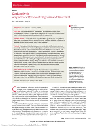
- 5. Copyright 2013 American Medical Association. All rights reserved. of viral conjunctivitis cases and is more prevalent in viral conjuncti- vitis compared with bacterial conjunctivitis.49 Prevention and Treatment Viral conjunctivitis secondary to adenoviruses is highly contagious, andtheriskoftransmissionhasbeenestimatedtobe10%to50%.6,14 The virus spreads through direct contact via contaminated fingers, medicalinstruments,swimmingpoolwater,orpersonalitems;inone study, 46% of infected people had positive cultures grown from swabs of their hands.69 Because of the high rates of transmission, handwashing,strictinstrumentdisinfection,andisolationofthein- fectedpatientsfromtherestoftheclinichasbeenadvocated.70 In- cubation and communicability are estimated to be 5 to 12 days and 10 to 14 days, respectively.14 Although no effective treatment exists, artificial tears, topical antihistamines,orcoldcompressesmaybeusefulinalleviatingsome of the symptoms (Table 2).16,50 Available antiviral medications are notuseful16,50 andtopicalantibioticsarenotindicated.18 Topicalan- tibiotics do not protect against secondary infections, and their use maycomplicatetheclinicalpresentationbycausingallergyandtox- icity,leadingtodelayindiagnosisofotherpossibleoculardiseases.49 Use of antibiotic eyedrops can increase the risk of spreading the in- fection to the other eye from contaminated droppers.49 Increased resistance is also of concern with frequent use of antibiotics.6 Pa- tientsshouldbereferredtoanophthalmologistifsymptomsdonot resolve after 7 to 10 days because of the risk of complications.1 Herpes Conjunctivitis Herpes simplex virus comprises 1.3% to 4.8% of all cases of acute conjunctivitis.9-12 Conjunctivitis caused by the virus is usually uni- lateral.Thedischargeisthinandwatery,andaccompanyingvesicu- lar eyelid lesions may be present. Topical and oral antivirals are rec- ommended(Table2)toshortenthecourseofthedisease.16 Topical corticosteroids should be avoided because they potentiate the vi- rus and may cause harm.16,71 Herpes zoster virus, responsible for shingles, can involve ocu- lar tissue, especially if the first and second branches of the trigemi- nal nerve are involved. Eyelids (45.8%) are the most common site of ocular involvement, followed by the conjunctiva (41.1%).72 Cor- neal complication and uveitis may be present in 38.2% and 19.1% of cases, respectively.72 Patients with suspected eyelid or eye in- volvementorthosepresentingwithHutchinsonsign(vesiclesatthe tip of the nose, which has high correlations with corneal involve- ment) should be referred for a thorough ophthalmic evaluation. Treatmentusuallyconsistsofacombinationoforalantiviralsandtopi- cal steroids.73 Bacterial Conjunctivitis Epidemiology, Cause, and Presentation The incidence of bacterial conjunctivitis was estimated to be 135 in 10 000inonestudy.3 Bacterialconjunctivitiscanbecontracteddi- rectly from infected individuals or can result from abnormal prolif- erationofthenativeconjunctivalflora.17 Contaminatedfingers,14 ocu- logenitalspread,16 andcontaminatedfomites48 arecommonroutes of transmission. In addition, certain conditions such as compro- mised tear production, disruption of the natural epithelial barrier, abnormality of adnexal structures, trauma, and immunosup- pressed status predispose to bacterial conjunctivitis.16 The most commonpathogensforbacterialconjunctivitisinadultsarestaphy- lococcal species, followed by Streptococcus pneumoniae and Hae- mophilus influenzae.41 In children, the disease is often caused by H influenzae, S pneumoniae, and Moraxella catarrhalis.41 The course of the disease usually lasts 7 to 10 days (Figure 3).62 Table 3. Evidence-Based Recommendations in Conjunctivitis Recommendation Level of Evidence Topical antibiotics are effective in reducing the duration of conjunctivitis. A19 Observation is reasonable in most cases of bacterial conjunctivitis (suspected or confirmed) because they often resolve spontane- ously and no treatment is necessary. A41 It is reasonable to use any broad-spectrum antibiotics for treating bacterial conjunctivitis. A19,41 In allergic conjunctivitis, use of topical antihistamines and mast cell stabilizers is recommended. A52 Good hand hygiene can be used to decrease the spread of acute viral conjunctivitis. C16 Bacterial cultures can be useful in cases of severely purulent conjunctivitis or cases that are recalcitrant to therapy. C16 It may be helpful to treat viral conjunctivitis with artificial tears, topical antihistamines, or cold compresses. C16 Topical steroids are not recommended for bacterial conjunctivitis. C65 Figure 3. Characteristic Appearance of Bacterial and Viral Conjunctivitis A Bacterial conjunctivitis B Hyperacute bacterial conjunctivitis C Viral conjunctivitis A, Bacterial conjunctivitis characterized by mucopurulent discharge and conjunctival hyperemia. B, Severe purulent discharge seen in hyperacute bacterial conjunctivitis secondary to gonorrhea. C, Intensely hyperemic response with thin, watery discharge characteristic of viral conjunctivitis. Images reproduced with permission: © 2013 American Academy of Ophthalmology. Review of Conjunctivitis Diagnosis and Treatment Review Clinical Review & Education jama.com JAMA October 23/30, 2013 Volume 310, Number 16 1725 Copyright 2013 American Medical Association. All rights reserved. Downloaded From: http://jama.jamanetwork.com/ by Rachel Donihoo on 05/13/2015
- 6. Copyright 2013 American Medical Association. All rights reserved. Hyperacute bacterial conjunctivitis presents with a severe copious purulent discharge and decreased vision (Figure 3). There is often accompanying eyelid swelling, eye pain on palpation, and preauricular adenopathy. It is often caused by Neisseria gonor- rhoeae and carries a high risk for corneal involvement and subse- quent corneal perforation.17 Treatment for hyperacute conjunctivi- tis secondary to N gonorrhoeae consists of intramuscular ceftriaxone, and concurrent chlamydial infection should be man- aged accordingly.47 Chronicbacterialconjunctivitisisusedtodescribeanyconjunc- tivitis lasting more than 4 weeks, with Staphylococcus aureus, Mo- raxellalacunata,andentericbacteriabeingthemostcommoncauses in this setting62 ; ophthalmologic consultation should be sought for management. Signs and symptoms include red eye, purulent or mucopuru- lent discharge, and chemosis (Figure 3).17 The period of incubation and communicability is estimated to be 1 to 7 days and 2 to 7 days, respectively.14 Bilateral mattering of the eyelids and adherence of theeyelids,lackofitching,andnohistoryofconjunctivitisarestrong positivepredictorsofbacterialconjunctivitis.64 Severepurulentdis- charge should always be cultured and gonococcal conjunctivitis should be considered (Figure 3B).16 Conjunctivitis not responding tostandardantibiotictherapyinsexuallyactivepatientswarrantsa chlamydial evaluation.18 The possibility of bacterial keratitis is high in contact lens wearers, who should be treated with topical antibiotics14 and referred to an ophthalmologist. A patient wearing contact lenses should be asked to immediately remove them.65 Use of Antibiotics in Bacterial Conjunctivitis At least 60% of cases of suspected or culture-proven acute bacte- rial conjunctivitis are self-limiting within 1 to 2 weeks of presentation.14 Although topical antibiotics reduce the duration of the disease, no differences have been observed in outcomes be- tweentreatmentandplacebogroups.Inalargemeta-analysis,19 con- sisting of a review of 3673 patients in 11 randomized clinical trials, there was an approximately 10% increase in the rate of clinical im- provement compared with that for placebo for patients who re- ceivedeither2to5daysor6to10daysofantibiotictreatmentcom- paredwiththeplacebo.Noserioussight-threateningoutcomeswere reported in any of the placebo groups.74 Some highly virulent bac- teria, such as S pneumoniae, N gonorrhoeae, and H influenzae, can penetrate an intact host defense more easily and cause more seri- ous damage.17 Topical antibiotics seem to be more effective in patients who havepositivebacterialcultureresults.Inalargesystemicreview,they werefoundtobeeffectiveatincreasingboththeclinicalandmicro- biologicalcurerateinthegroupofpatientswithculture-provenbac- terial conjunctivitis, whereas only an improved microbial cure rate wasobservedinthegroupofpatientswithclinicallysuspectedbac- terial conjunctivitis.67 Other studies found no significant differ- ence in clinical cure rate when the frequencies of the administered antibiotics were slightly changed.41,75 ChoicesofAntibiotics|Allbroad-spectrumantibioticeyedropsseem in general to be effective in treating bacterial conjunctivitis. There arenosignificantdifferencesinachievingclinicalcurebetweenany ofthebroad-spectrumtopicalantibiotics.Factorsthatinfluencean- tibiotic choice are local availability, patient allergies, resistance pat- terns, and cost. Initial therapy for acute nonsevere bacterial con- junctivitis is listed in Table 2. AlternativestoImmediateAntibioticTherapy|Toourknowledge,no studies have been conducted to evaluate the efficacy of ocular de- congestant, topical saline, or warm compresses for treating bacte- rial conjunctivitis.41 Topical steroids should be avoided because of the risk of potentially prolonging the course of the disease and po- tentiating the infection.16 Summary of Recommendations for Managing Bacterial Conjunctivitis In conclusion, benefits of antibiotic treatment include quicker re- covery, decrease in transmissibility,49 and early return to school.4 Simultaneously,adverseeffectsareabsentifantibioticsarenotused in uncomplicated cases of bacterial conjunctivitis. Therefore, no treatment, a wait-and-see policy, and immediate treatment all ap- pear to be reasonable approaches in cases of uncomplicated con- junctivitis. Antibiotic therapy should be considered in cases of pu- rulent or mucopurulent conjunctivitis and for patients who have distinct discomfort, who wear contact lenses,14,18 who are immu- nocompromised, and who have suspected chlamydial and gono- coccal conjunctivitis. Special Topics in Bacterial Conjunctivitis Methicillin-Resistant S aureus Conjunctivitis It is estimated that 3% to 64% of ocular staphylococcal infections are due to methicillin-resistant S aureus conjunctivitis; this condi- tion is becoming more common and the organisms are resistant to many antibiotics.76 Patients with suspected cases need to be re- ferredtoanophthalmologistandtreatedwithfortifiedvancomycin.77 Chlamydial Conjunctivitis Itisestimatedthat1.8%to5.6%ofallacuteconjunctivitisiscaused by chlamydia,5,8-11 and the majority of cases are unilateral and have concurrent genital infection.1 Conjunctival hyperemia, mucopuru- lentdischarge,andlymphoidfollicleformation51 arehallmarksofthis condition. Discharge is often purulent or mucopurulent.18 How- ever,patientsmoreoftenpresentwithmildsymptomsforweeksto months.Upto54%ofmenand74%ofwomenhaveconcurrentgeni- tal chlamydial infection.78 The disease is often acquired via oculo- genital spread or other intimate contact with infected individuals; in newborns the eyes can be infected after vaginal delivery by in- fected mothers.16 Treatment with systemic antibiotics such as oral azithromycin and doxycycline is efficacious (Table 2); patients and their sexual partners must be treated and a coinfection with gon- orrhea must be investigated. No data support the use of topical an- tibiotictherapyinadditiontosystemictreatment.16 Infantswithchla- mydial conjunctivitis require systemic therapy because more than 50% can have concurrent lung, nasopharynx, and genital tract infection.16 Gonococcal Conjunctivitis Conjunctivitis caused by N gonorrhoeae is a frequent source of hy- peracute conjunctivas in neonates and sexually active adults and youngadolescents.17 Treatmentconsistsofbothtopicalandoralan- tibiotics.Neisseriagonorrhoeaeisassociatedwithahighriskofcor- neal perforation.65 Clinical Review & Education Review Review of Conjunctivitis Diagnosis and Treatment 1726 JAMA October 23/30, 2013 Volume 310, Number 16 jama.com Copyright 2013 American Medical Association. All rights reserved. Downloaded From: http://jama.jamanetwork.com/ by Rachel Donihoo on 05/13/2015
- 7. Copyright 2013 American Medical Association. All rights reserved. Conjunctivitis Secondary to Trachoma Trachoma is caused by Chlamydia trachomatis subtypes A through C and is the leading cause of blindness, affecting 40 million people worldwide in areas with poor hygiene.79,80 Mucopurulent dis- chargeandoculardiscomfortmaybethepresentingsignsandsymp- tomsinthiscondition.Latecomplicationssuchasscarringoftheeye- lid,conjunctiva,andcorneamayleadtolossofvision.Treatmentwith a single dose of oral azithromycin (20 mg/kg) is effective. Patients may also be treated with topical antibiotic ointments for 6 weeks (ie, tetracycline or erythromycin). Systemic antibiotics other than azithromycin,suchastetracyclineorerythromycinfor3weeks,may be used alternatively.79,80 Noninfectious Conjunctivitis Allergic Conjunctivitis Prevalence and Cause Allergic conjunctivitis is the inflammatory response of the conjunc- tiva to allergens such as pollen, animal dander, and other environ- mental antigens15 and affects up to 40% of the population in the UnitedStates15 ;onlyabout10%ofindividualswithallergicconjunc- tivitis seek medical attention, and the entity is often underdiagnosed.81 Redness and itching are the most consistent symptoms.15 Seasonal allergic conjunctivitis comprises 90% of all allergic conjunctivitis in the United States.82 Treatment Treatment consists of avoidance of the offending antigen52 and use of saline solution or artificial tears to physically dilute and remove the allergens.15 Topical decongestants, antihistamines,52 mast cell stabilizers,52 nonsteroidal anti-inflammatory drugs,53,54 and corticosteroids82 may be indicated. In a large systemic review, both antihistamines and mast cell stabilizers were superior to placebo in reducing the symptoms of allergic conjunc- tivitis; researchers also found that antihistamines were superior to mast cell stabilizers in providing short-term benefits.52 Long-term use of the antihistamine antazoline and the vasocon- strictor naphazoline should be avoided because they both can cause rebound hyperemia.52 Steroids should be used with cau- tion and judiciously. Topical steroids are associated with forma- tion of cataract and can cause an increase in eye pressure, leading to glaucoma. Drug-, Chemical-, and Toxin-Induced Conjunctivitis A variety of topical medications such as antibiotic eyedrops, topi- calantiviralmedications,andlubricatingeyedropscaninducealler- gic conjunctival responses largely because of the presence of ben- zalkoniumchlorideineyedroppreparations.83 Cessationofreceiving the offending agent leads to resolution of symptoms.16 Systemic Diseases Associated With Conjunctivitis A variety of systemic diseases, including mucous membrane pem- phigoid, Sjögren syndrome, Kawasaki disease,84 Stevens-Johnson syndrome,85 andcarotidcavernousfistula,86 canpresentwithsigns and symptoms of conjunctivitis, such as conjunctival redness and discharge.Therefore,theabovecausesshouldbeconsideredinpa- tientspresentingwithconjunctivitis.Forexample,patientswithlow- grade carotid cavernous fistula can present with chronic conjuncti- vitisrecalcitranttomedicaltherapy,which,ifleftuntreated,canlead to death. Ominous Signs As recommended by the American Academy of Ophthalmology,16 patientswithconjunctivitiswhoareevaluatedbynonophthalmolo- gisthealthcarepractitionersshouldbereferredpromptlytoanoph- thalmologist if any of the following develops: visual loss, moderate orseverepain,severepurulentdischarge,cornealinvolvement,con- junctivalscarring,lackofresponsetotherapy,recurrentepisodesof conjunctivitis, or history of herpes simplex virus eye disease. In ad- dition,thefollowingpatientsshouldbeconsideredforreferral:con- tact lens wearers, patients requiring steroids, and those with pho- tophobia.Patientsshouldbereferredtoanophthalmologistifthere is no improvement after 1 week.1 Importance of Not Using Antibiotic/Steroid Combination Drops Steroid drops or combination drops containing steroids should not be used routinely. Steroids can increase the latency of the adeno- viruses,thereforeprolongingthecourseofviralconjunctivitis.Inad- dition, if an undiagnosed corneal ulcer secondary to herpes, bacte- ria, or fungus is present, steroids can worsen the condition, leading to corneal melt and blindness. Conclusions Approximately 1% of all patient visits to a primary care clinician are conjunctivitis related, and the estimated cost of the bacterial con- junctivitis alone is $377 million to $857 million annually.3,5 Relying on the signs and symptoms often leads to an inaccurate diagnosis. Nonherpetic viral conjunctivitis followed by bacterial conjunctivi- tis is the most common cause for infectious conjunctivitis.7-13 Aller- gic conjunctivitis affects nearly 40% of the population, but only a small proportion seeks medical care.15,81 The majority of viral con- junctivitiscasesareduetoadenovirus.49 Thereisnorolefortheuse oftopicalantibioticsinviralconjunctivitis,andtheyshouldbeavoided becauseofadversetreatmenteffects.6,49 Usingarapidantigentest to diagnose viral conjunctivitis and avoid inappropriate use of anti- biotics is an appropriate strategy.66 Bacterial pathogens are iso- latedinonly50%ofcasesofsuspectedconjunctivitis,18 andatleast 60% of bacterial conjunctivitis (clinically suspected or culture proven) is self-limited without treatment.14 Cultures are useful in cases that do not respond to therapy, cases of hyperacute conjunc- tivitis, and suspected chlamydial conjunctivitis.16 Treatment with topical antibiotics is usually recommended for contact lens wear- ers, those with mucopurulent discharge and eye pain, suspected casesofchlamydialandgonococcalconjunctivitis,andpatientswith preexisting ocular surface disease.14,18 The advantages of antibi- oticuseincludeearlyresolutionofthedisease,19 earlyreturntowork or school,4,14 and the possibility of decreased complications from conjunctivitis.14 The majority of cases of allergic conjunctivitis are duetoseasonalallergies.82 Antihistamines,mastcellinhibitors,and topical steroids (in selected cases) are indicated for treating aller- gic conjunctivitis.82 Steroids must be used judiciously and only af- terathoroughophthalmologicexaminationhasbeenperformedto Review of Conjunctivitis Diagnosis and Treatment Review Clinical Review & Education jama.com JAMA October 23/30, 2013 Volume 310, Number 16 1727 Copyright 2013 American Medical Association. All rights reserved. Downloaded From: http://jama.jamanetwork.com/ by Rachel Donihoo on 05/13/2015
- 8. Copyright 2013 American Medical Association. All rights reserved. ruleoutherpeticinfectionorcornealinvolvement,bothofwhichcan worsen with steroids.16,71 Physicians must be vigilant to not overlook sight-threatening conditionswithsimilaritiestoconjunctivitis,assummarizedinTable1. ARTICLE INFORMATION Conflict of Interest Disclosures: All authors have completed and submitted the ICMJE Form for Disclosure of Potential Conflicts of Interest and none were reported. Funding/Support: This work was supported by National Institutes of Health (NIH) grant P30-EY016665 (Core Grant for Vision Research) and an unrestricted department award from Research to Prevent Blindness. The project was also supported by the Clinical and Translational Science Award program through the NIH National Center for Advancing Translational Sciences, grant UL1TR000427. Role of the Sponsor: The sponsors played no role in thedesignandconductofthestudy;collection,man- agement,analysis,andinterpretationofthedata; preparation,review,orapprovalofthemanuscript; anddecisiontosubmitthemanuscriptforpublication. Correction: This article was corrected on December 5, 2013, to correct the dosage of acyclovir for herpes in Table 2 and to update the algorithm in Figure 2 to include viral conjunctivitis. Submissions:We encourage authors to submit papers for consideration as a Review. Please contact Mary McGrae McDermott, MD, at mdm608 @northwestern.edu. REFERENCES 1. Leibowitz HM. The red eye. N Engl J Med. 2000;343(5):345-351. 2. Udeh BL, Schneider JE, Ohsfeldt RL. Cost effectiveness of a point-of-care test for adenoviral conjunctivitis. Am J Med Sci. 2008;336(3):254-264. 3. Smith AF, Waycaster C. Estimate of the direct and indirect annual cost of bacterial conjunctivitis in the United States. BMC Ophthalmol. 2009;9:13. 4. Ohnsman CM. Exclusion of students with conjunctivitis from school: policies of state departments of health. J Pediatr Ophthalmol Strabismus. 2007;44(2):101-105. 5. Shields T, Sloane PD. A comparison of eye problems in primary care and ophthalmology practices. Fam Med. 1991;23(7):544-546. 6. Kaufman HE. Adenovirus advances: new diagnostic and therapeutic options. Curr Opin Ophthalmol. 2011;22(4):290-293. 7. Hørven I. Acute conjunctivitis: a comparison of fusidic acid viscous eye drops and chloramphenicol. Acta Ophthalmol (Copenh). 1993;71(2):165-168. 8. Stenson S, Newman R, Fedukowicz H. Laboratory studies in acute conjunctivitis. Arch Ophthalmol. 1982;100(8):1275-1277. 9. Rönnerstam R, Persson K, Hansson H, Renmarker K. Prevalence of chlamydial eye infection in patients attending an eye clinic, a VD clinic, and in healthy persons. Br J Ophthalmol. 1985;69(5):385-388. 10. Harding SP, Mallinson H, Smith JL, Clearkin LG. Adult follicular conjunctivitis and neonatal ophthalmia in a Liverpool eye hospital, 1980-1984. Eye (Lond). 1987;1(pt 4):512-521. 11. Uchio E, Takeuchi S, Itoh N, et al. Clinical and epidemiological features of acute follicular conjunctivitis with special reference to that caused by herpes simplex virus type 1. Br J Ophthalmol. 2000;84(9):968-972. 12. Woodland RM, Darougar S, Thaker U, et al. Causes of conjunctivitis and keratoconjunctivitis in Karachi, Pakistan. Trans R Soc Trop Med Hyg. 1992;86(3):317-320. 13. Fitch CP, Rapoza PA, Owens S, et al. Epidemiology and diagnosis of acute conjunctivitis at an inner-city hospital. Ophthalmology. 1989;96(8):1215-1220. 14. Høvding G. Acute bacterial conjunctivitis. Acta Ophthalmol. 2008;86(1):5-17. 15. Bielory BP, O’Brien TP, Bielory L. Management of seasonal allergic conjunctivitis: guide to therapy. Acta Ophthalmol. 2012;90(5):399-407. 16. American Academy of Ophthalmology; Cornea/External Disease Panel. Preferred Practice Pattern Guidelines: Conjunctivitis-Limited Revision. San Francisco, CA: American Academy of Ophthalmology; 2011. 17. Mannis MJ, Plotnik RD. Bacterial conjunctivitis. In: Tasman W, Jaeger EA, eds. Duanes Ophthalmology on CD-ROM. Lippincott Williams & Wilkins; 2006. 18. Cronau H, Kankanala RR, Mauger T. Diagnosis and management of red eye in primary care. Am Fam Physician. 2010;81(2):137-144. 19. Sheikh A, Hurwitz B, van Schayck CP, McLean S, Nurmatov U. Antibiotics versus placebo for acute bacterial conjunctivitis. Cochrane Database Syst Rev. 2012;9:CD001211. 20. Montero J, Perea E. A double-blind double-dummy comparison of topical lomefloxacin 0.3% twice daily with topical gentamicin 0.3% four times daily in the treatment of acute bacterial conjunctivitis. J Clin Res. 1998;1:29-39. 21. Papa V, Aragona P, Scuderi AC, et al. Treatment of acute bacterial conjunctivitis with topical netilmicin. Cornea. 2002;21(1):43-47. 22. Lohr JA, Austin RD, Grossman M, Hayden GF, Knowlton GM, Dudley SM. Comparison of three topical antimicrobials for acute bacterial conjunctivitis. Pediatr Infect Dis J. 1988;7(9):626-629. 23. Huerva V, Ascaso FJ, Latre B. Tolerancia y eficacia de la tobramicina topica vs cloranfenicol en el tratamiento de las conjunctivitis bacterianas. Ciencia Pharmaceutica. 1991;1:221-224. 24. Alves MRKJ. Evaluation of the clinical and microbiological efficacy of 0.3% ciprofloxacin drops and 0.3% tobramycin drops in the treatment of acute bacterial conjunctivitis. Rev Bras Oftalmol. 1993;52:371-377. 25. Gallenga PE, Lobefalo L, Colangelo L, et al. Topical lomefloxacin 0.3% twice daily versus tobramycin 0.3% in acute bacterial conjunctivitis: a multicenter double-blind phase III study. Ophthalmologica. 1999;213(4):250-257. 26. Jackson WB, Low DE, Dattani D, Whitsitt PF, Leeder RG, MacDougall R. Treatment of acute bacterial conjunctivitis: 1% fusidic acid viscous drops vs 0.3% tobramycin drops. Can J Ophthalmol. 2002;37(4):228-237; discussion 237. 27. Bremond-Gignac D, Mariani-Kurkdjian P, Beresniak A, et al. Efficacy and safety of azithromycin 1.5% eye drops for purulent bacterial conjunctivitis in pediatric patients. Pediatr Infect Dis J. 2010;29(3):222-226. 28. Leibowitz HM. Antibacterial effectiveness of ciprofloxacin 0.3% ophthalmic solution in the treatment of bacterial conjunctivitis. Am J Ophthalmol. 1991;112(4)(suppl):29S-33S. 29. Gross RD, Hoffman RO, Lindsay RN. A comparison of ciprofloxacin and tobramycin in bacterial conjunctivitis in children. Clin Pediatr (Phila). 1997;36(8):435-444. 30. DenisF,ChaumeilC,GoldschmidtP,etal.Micro- biologicalefficacyof3-daytreatmentwithazithromy- cin1.5%eye-dropsforpurulentbacterialconjunctivi- tis.EurJOphthalmol.2008;18(6):858-868. 31. Silverstein BE, Allaire C, Bateman KM, et al. Efficacy and tolerability of besifloxacin ophthalmic suspension 0.6% administered twice daily for 3 days in the treatment of bacterial conjunctivitis: a multicenter, randomized, double-masked, vehicle-controlled, parallel-group study in adults and children. Clin Ther. 2011;33(1):13-26. 32. Karpecki P, Depaolis M, Hunter JA, et al. Besifloxacin ophthalmic suspension 0.6% in patients with bacterial conjunctivitis: a multicenter, prospective, randomized, double-masked, vehicle-controlled, 5-day efficacy and safety study. Clin Ther. 2009;31(3):514-526. 33. Tepedino ME, Heller WH, Usner DW, et al. Phase III efficacy and safety study of besifloxacin ophthalmic suspension 0.6% in the treatment of bacterial conjunctivitis. Curr Med Res Opin. 2009;25(5):1159-1169. 34. McDonald MB, Protzko EE, Brunner LS, et al. Efficacy and safety of besifloxacin ophthalmic suspension 0.6% compared with moxifloxacin ophthalmic solution 0.5% for treating bacterial conjunctivitis. Ophthalmology. 2009;116(9):1615-1623; e1. 35. Gong L, Sun XH, Qiu XD, et al. Comparative research of the efficacy of the gatifloxacin and levofloxacin for bacterial conjunctivitis in human eyes [in Chinese]. Zhonghua Yan Ke Za Zhi. 2010;46(6):525-531. 36. Hwang DG, Schanzlin DJ, Rotberg MH, et al. A phase III, placebo controlled clinical trial of 0.5% levofloxacin ophthalmic solution for the treatment of bacterial conjunctivitis. Br J Ophthalmol. 2003;87(8):1004-1009. 37. Schwab IR, Friedlaender M, McCulley J, et al. A phase III clinical trial of 0.5% levofloxacin ophthalmic solution versus 0.3% ofloxacin ophthalmic solution for the treatment of bacterial conjunctivitis. Ophthalmology. 2003;110(3): 457-465. 38. Zhang M, Hu Y, Chen F. Clinical investigation of 0.3% levofloxacin eyedrops on the treatment of cases with acute bacterial conjunctivitis and bacterial keratitis [in Chinese]. Yan Ke Xue Bao. 2000;16(2):146-148. 39. Gross RD, Lichtenstein SJ, Schlech BA. Early clinical and microbiological responses in the treatment of bacterial conjunctivitis with moxifloxacin ophthalmic solution 0.5% (Vigamox) Clinical Review & Education Review Review of Conjunctivitis Diagnosis and Treatment 1728 JAMA October 23/30, 2013 Volume 310, Number 16 jama.com Copyright 2013 American Medical Association. All rights reserved. Downloaded From: http://jama.jamanetwork.com/ by Rachel Donihoo on 05/13/2015
- 9. Copyright 2013 American Medical Association. All rights reserved. using BID dosing. Todays Ther Trends. 2003;21:227-237. 40. Granet DB, Dorfman M, Stroman D, Cockrum P. A multicenter comparison of polymyxin B sulfate/trimethoprim ophthalmic solution and moxifloxacin in the speed of clinical efficacy for the treatment of bacterial conjunctivitis. J Pediatr Ophthalmol Strabismus. 2008;45(6):340-349. 41. Epling J, Smucny J. Bacterial conjunctivitis. Clin Evid. 2005;2(14):756-761. 42. Tabbara KF, El-Sheikh HF, Islam SM, Hammouda E. Treatment of acute bacterial conjunctivitis with topical lomefloxacin 0.3% compared to topical ofloxacin 0.3%. Eur J Ophthalmol. 1999;9(4):269-275. 43. Abelson MB, Heller W, Shapiro AM, et al. Clinical cure of bacterial conjunctivitis with azithromycin 1%: vehicle-controlled, double-masked clinical trial. Am J Ophthalmol. 2008;145(6):959-965. 44. Cochereau I, Meddeb-Ouertani A, Khairallah M, et al. 3-Day treatment with azithromycin 1.5% eye drops versus 7-day treatment with tobramycin 0.3% for purulent bacterial conjunctivitis: multicentre, randomised and controlled trial in adults and children. Br J Ophthalmol. 2007;91(4):465-469. 45. Hallett JW, Leopold IH. Clinical trial of erythromycin ophthalmic ointment. Am J Ophthalmol. 1957;44(4 pt 1):519-522. 46. Trimethoprim-Polymyxin B Sulphate Ophthalmic Ointment Study Group. Trimethoprim-polymyxin B sulphate ophthalmic ointment versus chloramphenicol ophthalmic ointment in the treatment of bacterial conjunctivitis. J Antimicrob Chemother. 1989;23(2):261-266. 47. Workowski KA, Berman S; Centers for Disease Control and Prevention (CDC). Sexually transmitted diseases treatment guidelines, 2010. MMWR Recomm Rep. 2010;59(RR-12):1-110. 48. Sattar SA, Dimock KD, Ansari SA, Springthorpe VS. Spread of acute hemorrhagic conjunctivitis due to enterovirus-70: effect of air temperature and relative humidity on virus survival on fomites. J Med Virol. 1988;25(3):289-296. 49. O’Brien TP, Jeng BH, McDonald M, Raizman MB. Acute conjunctivitis: truth and misconceptions. Curr Med Res Opin. 2009;25(8):1953-1961. 50. Skevaki CL, Galani IE, Pararas MV, et al. Treatment of viral conjunctivitis with antiviral drugs. Drugs. 2011;71(3):331-347. 51. Katusic D, Petricek I, Mandic Z, et al. Azithromycin vs doxycycline in the treatment of inclusion conjunctivitis. Am J Ophthalmol. 2003;135(4):447-451. 52. Owen CG, Shah A, Henshaw K, et al. Topical treatments for seasonal allergic conjunctivitis: systematic review and meta-analysis of efficacy and effectiveness. Br J Gen Pract. 2004;54(503): 451-456. 53. Yaylali V, Demirlenk I, Tatlipinar S, et al. Comparative study of 0.1% olopatadine hydrochloride and 0.5% ketorolac tromethamine in the treatment of seasonal allergic conjunctivitis. Acta Ophthalmol Scand. 2003;81(4):378-382. 54. Donshik PC, Pearlman D, Pinnas J, et al. Efficacy and safety of ketorolac tromethamine 0.5% and levocabastine 0.05%: a multicenter comparison in patients with seasonal allergic conjunctivitis. Adv Ther. 2000;17(2):94-102. 55. Greiner JV, Udell IJ. A comparison of the clinical efficacy of pheniramine maleate/naphazoline hydrochloride ophthalmic solution and olopatadine hydrochloride ophthalmic solution in the conjunctival allergen challenge model. Clin Ther. 2005;27(5):568-577. 56. Greiner JV, Minno G. A placebo-controlled comparison of ketotifen fumarate and nedocromil sodium ophthalmic solutions for the prevention of ocular itching with the conjunctival allergen challenge model. Clin Ther. 2003;25(7):1988-2005. 57. Greiner JV, Michaelson C, McWhirter CL, Shams NB. Single dose of ketotifen fumarate 025% vs 2 weeks of cromolyn sodium 4% for allergic conjunctivitis. Adv Ther. 2002;19(4):185-193. 58. Butrus S, Greiner JV, Discepola M, Finegold I. Comparison of the clinical efficacy and comfort of olopatadine hydrochloride 0.1% ophthalmic solution and nedocromil sodium 2% ophthalmic solution in the human conjunctival allergen challenge model. Clin Ther. 2000;22(12):1462-1472. 59. Deschenes J, Discepola M, Abelson M. Comparative evaluation of olopatadine ophthalmic solution (0.1%) versus ketorolac ophthalmic solution (0.5%) using the provocative antigen challenge model. Acta Ophthalmol Scand Suppl. 1999;(228):47-52. 60. Gibbons RJ, Smith S, Antman E; American College of Cardiology; American Heart Association. American College of Cardiology/American Heart Association clinical practice guidelines, part I. Circulation. 2003;107(23):2979-2986. 61. Rietveld RP, van Weert HC, ter Riet G, Bindels PJ. Diagnostic impact of signs and symptoms in acute infectious conjunctivitis: systematic literature search. BMJ. 2003;327(7418):789. 62. Disorders of the conjunctiva and limbus. In: Yannof J, Duker JS, eds. Ophthalmology. 2nd ed. Spain: Mosby; 2004:397-412. 63. Morrow GL, Abbott RL. Conjunctivitis. Am Fam Physician. 1998;57(4):735-746. 64. Rietveld RP, ter Riet G, Bindels PJ, Sloos JH, van Weert HC. Predicting bacterial cause in infectious conjunctivitis. BMJ. 2004;329(7459):206-210. 65. Tarabishy AB, Jeng BH. Bacterial conjunctivitis: a review for internists. Cleve Clin J Med. 2008;75(7):507-512. 66. Sambursky R, Tauber S, Schirra F, et al. The RPS adeno detector for diagnosing adenoviral conjunctivitis. Ophthalmology. 2006;113(10):1758- 1764. 67. Epling J. Bacterial conjunctivitis. Clin Evid (Online). 2010;2010. 68. Mahmood AR, Narang AT. Diagnosis and management of the acute red eye. Emerg Med Clin North Am. 2008;26(1):35-55; vi. 69. Azar MJ, Dhaliwal DK, Bower KS, et al. Possible consequences of shaking hands with your patients with epidemic keratoconjunctivitis. Am J Ophthalmol. 1996;121(6):711-712. 70. Warren D, Nelson KE, Farrar JA, et al. A large outbreak of epidemic keratoconjunctivitis: problems in controlling nosocomial spread. J Infect Dis. 1989;160(6):938-943. 71. Wilhelmus KR. Diagnosis and management of herpes simplex stromal keratitis. Cornea. 1987;6(4):286-291. 72. Puri LR, Shrestha GB, Shah DN, Chaudhary M, Thakur A. Ocular manifestations in herpes zoster ophthalmicus. Nepal J Ophthalmol. 2011;3(2):165-171. 73. Sy A, McLeod SD, Cohen EJ, et al. Practice patterns and opinions in the management of recurrent or chronic herpes zoster ophthalmicus. Cornea. 2012;31(7):786-790. 74. Sheikh A, Hurwitz B. Topical antibiotics for acute bacterial conjunctivitis: Cochrane systematic review and meta-analysis update. Br J Gen Pract. 2005;55(521):962-964. 75. Szaflik J, Szaflik JP, Kaminska A; Levofloxacin Bacterial Conjunctivitis Dosage Study Group. Clinical and microbiological efficacy of levofloxacin administered three times a day for the treatment of bacterial conjunctivitis. Eur J Ophthalmol. 2009;19(1):1-9. 76. Shanmuganathan VA, Armstrong M, Buller A, Tullo AB. External ocular infections due to methicillin-resistant Staphylococcus aureus (MRSA). Eye (Lond). 2005;19(3):284-291. 77. Freidlin J, Acharya N, Lietman TM, et al. Spectrum of eye disease caused by methicillin-resistant Staphylococcus aureus. Am J Ophthalmol. 2007;144(2):313-315. 78. Postema EJ, Remeijer L, van der Meijden WI. Epidemiology of genital chlamydial infections in patients with chlamydial conjunctivitis. Genitourin Med. 1996;72(3):203-205. 79. Kumaresan JA, Mecaskey JW. The global elimination of blinding trachoma: progress and promise. Am J Trop Med Hyg. 2003;69(5)(suppl): 24-28. 80. Avery RK, Baker AS. Chlamydial disease. In: Albert and Jakobiec's Principle and Practice of Ophthalmology. 3rd ed. Philadelphia, PA: Saunders Elsevier; 2008:4791-4801. 81. Rosario N, Bielory L. Epidemiology of allergic conjunctivitis. Curr Opin Allergy Clin Immunol. 2011;11(5):471-476. 82. Bielory L. Allergic conjunctivitis: the evolution of therapeutic options. Allergy Asthma Proc. 2012;33(2):129-139. 83. Baudouin C. Allergic reaction to topical eyedrops. Curr Opin Allergy Clin Immunol. 2005;5(5):459-463. 84. Newburger JW, Takahashi M, Gerber MA, et al. Diagnosis, treatment, and long-term management of Kawasaki disease: a statement for health professionals from the Committee on Rheumatic Fever, Endocarditis, and Kawasaki Disease, Council on Cardiovascular Disease in the Young, American Heart Association. Pediatrics. 2004;114(6):1708- 1733. 85. Gregory DG. The ophthalmologic management of acute Stevens-Johnson syndrome. Ocul Surf. 2008;6(2):87-95. 86. Miller NR. Diagnosis and management of dural carotid-cavernous sinus fistulas. Neurosurg Focus. 2007;23(5):13. Review of Conjunctivitis Diagnosis and Treatment Review Clinical Review & Education jama.com JAMA October 23/30, 2013 Volume 310, Number 16 1729 Copyright 2013 American Medical Association. All rights reserved. Downloaded From: http://jama.jamanetwork.com/ by Rachel Donihoo on 05/13/2015