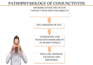
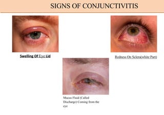
CONJUNCTIVITIS
PRESENTED BY
MD AJMAL SIDDIQUI
DR. OF PHARMACY 2nd
YEAR
Student PU, Gujarat, India
PRESENTATION ON
Inflammation of conjunctiva
Conjunctiva: Thin, translucent, elastic tissue layer with bulbar and palpebral portions
Bulbar: Lines the outer surfaces of the globe to the limbus (junction of sclera and cornea)
Palbepral: Covers the inside of the eyelids
Two layers: Epithelium and Substantia Propria
CONJUNCTIVITIS
1.Infective conjunctivitis: Bacterial, Chlamydial, Viral, Fungal, Rickettsial, Spiro,
Chaetal, Protozoal, Parasitic etc
2.Allergic Conjunctivitis 3.Irritative Conjunctivitis
4.Keratoconjunctivitis associated with diseases of skin and mucous membrane.
5.Traumatic Conjuctivitis
6.Keratoconjunctivitis of unknown etiology (eg: Trachoma)
CONJUNCTIVITIS CLASSIFICATION
Viral conjunctivitis - Adenovirus, HSV
Bacterial conjunctivitis - Staphylococcus aureus, Streptococcus
pneumoniae, Haemophilus influenza, Chlamydia trachomatis
Allergic conjunctivitis - Pollen, perfumes, cosmetics, smoke, dust
Chemical conjunctivitis - Acid or alkali's
CAUSES OF CONJUNCTIVITIS
There are many emergent and non-emergent causes of eye redness. When considering a
diagnosis of conjunctivitis, it is essential to rule out the emergent causes of vision loss.
The differentials for conjunctivitis include:
Glaucoma
Iritis
Keratitis
Episcleritis
Scleritis
Pterygium
Corneal ulcer
Corneal abrasion
Corneal foreign body
Subconjunctival hemorrhage
Blepharitis
Hordeolum
Chalazion
Contact lens overwear
Dry eye
DIFFERENTIAL DIAGNOSIS FOR CONJUNCTIVITIS
SLIT LAMP EXAM
Most of the time, your doctor can diagnose conjunctivitis by using a slit lamp—an instrument that
consists of a microscope and a high-energy beam of light. During a slit-lamp exam, your
ophthalmologist shines a thin beam of light into your eye. This beam allows your doctor to
examine the entire eye, including the conjunctiva; the sclera, or the white of the eye; the iris; and
the cornea.
VISUAL ACUITY TEST
Doctors also check to see if conjunctivitis has affected your vision by conducting a visual acuity
test. This test checks to see how well you can read letters or symbols from 20 feet away, while
covering one eye at a time.
EYE CULTURE
If you have had conjunctivitis for more than two or three weeks and it has not gone away on its
own or with the help of home treatments, your doctor may want to perform an eye culture.
During this test, your doctor takes a sample of the cells on the inside of your eyelids with a cotton
swab and sends it to a laboratory to be examined by a pathologist.
A pathologist, who studies diseases under a microscope, can determine whether your
conjunctivitis is caused by viruses or bacteria.
DIAGNOSIS METHOD FOR CONJUNCTIVITIS
Treatment of both viral and bacterial conjunctivitis should include patient education
to decrease the rate of transmission.
Bacterial conjunctivitis, while typically self-limiting, can be treated to help reduce
the duration of symptoms.
While ointments typically last longer than drops, they tend to interfere with vision.
Initial treatment for acute, non-severe bacterial conjunctivitis varies depending on the
antimicrobial agent but generally is administered to the affected eye every two to 6
hours for 5 to 7 days.
For mild bacterial conjunctivitis, older-generation antibiotics are generally advised.
Later-generation antibiotics are reserved for more grave infections to minimize the
development of resistance in the ocular surface flora.
TREATMEN FOR CONJUNCTIVITIS
Conjunctivitis results from inflammation of the conjunctiva.
The cause of this inflammation can be due to infectious pathogens or non-
infectious irritants.
The result of this irritation or infection is injection or dilation of the conjunctival
vessels; this results in the classic redness or hyperemia and edema of the
conjunctiva.
The entire conjunctiva is involved, and there is often discharge as well.
The quality of discharge varies depending on the causative agent.
 In bacterial conjunctivitis, the surface tissues of the eye are colonized by normal
flora, such as staphylococci, streptococci, and corynebacteria.
The primary defense mechanism against infection is the epithelial covering of the
conjunctiva. Any disruption in this barrier can cause infection.
Secondary defense mechanisms include immune reactions carried out by the tear
film immunoglobulins and lysozyme, conjunctival vasculature, and the rinsing
action of blinking and lacrimation.
PATHOPHYSIOLOGY OF CONJUNCTIVITIS
MICROBES ENTER THE EYE ON
CONTACT WITH INFECTED OBJECTS
INFLAMMATION OF EYE
CONGESTION AND
INCREASED PERMEABILITY
OF BLOOD VESSELS
SWELLING, REDNESS,
EXUDATES AND
DISCHARGE
PATHOPHYSIOLOGY OF CONJUNCTIVITIS
Swelling Of Eye Lid Redness On Sclera(white Part)
Mucus Fluid (Called
Discharge) Coming from the
eye
SIGNS OF CONJUNCTIVITIS
 Redness in one or both eyes.
 Itchiness in one or both eyes.
 A gritty feeling in one or both eyes.
 A discharge in one or both eyes that forms a
crust during the night that may prevent your
eye or eyes from opening in the morning.
 Tearing.
 Sensitivity to light, called photophobia.
 Blurring or reduction of vision in
conjunctivitis is rare(and if at all present,
typically clears with blinking, or cleaning
discharge
Symptoms
TREATMENT FOR CONJUNCTIVITIS
In moderate to severe cases of bacterial conjunctivitis, the
latest- generation fluoroquinolones are more suitable as they provide
strong gram-negative and some gram-positive coverage.
Antibiotic options are available as liquid
ointments. Liquid suspension/solutions
solutions and topical
include polymyxin
levofloxacin,
B/trimethoprim, ciprofloxacin, ofloxacin,
moxifloxacin, gatifloxacin or azithromycin, while bacitracin,
erythromycin or ciprofloxacin can be administered as an ointment.
Fluoroquinolones should be prescribed for contact lens
wearers to provide empiric coverage for Pseudomonas.
The recommended treatment for gonococcal conjunctivitis
is ceftriaxone 1 gram intramuscular (IM), and it is
recommended to treat concurrent chlamydial infection with 1 gm
azithromycin PO as well.
The neonatal dosing for gonococcal conjunctivitis is 25 to 50
mg/kg ceftriaxone intravenous (IV)/IM with a max dose of 125 mg,
with 20 mg/kg azithromycin PO once daily for three days.
REFRENCES
REEFRENCES FOR
Etiology: Statpearls book of author Muhammad F. Hashmi; Bharat Gurnani and
Scarlet Benson. Updated in December 6, 2022.
Sign and Symptoms: Statpearls book of author Muhammad F.
Hashmi; Bharat Gurnani and Scarlet Benson. Updated in December 6, 2022.
Pathophysiology: Statpearls book of author Muhammad F. Hashmi; Bharat
Gurnani and Scarlet Benson. Updated in December 6, 2022..
Differential Diagnosis: Statpearls book of author Muhammad F.
Hashmi; Bharat Gurnani and Scarlet Benson. Updated in December 6, 2022.
Diagnosis Methods: Statpearls book of author Muhammad F.
Hashmi; Bharat Gurnani and Scarlet Benson. Updated in December 6, 2022.
Treatment: Statpearls book of author Muhammad F. Hashmi; Bharat Gurnani
and Scarlet Benson. Updated in December 6, 2022.
https://www.ncbi.nlm.nih.gov/books/NBK541034/#:~:text=Infectious%20conjunct
ivitis%20can%20result%20from,of%20cases%20of%20viral%20conjunctivitis.

Conjunctivatis presentation.AJ.pptx

  • 1.
    CONJUNCTIVITIS PRESENTED BY MD AJMALSIDDIQUI DR. OF PHARMACY 2nd YEAR Student PU, Gujarat, India PRESENTATION ON
  • 2.
    Inflammation of conjunctiva Conjunctiva:Thin, translucent, elastic tissue layer with bulbar and palpebral portions Bulbar: Lines the outer surfaces of the globe to the limbus (junction of sclera and cornea) Palbepral: Covers the inside of the eyelids Two layers: Epithelium and Substantia Propria CONJUNCTIVITIS
  • 3.
    1.Infective conjunctivitis: Bacterial,Chlamydial, Viral, Fungal, Rickettsial, Spiro, Chaetal, Protozoal, Parasitic etc 2.Allergic Conjunctivitis 3.Irritative Conjunctivitis 4.Keratoconjunctivitis associated with diseases of skin and mucous membrane. 5.Traumatic Conjuctivitis 6.Keratoconjunctivitis of unknown etiology (eg: Trachoma) CONJUNCTIVITIS CLASSIFICATION
  • 4.
    Viral conjunctivitis -Adenovirus, HSV Bacterial conjunctivitis - Staphylococcus aureus, Streptococcus pneumoniae, Haemophilus influenza, Chlamydia trachomatis Allergic conjunctivitis - Pollen, perfumes, cosmetics, smoke, dust Chemical conjunctivitis - Acid or alkali's CAUSES OF CONJUNCTIVITIS
  • 5.
    There are manyemergent and non-emergent causes of eye redness. When considering a diagnosis of conjunctivitis, it is essential to rule out the emergent causes of vision loss. The differentials for conjunctivitis include: Glaucoma Iritis Keratitis Episcleritis Scleritis Pterygium Corneal ulcer Corneal abrasion Corneal foreign body Subconjunctival hemorrhage Blepharitis Hordeolum Chalazion Contact lens overwear Dry eye DIFFERENTIAL DIAGNOSIS FOR CONJUNCTIVITIS
  • 6.
    SLIT LAMP EXAM Mostof the time, your doctor can diagnose conjunctivitis by using a slit lamp—an instrument that consists of a microscope and a high-energy beam of light. During a slit-lamp exam, your ophthalmologist shines a thin beam of light into your eye. This beam allows your doctor to examine the entire eye, including the conjunctiva; the sclera, or the white of the eye; the iris; and the cornea. VISUAL ACUITY TEST Doctors also check to see if conjunctivitis has affected your vision by conducting a visual acuity test. This test checks to see how well you can read letters or symbols from 20 feet away, while covering one eye at a time. EYE CULTURE If you have had conjunctivitis for more than two or three weeks and it has not gone away on its own or with the help of home treatments, your doctor may want to perform an eye culture. During this test, your doctor takes a sample of the cells on the inside of your eyelids with a cotton swab and sends it to a laboratory to be examined by a pathologist. A pathologist, who studies diseases under a microscope, can determine whether your conjunctivitis is caused by viruses or bacteria. DIAGNOSIS METHOD FOR CONJUNCTIVITIS
  • 7.
    Treatment of bothviral and bacterial conjunctivitis should include patient education to decrease the rate of transmission. Bacterial conjunctivitis, while typically self-limiting, can be treated to help reduce the duration of symptoms. While ointments typically last longer than drops, they tend to interfere with vision. Initial treatment for acute, non-severe bacterial conjunctivitis varies depending on the antimicrobial agent but generally is administered to the affected eye every two to 6 hours for 5 to 7 days. For mild bacterial conjunctivitis, older-generation antibiotics are generally advised. Later-generation antibiotics are reserved for more grave infections to minimize the development of resistance in the ocular surface flora. TREATMEN FOR CONJUNCTIVITIS
  • 8.
    Conjunctivitis results frominflammation of the conjunctiva. The cause of this inflammation can be due to infectious pathogens or non- infectious irritants. The result of this irritation or infection is injection or dilation of the conjunctival vessels; this results in the classic redness or hyperemia and edema of the conjunctiva. The entire conjunctiva is involved, and there is often discharge as well. The quality of discharge varies depending on the causative agent.  In bacterial conjunctivitis, the surface tissues of the eye are colonized by normal flora, such as staphylococci, streptococci, and corynebacteria. The primary defense mechanism against infection is the epithelial covering of the conjunctiva. Any disruption in this barrier can cause infection. Secondary defense mechanisms include immune reactions carried out by the tear film immunoglobulins and lysozyme, conjunctival vasculature, and the rinsing action of blinking and lacrimation. PATHOPHYSIOLOGY OF CONJUNCTIVITIS
  • 9.
    MICROBES ENTER THEEYE ON CONTACT WITH INFECTED OBJECTS INFLAMMATION OF EYE CONGESTION AND INCREASED PERMEABILITY OF BLOOD VESSELS SWELLING, REDNESS, EXUDATES AND DISCHARGE PATHOPHYSIOLOGY OF CONJUNCTIVITIS
  • 10.
    Swelling Of EyeLid Redness On Sclera(white Part) Mucus Fluid (Called Discharge) Coming from the eye SIGNS OF CONJUNCTIVITIS
  • 11.
     Redness inone or both eyes.  Itchiness in one or both eyes.  A gritty feeling in one or both eyes.  A discharge in one or both eyes that forms a crust during the night that may prevent your eye or eyes from opening in the morning.  Tearing.  Sensitivity to light, called photophobia.  Blurring or reduction of vision in conjunctivitis is rare(and if at all present, typically clears with blinking, or cleaning discharge Symptoms
  • 12.
    TREATMENT FOR CONJUNCTIVITIS Inmoderate to severe cases of bacterial conjunctivitis, the latest- generation fluoroquinolones are more suitable as they provide strong gram-negative and some gram-positive coverage. Antibiotic options are available as liquid ointments. Liquid suspension/solutions solutions and topical include polymyxin levofloxacin, B/trimethoprim, ciprofloxacin, ofloxacin, moxifloxacin, gatifloxacin or azithromycin, while bacitracin, erythromycin or ciprofloxacin can be administered as an ointment. Fluoroquinolones should be prescribed for contact lens wearers to provide empiric coverage for Pseudomonas. The recommended treatment for gonococcal conjunctivitis is ceftriaxone 1 gram intramuscular (IM), and it is recommended to treat concurrent chlamydial infection with 1 gm azithromycin PO as well. The neonatal dosing for gonococcal conjunctivitis is 25 to 50 mg/kg ceftriaxone intravenous (IV)/IM with a max dose of 125 mg, with 20 mg/kg azithromycin PO once daily for three days.
  • 13.
    REFRENCES REEFRENCES FOR Etiology: Statpearlsbook of author Muhammad F. Hashmi; Bharat Gurnani and Scarlet Benson. Updated in December 6, 2022. Sign and Symptoms: Statpearls book of author Muhammad F. Hashmi; Bharat Gurnani and Scarlet Benson. Updated in December 6, 2022. Pathophysiology: Statpearls book of author Muhammad F. Hashmi; Bharat Gurnani and Scarlet Benson. Updated in December 6, 2022.. Differential Diagnosis: Statpearls book of author Muhammad F. Hashmi; Bharat Gurnani and Scarlet Benson. Updated in December 6, 2022. Diagnosis Methods: Statpearls book of author Muhammad F. Hashmi; Bharat Gurnani and Scarlet Benson. Updated in December 6, 2022. Treatment: Statpearls book of author Muhammad F. Hashmi; Bharat Gurnani and Scarlet Benson. Updated in December 6, 2022. https://www.ncbi.nlm.nih.gov/books/NBK541034/#:~:text=Infectious%20conjunct ivitis%20can%20result%20from,of%20cases%20of%20viral%20conjunctivitis.