1. The study examined 50 patients undergoing surgery for diffuse sinonasal polyposis to determine the incidence and impact of osteitis and bacterial biofilms.
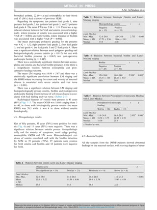
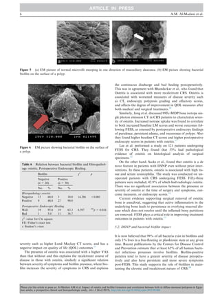
2. Histopathology found osteitis in 70% of patients. Scanning electron microscopy detected biofilms in 78% of patients, significantly higher than the 20% detected in controls.
3. Higher Lund-Mackay staging scores and osteitis scores correlated with higher rates of osteitis and biofilms histologically. Worse tissue disease and osteitis/biofilm were also linked to poorer postoperative healing.
![of the Faculty of Medicine, Alexandria University with the fol-
lowing ID: [IRB No. 00007555-FWA No. 0015712, June 2013].
3. Methods
All patients were subjected to the following. (1) Full History
taking about DSNP including history of bronchial asthma,
allergic rhinitis, aspirin intolerance, previous FESS, topical
and systemic steroid treatment and systemic antimicrobial
therapy, smoking, family history of polyposis and systemic dis-
eases. Subjective assessment of symptoms2
:
Major symptoms:
– Facial painnpressure (not considered if no other CRS com-
plaints present)
– Facial congestionnfullness
– Nasal obstructionnblockage
– Nasal dischargenpurulencendiscolored postnasal discharge
– Hyposmiananosmia
– Purulence of nasal cavity during examination
Minor symptoms:
Headache, fever, halitosis, fatigue, dental pain, cough and
ear painnpressure fullness.
Symptoms were scored by the visual analogue scale (0–4
scale) for the major and minor symptoms (0 = absent,
1 = mild, 2 = moderate, 3 = moderately severe, 4 = severe).
(2) Complete general Otorhinolarnygoscopic examination.
(3) Nasal endoscopy and reporting the extent of polyposis
by using Johansen endoscopic grading system.3
(4) Multislice CT scan with no contrast on the nose and
paranasal sinuses; coronal, axial, and sagittal CT scans were
examined and scored by the Lund–Mackay staging protocol
(maximum score is 24) and Global Osteitis Scoring scale.14
(Table 1)
(5) Complete blood count to record eosinophilia and for
preparation for surgery.
(6) During FESS a tissue biopsy was taken from the eth-
moid polypoidal tissue and divided into two samples as
follows:
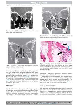
1. Histopathology for detection of osteitis: The samples of tis-
sue biopsy were fixed in 10% formol saline and sent to the
laboratory then prepared with xylon; Paraffin blocks will be
prepared and 5l sections will be stained using routine
hematoxylin and eosin (H&E) stain. Bony biopsy was pro-
cessed in acids then after softening and decalcification, the
bony specimen was processed as for soft tissue.
2. Biofilm detection by scanning EM: The specimens were
examined and photographed with JEOL, JSM-53009 scan-
ning electron microscope in EM unit, Faculty of Science,
Alexandria University. All specimens were prepared for
SEM using the following techniques. Tissue was initially
fixed for 2 h in 2.5% glutaraldehyde in phosphate-
buffered saline (PBS, pH 7.4) at 4–8 °C. Two rinses of
15 min each were then carried out using PBS. Next, the
specimens were fixed with 1% osmium tetroxide for 1 h.
They were then dehydrated through a graded ethanol series
as follows: 50% for 15 min, 70% for 15 min, 80% for
15 min, 90% for 15 min, and 100% twice for 15 min each
time. The tissue was immersed in 100% acetone for
15 min and washed in 100% isoamyl acetate for 15 min, fol-
lowed by critical point drying. Finally, specimens were
mounted on metal stubs and subsequently sputter coated
with gold preparation for imaging.
Structures categorized by water channels, 3D structure, and
matrix set in spherical or elliptical bodies were identified as evi-
dence of biofilms. It differs from viscous mucus, the latter is a
flat blanket, under which sometimes the comparative orderly
cilia could be seen, and irregular foreign granule might be
found. The entire area of each specimen was scanned for the
presence of biofilm structures. Images were taken at various
angles to effectively display the specimens and to minimize
errors and artifacts.
In the control group: Tissue specimens of approximately
0.5 cm3
were obtained 1 cm behind the anterior end of the infe-
rior turbinate and processed in the same manner for detection
of biofilm on the surface of mucosa.
(7) Postoperative follow-up: The patients were followed for
subjective satisfaction and objective healing of the cavity clin-
ically by endoscopy after surgery. A well healed cavity had
healthy mucosal lining with no evidence of inflammation,
mucosal swelling, polyposis or scarring.15
Data were analyzed using the Statistical Package for Social
Sciences (SPSS ver. 20, Chicago, IL, USA). The data were
reported as mean and standard deviation. T-tests and chi-
square tests were used to compare differences in means and
proportions where appropriate. The data were compared
between the patients and controls using paired t-test. Compar-
ison between two independent changes was done using inde-
pendent two-sample t-test. Statistical significance level was
set at 0.05.
4. Results
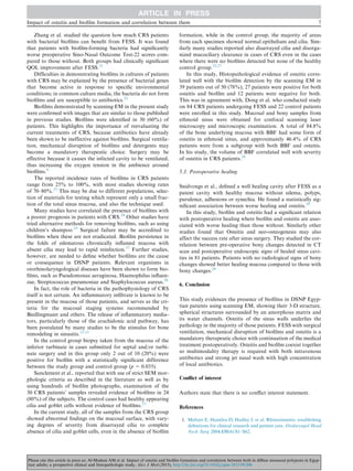
The patients group included 22 males (44%) and 28 females
with age ranged from 14 to 50 years with an average of
30.68 ± 7.24 years. Out of fifty patients, 15 (30%) had a
family history of DSNP, 18 (36%) were smokers, 25
(50%) had a history of aspirin intolerance, 29 (58%) had
Table 1 Determination of the severity of Osteitis in patients
with chronic rhinosinusitis using a new Global Osteitis Scoring
Scale.
Score Global Osteitis Scoring Scale
1 Less than 50% of sinus walls involved less than 3 mm thick
2 Less than 50% of sinus walls involved 3–5 mm thick
3 Less than 50% of sinus walls involved more than 5 mm
thick or more than 50% less than 3 mm thick
4 More than 50% of sinus walls involved 3–5 mm thick
5 More than 50% of sinus walls involved more than 5 mm
thick
The maximum thickness from each sinus wall is measured either by
the computer program on the computerized scan or from the scale
on the side of each cut. Each sinus to be given 0–5 score of 10
paranasal sinuses, 2 maxillary, 2 anterior ethmoid, 2 posterior
ethmoid, 2 frontal, 2 sphenoid. Total score from 0 to 50.
Non-significant less than 5, mild 5–20, moderate 21–35, severe
more than 35.
Impact of osteitis and biofilm formation and correlation between them 3
Please cite this article in press as: Al-Madani AM et al. Impact of osteitis and biofilm formation and correlation between both in diffuse sinonasal polyposis in Egyp-
tian adults; a prospective clinical and histopathologic study, Alex J Med (2015), http://dx.doi.org/10.1016/j.ajme.2015.09.006](https://image.slidesharecdn.com/d787f463-8485-4411-b76b-05572ec52b38-151205050213-lva1-app6891/85/puplished-paper-3-320.jpg)