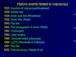
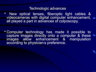
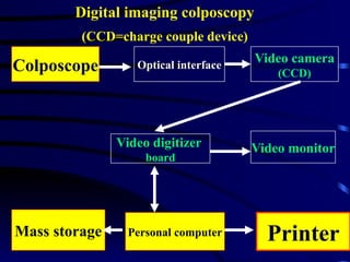
Colposcopy
Prof. Aboubakr Elnashar
Benha University Hospital. EGYPT
E-mail: elnashar@hotmail.com
• The colposcope was first developed in 1925 & is
well established in gynecologic practice for defining
& delineating cytologically detected lesions mainly
of the cervix but also the vagina & vulva.
• Colpscopy is now gradually spreading allover the
world & postgraduate training courses is now being
given in many centers.
Historic events related to colposcopy
1925: Invention of colposcope(Hinselman)
1928: Schiller test
1938: Acetic acid test (Hinselman)
1939: Green filter (Kratz)
1940: Pap test
1942: First photographs of cervix (Treite)
1960: Cryosurgery
1980: Laser surgery
1988: Computer-aided colposcope
1989: LLETZ (Prendiville & Cullimore)
1991: Pap Net
2000: Telecolposcopy ( Harper et al)
Technologic advances
• New optical lenses, fiberoptic light cables &
videocameras with digital computer enhancement,
all played a part in advances of colposcopy.
• Computer technology has made it possible to
capture images directly onto a computer & these
images allow enhancement & manipulation
according to physician,
s preference.
Colposcope Video camera
(CCD)
Optical interface
Video monitor
Video digitizer
board
Printer
Personal computer
Mass storage
Digital imaging colposcopy
(CCD=charge couple device)
Telecolposcopy
(Harper et al,2000)
*System incorporating a custom software package.
*All images were received without distortion in color,
size, or orientation.
*technically feasible,
can be implemented in an office system with limited
technical support
preferred by women who have to travel many miles to
receive referral health care.
Current indications of colposcopy
1. Part of any gynecologic examination
2. Primary screening for cervical cancer.
3. Clinically suspicious cervix.
4. Abnormal Pap smear.
5. Evaluation & treatment of CIN.
6. Follow up after conservative therapy of CIN.
7. Postcoital bleeding.
8. Patients with external vulval warts
9. Evaluation of sexual assault victims.
10. Patients with history of DES exposure
Uses
• Screening colposcopy is a feasible procedure &
more sensitive & more cost effective than
cytological screening. When access to
cytopathology is difficult, screening colposcopy is
an alternative
(Cecchini et al,1997).
• Portable colposcopy in rural areas is cost effective
& highly acceptable
(Martin et al,1998).
• The colposcopy improved detection of genital
trauma in adult female sexual assault victims as
compared with gross visual examination alone
(Lenahan,1998).
Recent recommendations of FIGO for management of abnormal
smear( Benedet,2000)
Persistent inflam., persistent ASCUS, LSIL, HSIL, AGCUS,Invasive
Colposcopy±biopsy
Normal or LSIL HGSIL Invasive
6 mo smear x 2 LEEP Appropriate TT
Normal Persistent
Annual screening
Steps
• Lugols’iodine test: beneficial test..
• ECB has replaced ECC: easier to use,
malleable & less expensive.
Its specificity 92%, sensitivity 90% & positive
predictive value 88%
(Martin et al, 1995).
• Punch biopsy: False negative rate up to
54%
(Buxton et al,1991)
Multiple biopsies
Excisional techniques are superior to
destructive techniques
Diagnostic criteria
1. Vascular pattern
2. Inercapillary distance
3. Contour.
4. Color
5. Clarity of demarcation
6. Appearance of gland opening.
7. Negativity after iodine test
8. Whiteness after acetic acid:
Density of whiteness, time needed to appear &
disappear, demarcation.
Changes >35 yr are thinner & less demarcated.,
punch biopsy
(Zahm et al, 1998).
9. Surface extent of the lesion:
more important prognostic indicator for invasion than
histological gradin
(Tidbury et al,1992)
International Federation of Cervical Pathology &
Colposcopy(1991)
Normal: Original squamous epithelium
Columnar epithelium
Normal transformation zone
Abnormal: Acetowhite epithelium Punctation
Mosaicism Leukoplakia
Iodine negative Atypical vessels
Suspect invasive cancer:
Unsatisfactory:SCJ not visible, severe inflam or atrophy, invisible cervix
Miscellaneous:Nonacetowhite micropapillary surface,
exophytic condyloma, inflammation, atrophy, ulcer
Niekerk (1998)
Low grade High grade
• Acetowhite epithelium: shiny or snow dull, oyster white color
white,semitransparent
• Surface: flat irregular contour, microexophytic
• Demarcation: diffuse, irregular, sharp, straight line,
flocculated, feathered,
internal demarcation absent internal demarcation present
• Vessels: fine, regular shape, uniform coarse, dilated, increased ICD,
caliber, normal arborization, spaghetti bizarre, commas, corkscrews
changing calibers sharp bends
• Iodine: uniform mahogany brown mustard yellow, yellow or iodine -
ve
Update of colposcopy of genital HPV
Meisels et al (1982): Florid, spiked, flat, condylomatous .
. vaginitis.
Flat condyloma & mild dysplasia represent the same biologic
phenomenon, namely, productive HPV infection (Reid,1993).
The expression of viral activity may be clinical or subclinical
when it is recognizable only on colposcopy.
Exophytic & flat condylomata are not homologous diseases.
Exophytic is usually caused by cutaneotropic viruses (6,11). Flat
are more likely to contain medium(31,33) or high risk(16,18) HPV
types.
Micropapillary condyloma should not be confused with
micropapillomatous labialis.
Colposcopy of the vulva
*Steps:
1. Examination after smearing with a water soluble
lubricant.
2. Prolonged acetic acid test
3. Toludine blue test: little clinical value.
* The junction between the glycogen bearing vaginal
epithelium & keratin producing vulval epithelium: high
risk for intraepithelial neoplasia.
*Abnormalities: diffuse acetowhite, localized
acetowhite, leukoplakia, micropapillae, papules.
Update on colposcopy in pregnancy
Difficult. & reserved for the most experienced
colposcopist.
Reassurance of the patient.
ECC is contrindicated & one directed biopsy.
Large speculum is usually needed
Sponge forceps to remove the mucous & acetic a as a
mucolytic
Unsatisfactory colposcopy: repeat after 8 w
The aim is to exclude cancer
CIN: follow up & definitive treatment 1-2 mo
postpartum.
Pitfalls in practice of colposcopy
A. In the technique
1. Failure to use a diagnostic protocol
2. Deviation from a diagnostic protocol.
3. Failure to visualize TZ.
B. In diagnosis
1. Misinterpretation of exagerated patterns of
pregnancy, previously treated cervix, carvical cancer.
2. Failure to select appropriate biopsy sites, enough
biopsies, sufficient volume of tissue.
3. Failure to accurately record colposcopic findings
C. In management
1. Miscommunication with the pathologist.
2. Failure to correlate cytology, colposcopy &
histopathology.
3.Destructive therapy without biopsy, for invasive or
glandular lesions.
D. In the colposcopist
1. Inadequate training.
2. Inadequate experience.
3. Inadequate understanding of the
disease. 4. Failure to keep up with
scientific developments 5. Failure to maintain
skills. 6. Failure to seek
consultation.
Diploma of colposcopy
• No one should be allowed to practice colposcopy
without having proper training or without a
diploma in colposcopy
(Jordan,1995).
• It would be a legal document that would safeguard
the public & raise the status of the colposcopist.
Future research in colposcopy
(Hilgarth,1998)
1. Computerized colposcopic documentation &
consecutive analysis of colposcopic findings.
2. Clinical significance & biologic behavior of minor
lesions visible with colposcopy in the presence of
different HPV types.
3. Clinical significance & relation to HPV infection of
minor lesions beyond the TZ.
4. Vulvar lesions in vulvodynia related to HPV
infection.
Future of colposcopy
(Niekerk,1998)
1. There are going increasing costs of medical care
& the demand for better quality control will intensify.
2. Technical advances will revolutionize this area &
digital imaging, the storage of up to 4.500 images on
an optical disk & rapid teletransmission of images
will become practical..
The use of these new technologies for better & more
cost effective patient care is the challenge we will
have to meet in the 21st century.
Benha University Hospital. EGYPT

colposcopy-today.pptx,,,,,,,,,,,,,,,,,,,,,,,,

  • 1.
    Colposcopy Prof. Aboubakr Elnashar BenhaUniversity Hospital. EGYPT E-mail: elnashar@hotmail.com
  • 2.
    • The colposcopewas first developed in 1925 & is well established in gynecologic practice for defining & delineating cytologically detected lesions mainly of the cervix but also the vagina & vulva. • Colpscopy is now gradually spreading allover the world & postgraduate training courses is now being given in many centers.
  • 3.
    Historic events relatedto colposcopy 1925: Invention of colposcope(Hinselman) 1928: Schiller test 1938: Acetic acid test (Hinselman) 1939: Green filter (Kratz) 1940: Pap test 1942: First photographs of cervix (Treite) 1960: Cryosurgery 1980: Laser surgery 1988: Computer-aided colposcope 1989: LLETZ (Prendiville & Cullimore) 1991: Pap Net 2000: Telecolposcopy ( Harper et al)
  • 4.
    Technologic advances • Newoptical lenses, fiberoptic light cables & videocameras with digital computer enhancement, all played a part in advances of colposcopy. • Computer technology has made it possible to capture images directly onto a computer & these images allow enhancement & manipulation according to physician, s preference.
  • 5.
    Colposcope Video camera (CCD) Opticalinterface Video monitor Video digitizer board Printer Personal computer Mass storage Digital imaging colposcopy (CCD=charge couple device)
  • 6.
    Telecolposcopy (Harper et al,2000) *Systemincorporating a custom software package. *All images were received without distortion in color, size, or orientation. *technically feasible, can be implemented in an office system with limited technical support preferred by women who have to travel many miles to receive referral health care.
  • 7.
    Current indications ofcolposcopy 1. Part of any gynecologic examination 2. Primary screening for cervical cancer. 3. Clinically suspicious cervix. 4. Abnormal Pap smear. 5. Evaluation & treatment of CIN. 6. Follow up after conservative therapy of CIN. 7. Postcoital bleeding. 8. Patients with external vulval warts 9. Evaluation of sexual assault victims. 10. Patients with history of DES exposure
  • 8.
    Uses • Screening colposcopyis a feasible procedure & more sensitive & more cost effective than cytological screening. When access to cytopathology is difficult, screening colposcopy is an alternative (Cecchini et al,1997). • Portable colposcopy in rural areas is cost effective & highly acceptable (Martin et al,1998). • The colposcopy improved detection of genital trauma in adult female sexual assault victims as compared with gross visual examination alone (Lenahan,1998).
  • 9.
    Recent recommendations ofFIGO for management of abnormal smear( Benedet,2000) Persistent inflam., persistent ASCUS, LSIL, HSIL, AGCUS,Invasive Colposcopy±biopsy Normal or LSIL HGSIL Invasive 6 mo smear x 2 LEEP Appropriate TT Normal Persistent Annual screening
  • 10.
    Steps • Lugols’iodine test:beneficial test.. • ECB has replaced ECC: easier to use, malleable & less expensive. Its specificity 92%, sensitivity 90% & positive predictive value 88% (Martin et al, 1995). • Punch biopsy: False negative rate up to 54% (Buxton et al,1991) Multiple biopsies Excisional techniques are superior to destructive techniques
  • 11.
    Diagnostic criteria 1. Vascularpattern 2. Inercapillary distance 3. Contour. 4. Color 5. Clarity of demarcation 6. Appearance of gland opening. 7. Negativity after iodine test
  • 12.
    8. Whiteness afteracetic acid: Density of whiteness, time needed to appear & disappear, demarcation. Changes >35 yr are thinner & less demarcated., punch biopsy (Zahm et al, 1998). 9. Surface extent of the lesion: more important prognostic indicator for invasion than histological gradin (Tidbury et al,1992)
  • 13.
    International Federation ofCervical Pathology & Colposcopy(1991) Normal: Original squamous epithelium Columnar epithelium Normal transformation zone Abnormal: Acetowhite epithelium Punctation Mosaicism Leukoplakia Iodine negative Atypical vessels Suspect invasive cancer: Unsatisfactory:SCJ not visible, severe inflam or atrophy, invisible cervix Miscellaneous:Nonacetowhite micropapillary surface, exophytic condyloma, inflammation, atrophy, ulcer
  • 14.
    Niekerk (1998) Low gradeHigh grade • Acetowhite epithelium: shiny or snow dull, oyster white color white,semitransparent • Surface: flat irregular contour, microexophytic • Demarcation: diffuse, irregular, sharp, straight line, flocculated, feathered, internal demarcation absent internal demarcation present • Vessels: fine, regular shape, uniform coarse, dilated, increased ICD, caliber, normal arborization, spaghetti bizarre, commas, corkscrews changing calibers sharp bends • Iodine: uniform mahogany brown mustard yellow, yellow or iodine - ve
  • 15.
    Update of colposcopyof genital HPV Meisels et al (1982): Florid, spiked, flat, condylomatous . . vaginitis. Flat condyloma & mild dysplasia represent the same biologic phenomenon, namely, productive HPV infection (Reid,1993). The expression of viral activity may be clinical or subclinical when it is recognizable only on colposcopy. Exophytic & flat condylomata are not homologous diseases. Exophytic is usually caused by cutaneotropic viruses (6,11). Flat are more likely to contain medium(31,33) or high risk(16,18) HPV types. Micropapillary condyloma should not be confused with micropapillomatous labialis.
  • 16.
    Colposcopy of thevulva *Steps: 1. Examination after smearing with a water soluble lubricant. 2. Prolonged acetic acid test 3. Toludine blue test: little clinical value. * The junction between the glycogen bearing vaginal epithelium & keratin producing vulval epithelium: high risk for intraepithelial neoplasia. *Abnormalities: diffuse acetowhite, localized acetowhite, leukoplakia, micropapillae, papules.
  • 17.
    Update on colposcopyin pregnancy Difficult. & reserved for the most experienced colposcopist. Reassurance of the patient. ECC is contrindicated & one directed biopsy. Large speculum is usually needed Sponge forceps to remove the mucous & acetic a as a mucolytic Unsatisfactory colposcopy: repeat after 8 w The aim is to exclude cancer CIN: follow up & definitive treatment 1-2 mo postpartum.
  • 18.
    Pitfalls in practiceof colposcopy A. In the technique 1. Failure to use a diagnostic protocol 2. Deviation from a diagnostic protocol. 3. Failure to visualize TZ. B. In diagnosis 1. Misinterpretation of exagerated patterns of pregnancy, previously treated cervix, carvical cancer. 2. Failure to select appropriate biopsy sites, enough biopsies, sufficient volume of tissue. 3. Failure to accurately record colposcopic findings
  • 19.
    C. In management 1.Miscommunication with the pathologist. 2. Failure to correlate cytology, colposcopy & histopathology. 3.Destructive therapy without biopsy, for invasive or glandular lesions. D. In the colposcopist 1. Inadequate training. 2. Inadequate experience. 3. Inadequate understanding of the disease. 4. Failure to keep up with scientific developments 5. Failure to maintain skills. 6. Failure to seek consultation.
  • 20.
    Diploma of colposcopy •No one should be allowed to practice colposcopy without having proper training or without a diploma in colposcopy (Jordan,1995). • It would be a legal document that would safeguard the public & raise the status of the colposcopist.
  • 21.
    Future research incolposcopy (Hilgarth,1998) 1. Computerized colposcopic documentation & consecutive analysis of colposcopic findings. 2. Clinical significance & biologic behavior of minor lesions visible with colposcopy in the presence of different HPV types. 3. Clinical significance & relation to HPV infection of minor lesions beyond the TZ. 4. Vulvar lesions in vulvodynia related to HPV infection.
  • 22.
    Future of colposcopy (Niekerk,1998) 1.There are going increasing costs of medical care & the demand for better quality control will intensify. 2. Technical advances will revolutionize this area & digital imaging, the storage of up to 4.500 images on an optical disk & rapid teletransmission of images will become practical.. The use of these new technologies for better & more cost effective patient care is the challenge we will have to meet in the 21st century.
  • 23.