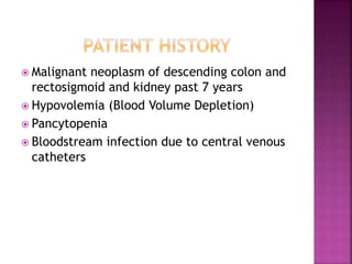
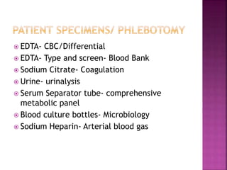
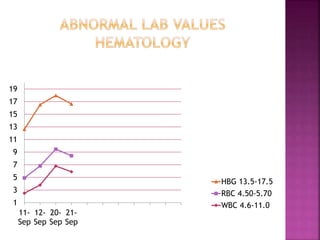
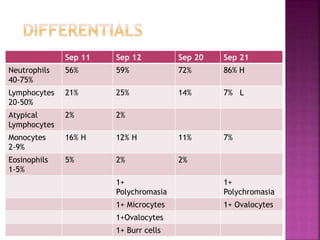
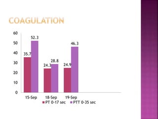
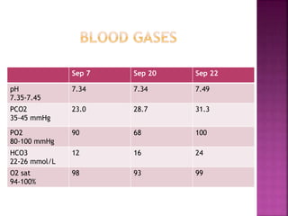
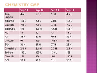
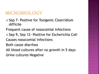
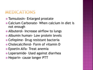
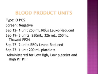
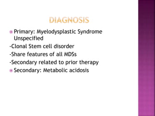
Sunny Schemery
Professor Tiffany Gill
MLT 2015
Decemeber 15, 2014
86 year old male came to ER
 Dizziness
Patient was admitted to ICU
 Dehydration
 Malignant neoplasm of descending colon and
rectosigmoid and kidney past 7 years
 Hypovolemia (Blood Volume Depletion)
 Pancytopenia
 Bloodstream infection due to central venous
catheters
 EDTA- CBC/Differential
 EDTA- Type and screen- Blood Bank
 Sodium Citrate- Coagulation
 Urine- urinalysis
 Serum Separator tube- comprehensive
metabolic panel
 Blood culture bottles- Microbiology
 Sodium Heparin- Arterial blood gas
1
3
5
7
9
11
13
15
17
19
11-
Sep
12-
Sep
20-
Sep
21-
Sep
HBG 13.5-17.5
RBC 4.50-5.70
WBC 4.6-11.0
Sep 11 Sep 12 Sep 20 Sep 21
Neutrophils
40-75%
56% 59% 72% 86% H
Lymphocytes
20-50%
21% 25% 14% 7% L
Atypical
Lymphocytes
2% 2%
Monocytes
2-9%
16% H 12% H 11% 7%
Eosinophils
1-5%
5% 2% 2%
1+
Polychromasia
1+
Polychromasia
1+ Microcytes 1+ Ovalocytes
1+Ovalocytes
1+ Burr cells
35.7
24.3 24.9
52.3
28.8
46.3
0
10
20
30
40
50
60
15-Sep 18-Sep 19-Sep
PT 0-17 sec PTT 0-35 sec
Sep 7 Sep 20 Sep 22
pH
7.35-7.45
7.34 7.34 7.49
PCO2
35-45 mmHg
23.0 28.7 31.3
PO2
80-100 mmHg
90 68 100
HCO3
22-26 mmol/L
12 16 24
O2 sat
94-100%
98 93 99
Sep 11 Sep 12 Sep 13 Sep 14
Total
Protein
4.6 L 5.0 L 5.3 L 4.6 L
Albumin 1.8 L 2.1 L 2.0 L 1.9 L
Calcium 7.0 L 7.3 L 7.4 L 7.6 L
TBilirubin 1.0 1.5 H 1.5 H 1.3 H
ALT 13 13 13 11 L
AST 35 H 37 H 40 H 35 H
Glucose 94 105 148 H 82
BUN 32 H 29 H 27 H 28 H
Creatinine 2.4 H 2.4 H 2.3 H 2.5 H
Sodium 135 L 135 L 134 L 138
Chloride 101 104 104 110 H
CO2 27.9 25.5 21.1 20.5 L
Total Protein Low – malabsorption
Total Bilirubin High- liver disease
Calcium Low- Vitamin D deficinecy
Chloride High- Intake IV
Sodium Low- renal loss
Albumin low- liver disease, nephrotic syndrome
AST High- liver disease
BUN High- kidney disease
Creatinine High- kidney disease
Sep 6 Sep 8 Sep 18
Color Yellow Yellow Amber
Clarity Slightly cloudy Clear Slightly cloudy
Bilirubin
Negative
Negative Negative Small
Ketones
Negtaive
Negative Negative Trace
Blood Negative Large Moderate Moderate
Protein
Negative
Negative Negative 30 mg/dL
RBC
0-5 hpf
10-14 hpf 1-4 hpf 5-9 hpf
WBC
0-10 hpf
10-14 hpf 1-4 hpf 5-9 hpf
Bacteria
Negative
present OCC OCC
 Sep 7- Positive for Toxigenic Clostridium
difficile
Frequent cause of nosocomial infections
 Sep 9, Sep 12- Positive for Escherichia Coli
Causes nosocomial infections
Both cause diarrhea
All blood cultures after no growth in 5 days
Urine cultures Negative
 Tamsulosin- Enlarged prostate
 Calcium Carbonate- When calcium in diet is
not enough
 Albuterol- Increase airflow to lungs
 Albumin human- Low protein levels
 Cefepime- Drug resistant bacteria
 Cholecalciferol- Form of vitamin D
 Epoetin Alfa- Treat anemia
 Loperamide- Used against diarrhea
 Heparin- cause longer PTT
Type: O POS
Screen: Negative
Sep 12- 1 unit 250 mL RBCs Leuko-Reduced
Sep 19- 3 units; 230mL, 326 mL, 250mL
Thawed FP24
Sep 22- 2 units RBCs Leuko-Reduced
Sep 22- 1 unit 200 mL platelets
Administered for Low Hgb, Low platelet and
High PT PTT
 Primary: Myelodysplastic Syndrome
Unspecified
-Clonal Stem cell disorder
-Share features of all MDSs
-Secondary related to prior therapy
 Secondary: Metabolic acidosis
 Transfusions to EPO
RBCs
 30 to 40% of cases lead to acute leukemia
 Blaney, K., & Howard, P. (2013). Blood Component
Preparation and Therapy. In Basic & applied concepts of
blood banking and transfusion practices (3rd ed., p.
312,313,316,317). St. Louis, Mo.: Mosby.
 Carr, J., & Rodak, B. (2009). Myelodysplastic Syndromes,
Diseases affecting Erythrocytes. In Clinical hematology
atlas (3rd ed., pp. 177-185, 119-134). St. Louis, Mo.:
Saunders Elsevier.
 Ciesla, B. (2007). The Myleodysplastic Syndromes,
Abnormalities of White Blood Cells:
Quantative,Qualitative, and the Lipid Storage Diseases. In
Hematology in practice (2nd ed., pp. 223-227, 143-155).
Philadelphia: F.A. Davis.
 Engelkirk, P., & Engelkirk, J. (2008). Gram-negative Bacilli:
The Family Enterobateracea, Anaerobic Bacteria. In
Laboratory diagnosis of infectious diseases: Essentials of
diagnostic microbiology (pp. 303-305, 421,422). Baltimore:
Wolters Kluwer Health/Lippincott Williams & Wilkins.
 Polansky, V. (2014). Clinical Chemistry Review. In
Quick review cards for medical laboratory
science (pp. 69-133). S.l.: F A Davis.
 RxList - The Internet Drug Index for prescription
drugs, medications and pill identifier. (2014,
January 1). Retrieved December 14, 2014, from
http://www.rxlist.com/script/main/hp.asp
 Strasinger, S., & Lorenzo, M. (2008). Chemical
examinaton of Urine. In Urinalysis and body
fluids (5th ed., pp. 53-75). Philadelphia: F.A.
Davis.
 Sunheimer, R., & Graves, L. (2011). Blood Gases,
pH and Acid-Base Balance. In Clinical laboratory
chemistry (1st ed., p. 296,297,300). Boston:
Pearson.

Clinical case study presentation

  • 1.
    Sunny Schemery Professor TiffanyGill MLT 2015 Decemeber 15, 2014
  • 2.
    86 year oldmale came to ER  Dizziness Patient was admitted to ICU  Dehydration
  • 3.
     Malignant neoplasmof descending colon and rectosigmoid and kidney past 7 years  Hypovolemia (Blood Volume Depletion)  Pancytopenia  Bloodstream infection due to central venous catheters
  • 4.
     EDTA- CBC/Differential EDTA- Type and screen- Blood Bank  Sodium Citrate- Coagulation  Urine- urinalysis  Serum Separator tube- comprehensive metabolic panel  Blood culture bottles- Microbiology  Sodium Heparin- Arterial blood gas
  • 6.
  • 7.
    Sep 11 Sep12 Sep 20 Sep 21 Neutrophils 40-75% 56% 59% 72% 86% H Lymphocytes 20-50% 21% 25% 14% 7% L Atypical Lymphocytes 2% 2% Monocytes 2-9% 16% H 12% H 11% 7% Eosinophils 1-5% 5% 2% 2% 1+ Polychromasia 1+ Polychromasia 1+ Microcytes 1+ Ovalocytes 1+Ovalocytes 1+ Burr cells
  • 8.
  • 10.
    Sep 7 Sep20 Sep 22 pH 7.35-7.45 7.34 7.34 7.49 PCO2 35-45 mmHg 23.0 28.7 31.3 PO2 80-100 mmHg 90 68 100 HCO3 22-26 mmol/L 12 16 24 O2 sat 94-100% 98 93 99
  • 11.
    Sep 11 Sep12 Sep 13 Sep 14 Total Protein 4.6 L 5.0 L 5.3 L 4.6 L Albumin 1.8 L 2.1 L 2.0 L 1.9 L Calcium 7.0 L 7.3 L 7.4 L 7.6 L TBilirubin 1.0 1.5 H 1.5 H 1.3 H ALT 13 13 13 11 L AST 35 H 37 H 40 H 35 H Glucose 94 105 148 H 82 BUN 32 H 29 H 27 H 28 H Creatinine 2.4 H 2.4 H 2.3 H 2.5 H Sodium 135 L 135 L 134 L 138 Chloride 101 104 104 110 H CO2 27.9 25.5 21.1 20.5 L
  • 12.
    Total Protein Low– malabsorption Total Bilirubin High- liver disease Calcium Low- Vitamin D deficinecy Chloride High- Intake IV Sodium Low- renal loss Albumin low- liver disease, nephrotic syndrome AST High- liver disease BUN High- kidney disease Creatinine High- kidney disease
  • 14.
    Sep 6 Sep8 Sep 18 Color Yellow Yellow Amber Clarity Slightly cloudy Clear Slightly cloudy Bilirubin Negative Negative Negative Small Ketones Negtaive Negative Negative Trace Blood Negative Large Moderate Moderate Protein Negative Negative Negative 30 mg/dL RBC 0-5 hpf 10-14 hpf 1-4 hpf 5-9 hpf WBC 0-10 hpf 10-14 hpf 1-4 hpf 5-9 hpf Bacteria Negative present OCC OCC
  • 16.
     Sep 7-Positive for Toxigenic Clostridium difficile Frequent cause of nosocomial infections  Sep 9, Sep 12- Positive for Escherichia Coli Causes nosocomial infections Both cause diarrhea All blood cultures after no growth in 5 days Urine cultures Negative
  • 17.
     Tamsulosin- Enlargedprostate  Calcium Carbonate- When calcium in diet is not enough  Albuterol- Increase airflow to lungs  Albumin human- Low protein levels  Cefepime- Drug resistant bacteria  Cholecalciferol- Form of vitamin D  Epoetin Alfa- Treat anemia  Loperamide- Used against diarrhea  Heparin- cause longer PTT
  • 18.
    Type: O POS Screen:Negative Sep 12- 1 unit 250 mL RBCs Leuko-Reduced Sep 19- 3 units; 230mL, 326 mL, 250mL Thawed FP24 Sep 22- 2 units RBCs Leuko-Reduced Sep 22- 1 unit 200 mL platelets Administered for Low Hgb, Low platelet and High PT PTT
  • 19.
     Primary: MyelodysplasticSyndrome Unspecified -Clonal Stem cell disorder -Share features of all MDSs -Secondary related to prior therapy  Secondary: Metabolic acidosis
  • 20.
     Transfusions toEPO RBCs  30 to 40% of cases lead to acute leukemia
  • 21.
     Blaney, K.,& Howard, P. (2013). Blood Component Preparation and Therapy. In Basic & applied concepts of blood banking and transfusion practices (3rd ed., p. 312,313,316,317). St. Louis, Mo.: Mosby.  Carr, J., & Rodak, B. (2009). Myelodysplastic Syndromes, Diseases affecting Erythrocytes. In Clinical hematology atlas (3rd ed., pp. 177-185, 119-134). St. Louis, Mo.: Saunders Elsevier.  Ciesla, B. (2007). The Myleodysplastic Syndromes, Abnormalities of White Blood Cells: Quantative,Qualitative, and the Lipid Storage Diseases. In Hematology in practice (2nd ed., pp. 223-227, 143-155). Philadelphia: F.A. Davis.  Engelkirk, P., & Engelkirk, J. (2008). Gram-negative Bacilli: The Family Enterobateracea, Anaerobic Bacteria. In Laboratory diagnosis of infectious diseases: Essentials of diagnostic microbiology (pp. 303-305, 421,422). Baltimore: Wolters Kluwer Health/Lippincott Williams & Wilkins.
  • 22.
     Polansky, V.(2014). Clinical Chemistry Review. In Quick review cards for medical laboratory science (pp. 69-133). S.l.: F A Davis.  RxList - The Internet Drug Index for prescription drugs, medications and pill identifier. (2014, January 1). Retrieved December 14, 2014, from http://www.rxlist.com/script/main/hp.asp  Strasinger, S., & Lorenzo, M. (2008). Chemical examinaton of Urine. In Urinalysis and body fluids (5th ed., pp. 53-75). Philadelphia: F.A. Davis.  Sunheimer, R., & Graves, L. (2011). Blood Gases, pH and Acid-Base Balance. In Clinical laboratory chemistry (1st ed., p. 296,297,300). Boston: Pearson.

Editor's Notes

  • #7 High monocytes indicate Bone marrow failure, malignancies High neutrophils; malignancies, physical conditions Ciesla, 2007, p. 224
  • #8 High monocytes indicate Bone marrow failure, malignancies High neutrophils; malignancies, physical conditions Ciesla, 2007, p. 144-145
  • #9 PT and PTT elevated (increased clotting times, slowly decreases Ciesla, 2007, p. 233-245
  • #11 Lab values indicate metabolic acidosis Sunheimer, & Graves, 2011 p. 296-297
  • #13 Polansky, 2014 p. 69-133
  • #15 Protein, Ketone increases: dehydration Strasinger, & Lorenzo,2008 p. 53-75
  • #17 Engelkirk, & Engelkirk, 2008 p. 303-305, 421,422
  • #18 RxList, 2014
  • #19 Blaney, & Howard, 2013 p. 312,313,316,317
  • #20 Carr, & Rodak, 2009 p. 177-185, Ciesla, 2007 p. 223-227, Sunheimer, & Graves, 2011 p. 296-297,300
  • #21 Carr, & Rodak, 2009 p. 177-185, Ciesla, 2007 p. 223-227, Sunheimer, & Graves, 2011 p. 296-297,300