
Recommended
More Related Content
Similar to cholelithiasis power point presentation1
Similar to cholelithiasis power point presentation1 (20)
Recently uploaded
Recently uploaded (20)
cholelithiasis power point presentation1
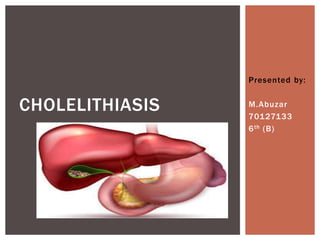
- 2. Cholelithiasis or gallstones are hardened deposits of digestive fluid that can form in your gallbladder. Your gall bladder is a small, pear-shaped organ of your abdomen, just beneath your liver. The gallbladder holds a digestive fluid called bile that’s released in your small intestine. Gallstones range is size from as small as grain to as large as a golf ball. Some people develop just one gallstone, while others develop many gallstones at the same time. INTRODUCTION
- 4. Gallstones are stones or lump that develop in the gallbladder or bile duct when certain substances harden. The gallbladder is a small sac located on the right-hand side of the body, on the underside of the liver. DEFINITION
- 5. Factors that may increase your risk of gallstones include: Being female Being age 40 or older Being overweight or obese Being sedentary Being pregnant Eating a high cholesterol diet Having diabetes Having a family history of gallstones Taking medications that contain estrogen, such as oral contraceptives or hormone therapy drugs RISK FACTORS
- 6. Too much cholesterol. Normally, your bile contains enough chemicals to dissolve the cholesterol excreted by your bile. But if your liver excretes more cholesterol than your bile can dissolve, the excess cholesterol may form into crystals and eventually into stones. Bile contains too much bilirubin. Bilirubin is a chemical that’s produced when your body breaks down red blood cells. Certain conditions cause your liver too much bilirubin, including liver cirrhosis, biliary tract infections and certain blood disorders. The excess bilirubin contributes to gallstone formation. Gallbladder doesn’t empty correctly. If gallbladder doesn’t empty completely or often enough, bile may become very concentrated, contributing to the formation of gallstones. CAUSES
- 7. Types of gallstones that can form in the gallbladder include: Cholesterol gallstones. The most common type of gall stone, called a cholesterol gallstone, often appears yellow in color. These gallstones are composed mainly of undissolved cholesterol, but may contain other components. Pigment gallstones. These dark brown or black stones form when your bile contains too much bilirubin. TYPES OF GALLSTONES
- 8. Sudden and rapidly intensifying pain in the upper right portion of your abdomen. Sudden and rapidly intensifying pain in the center of your abdomen, just below your breastbone. Back pain between your shoulder blades. Pain in your right shoulder. Nausea or vomiting. SIGNS AND SYMPTOMS
- 9. Physical examination. History collection. Abdominal ultrasound. This test is the most commonly used to look for signs of gallstones. Abdominal ultrasound involves moving a device (transducer) back and forth across tour stomach area. Endoscopic ultrasound. This procedure can help identify smaller stones that may be missed on an abdominal ultrasound. During EUS your doctor passes a thin, flexible tube (endoscope) through your mouth and through your digestive tract. Blood test. Blood test may reveal infection, jaundice, pancreatitis or other complications caused by gallstones. DIAGNOSTIC EVALUATION
- 10. Don’t skip meals. Lose weight slowly. Eat more higher-fiber foods. Maintain a healthy weight. PREVENTION
- 11. Lifestyle modifications: Dietary changes, including a low-fat diet, can help manage symptoms and reduce the risk of gallstone formation . Weight management and gradual weight loss may be recommended for overweight individuals. Medications : Medications can be prescribed to dissolve certain types of cholesterol gallstones over time. It is most effective for small stones and may take several months to work. Extracorporeal Shock Wave Lithotripsy (ESWL): ESWL uses shock waves to break gallstones into smaller pieces, making them easier to pass. This method is typically used for patients who are not suitable candidates for surgery. Cholecystectomy: This minimally invasive surgery is the most common and effective treatment for cholelithiasis. It involves removing the gallbladder through small incisions. Postoperative care: Following surgery, patients are advised on diet modifications to prevent discomfort and digestive issues. Early mobilization and light physical activity are encouraged to aid recovery. . TREATMENT
- 12. A 42-year-old office manager, seeks medical attention for severe right upper abdominal pain, accompanied by nausea and vomiting. Her medical history reveals occasional indigestion, but she has never experienced pain of this intensity. The clinical examination and diagnostic investigations point towards cholelithiasis, highlighting the need for a comprehensive approach to manage her condition. CASE SCENARIO
- 13. • Name: Sara Alam • Age: 42 • Gender: Female • Occupation: Office manager • Diagnosis: Cholelithiasis DEMOGRAPHICS
- 14. Pulse rate Blood pressure Temperature Respiratory rate 90 beats/min 130/80 mmHg 98 F 18 breaths/min 60-100 beats/min 120/80 mmHg 98.6 F 12-20 breaths/min OBJECTIVE HISTORY
- 15. Presenting complain History of presenting illness Past medical history Past family history The patient presents with severe right upper abdominal pain that started a few hours ago. The pain is sharp and intermittent, radiating to her back. She also complains of nausea and has vomited once. She denies any fever, chills, or recent dietary changes. She mentions that she had a fatty meal at a restaurant the night before. The pain started as a dull ache but gradually intensified, leading her to seek medical attention. She recalls similar but less severe episodes in the past, which resolved spontaneously. No significant prior medical conditions, occasional episodes of indigestion. Non significant SUBJECTIVE HISTORY
- 16. Abdominal examination reveals tenderness and guarding over the right upper quadrant. Murphy's sign is positive, causing Sara pain when pressure is applied to the right subcostal area during inspiration. ASSESSMENT
- 17. Abdominal Ultrasound: Confirms the presence of gallstones in the gallbladder and identifies signs of inflammation, such as thickening of the gallbladder wall and pericholecystic fluid. Blood Tests: Elevated levels of ALT and alkaline phosphatase, indicating possible biliary obstruction. EXAMINATION
- 18. Sara is diagnosed with acute cholecystitis secondary to cholelithiasis, which is the formation of gallstones in the gallbladder. DIAGNOSIS
- 19. Medical treatment Surgical treatment Physiotherapy treatment (post operative) • NSAIDs • Antibiotics • Intravenous Fluids (fluid replacement is crucial to maintain hydration, especially if there has been fluid loss due to vomiting.) • Cholecystectomy(This surgery eliminates the source of gallstones and reduces the risk of recurrent cholelithiasis.) • Breathing exercises • Early ambulation • Core strengthening PLAN OF CARE
- 20. THANK YOU!