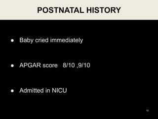
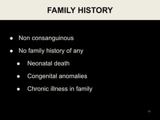
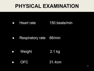
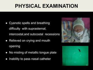
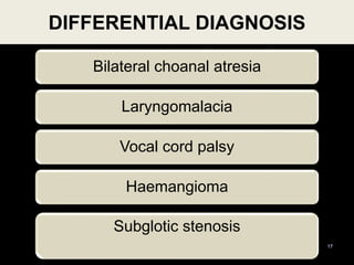
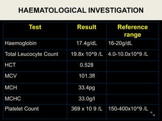
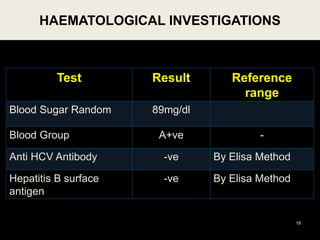
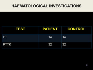
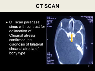
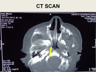
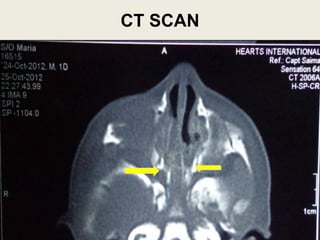
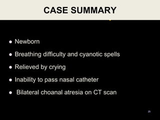
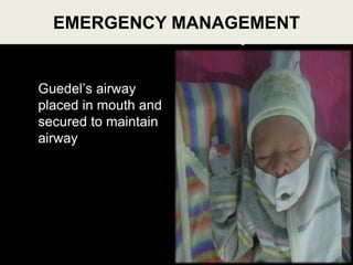
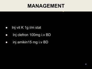
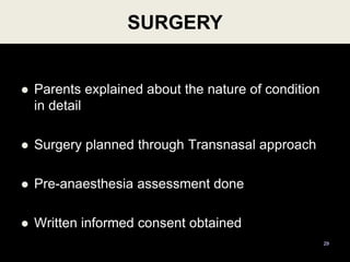
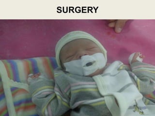
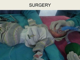
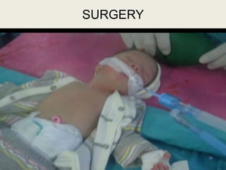
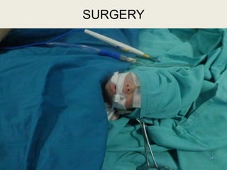
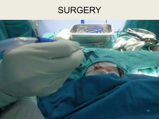
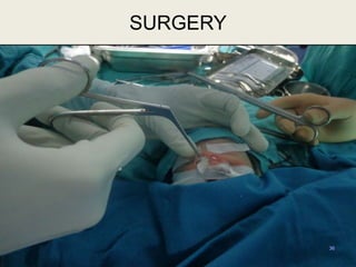
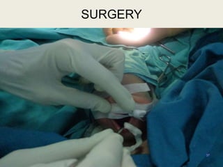
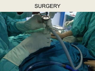
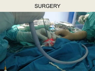
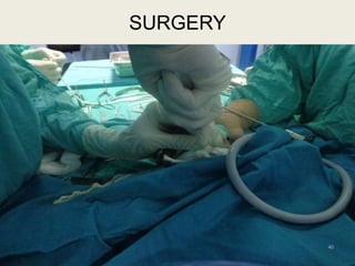
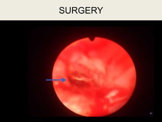
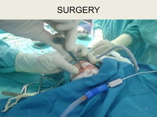
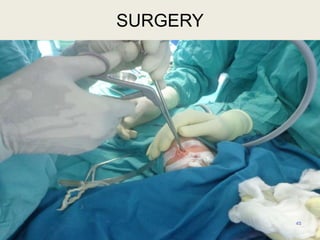
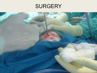
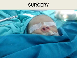
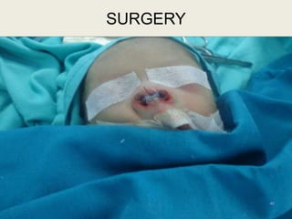
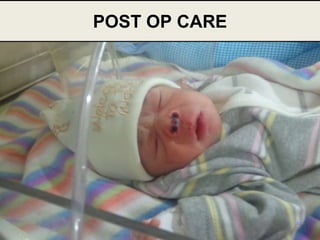
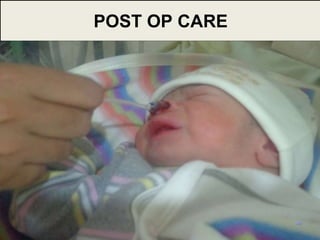
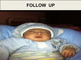
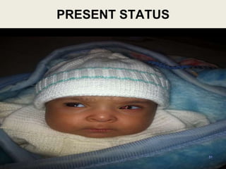
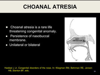
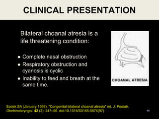
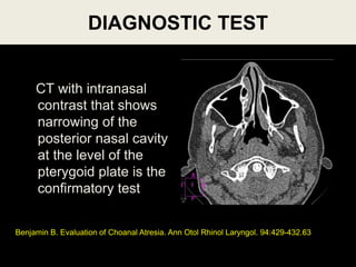
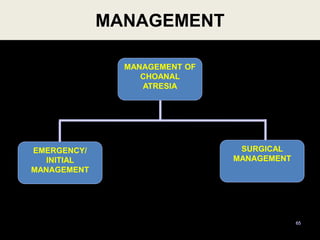
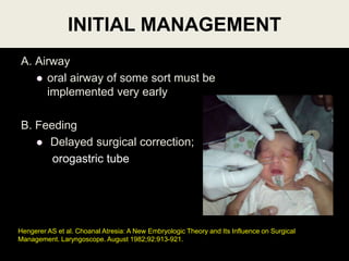
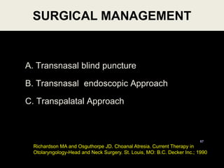
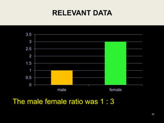
5
1
DR TARIQUE AHMED MAKA
REGISTRAR ENT
MANAGEMENT OF BILATERAL
CHOANAL ATRESIA
MANAGEMENT OF BILATERAL
CHOANAL ATRESIA
 SEQUENCE OF CPC
 Case presentation
 Literature review
 Statistical data
 Conclusion
3
4
CASE PRESENTATION
MANAGEMENT OF BILATERAL
CHOANAL ATRESIA
PATIENT PROFILE
 Name XYZ
 Age Newborn
 Gender Male
 Residence Rawalpindi
 Date of admission 25.10.12
5
PRESENTING COMPLAINTS
 Difficulty in breathing
 Cyanotic spells
6
since birth
HISTORY OF PRESENT ILLNES
 Difficulty in breathing
 Cyanotic spells
7
ANTENATAL HISTORY
 25yrs
 PG
 Planned pregnancy
 Regular antenatal visits
8
NATAL HISTORY
 39 weeks pregnancy
 Spontaneous labour
 Vertex presentation
 Emergency LSCS
 Liquor clear
9
POSTNATAL HISTORY
 Baby cried immediately
 APGAR score 8/10 ,9/10
 Admitted in NICU
10
FAMILY HISTORY
 Non consanguinous
 No family history of any
 Neonatal death
 Congenital anomalies
 Chronic illness in family
11
 Medical History
 Surgical History
 Drug History
 Socio economic history
Unremarkable
12
 Heart rate 150 beats/min
 Respiratory rate 66/min
 Weight 2.1 kg
 OFC 31.4cm
PHYSICAL EXAMINATION
13
 Cyanotic spells and breathing
difficulty with suprasternal,
intercostal,and subcostal recessions
 Relieved on crying and mouth
opening
 No misting of metallic tongue plate
 Inability to pass nasal catheter
PHYSICAL EXAMINATION
14
 No mass/cyst
 No laryngeal air way obstruction
 Vocal cords equally mobile
15
DIRECT LARYNGOSCOPY
 GIT system
 Central Nervous system
 Cardiovascular System
Unremarkable
16
SYSTEMIC EXAMINATION
Bilateral choanal atresia
Laryngomalacia
Vocal cord palsy
Haemangioma
DIFFERENTIAL DIAGNOSIS
17
Subglotic stenosis
Test Result Reference
range
Haemoglobin 17.4g/dL 16-20g/dL
Total Leucocyte Count 19.8x 10^9 /L 4.0-10.0x10^9 /L
HCT 0.528
MCV 101.3fl
MCH 33.4pg
MCHC 33.0g/l
Platelet Count 369 x 10 9 /L 150-400x10^9 /L
HAEMATOLOGICAL INVESTIGATION
18
18
Test Result Reference
range
Blood Sugar Random 89mg/dl
Blood Group A+ve -
Anti HCV Antibody -ve By Elisa Method
Hepatitis B surface
antigen
-ve By Elisa Method
HAEMATOLOGICAL INVESTIGATIONS
19
TEST PATIENT CONTROL
PT 14 14
PTTK 32 32
HAEMATOLOGICAL INVESTIGATIONS
20
CT SCAN
 CT scan paranasal
sinus with contrast for
delineation of
Choanal atresia
confirmed the
diagnosis of bilateral
choanal atresia of
bony type
22
CT SCAN
23
CT SCAN
24
Case Summary
 Newborn
 Breathing difficulty and cyanotic spells
 Relieved by crying
 Inability to pass nasal catheter
 Bilateral choanal atresia on CT scan
CASE SUMMARY
25
DIAGNOSIS
26
BILATERAL CHOANAL
ATRESIA
Case Summary
Guedel’s airway
placed in mouth and
secured to maintain
airway
27
EMERGENCY MANAGEMENT
Case Summary
 Inj vit K 1g i/m stat
 Inj clefron 100mg i.v BD
 inj amikin15 mg i.v BD
MANAGEMENT
28
 Parents explained about the nature of condition
in detail
 Surgery planned through Transnasal approach
 Pre-anaesthesia assessment done
 Written informed consent obtained
29
SURGERY
30
OPERATIVE STEPS
31
SURGERY
32
SURGERY
33
SURGERY
34
SURGERY
35
SURGERY
36
SURGERY
37
SURGERY
38
SURGERY
39
SURGERY
40
SURGERY
41
SURGERY
42
SURGERY
43
SURGERY
44
SURGERY
45
SURGERY
46
SURGERY
 Nursed at neonatal ICU for 2 days
 Administered
 Inj augmentin
 Inj dexamethasone
 I/V fluids as advised by Pediatrician
 Regular suction of tubes to prevent blockage
47
POST OP CARE
48
POST OP CARE
49
POST OP CARE
50
FOLLOW UP
51
PRESENT STATUS
LITERATURE
REVIEW
52
PART II
LITERATURE REVIEW
1755,
Roederer
18th Century
First recorded
case
19th Century
First successful
surgery
HISTORY OF
CHOANAL ATRESIA
53
1854, Emmert
BRIEF ANATOMY OF CHOANAE
• Medially: Free posterior edge of vomer
• Laterally: Medial pterygoid lamina
• Roof : Body of sphenoid
• Floor : Free posterior edge of horizontal plate
54
 Choanal atresia is a rare life
threatening congenital anomaly.
 Persistence of nasobuccal
membrane.
 Unilateral or bilateral
CHOANAL ATRESIA
55
Haddad J Jr. Congenital disorders of the nose. In: Kliegman RM, Behrman RE, Jenson
HB, Stanton BF, eds.
EPIDEMIOLOGY
 1 in 5,000 to 7,000 live births
 Females > males
 Atresias are bony,membranous or mixed
 Unilateral to bilateral cases is 2:1
 Unilateral presentation predominantly affects the
right nasal aperture
56
Hengerer AS, Brickman TM, Jeyakumar A. Choanal atresia: embryologic analysis and evolution
of treatment, a 30-year experience. Laryngoscope. May 2008;118(5):862-866.
ETIOLOGY
 Cause is unknown
 Smoking
 Coffee consumption
 High zinc and B-12 intake
 Prenatal use of antithyroid
(methimazole, carbimazole) medications
57
Barbero P, Valdez R, Rodríguez H, et al. Choanal atresia associated with maternal
hyperthyroidism treated with methimazole: a case-control study. Am J Med Genet A. Sep 15
2008;146A(18):2390-5.
PATHOGENESIS
58
 Persistence of the buccopharyngeal membrane
 Failure of the bucconasal membrane to rupture
 Medial outgrowth of vertical and horizontal processes
of the palatine bone
 Abnormal mesodermal adhesions forming in the
choanal area
Assanasen P, Metheetrairut C. Choanal atresia. J Med Assoc Thai. May 2009;92(5):699-706.
ASSOCIATED CONDITIONS
59
 Coloboma
 Heart defects
 Atresia choanae
 Retardation of growth
 Genital anomalies
 Ear abnormalities
(CHARGE)
Asher BF et al. Airway Complications in CHARGE Association. Arch Otolaryngol Head Neck
Surg. May 1990;116:594-595.
CLINICAL PRESENTATION
60
Bilateral choanal atresia is a
life threatening condition:
 Complete nasal obstruction
 Respiratory obstruction and
cyanosis is cyclic
 Inability to feed and breath at the
same time.
Sadek SA (January 1998). "Congenital bilateral choanal atresia" Int. J. Pediatr.
Otorhinolaryngol. 42 (3): 247–56. doi:10.1016/S0165-5876(97)
CLINICAL EXAMINATION
Pashley NRT. Choanal Atresia,(Chapter33). Clinical Pediatric Otolaryngology. St. Louis,
MO: C.V. Mosby Company; 1986 . 61v
 Failure to pass a # 6 to 8 french plastic catheter
through the nares
 Placing wisps of cotton in front of the nares and
absence of nasal movement of air.
 Absence of fog on a mirror when placed under the
nostrils
RECOMMENDED TESTS
62
 CT scan
 Endoscopy of the nose
 Cardiac echo
 Renal ultrasound
 Rhinogram
DIAGNOSTIC TEST
CT with intranasal
contrast that shows
narrowing of the
posterior nasal cavity
at the level of the
pterygoid plate is the
confirmatory test
Benjamin B. Evaluation of Choanal Atresia. Ann Otol Rhinol Laryngol. 94:429-432.63
64
MANAGEMENT OF
CHOANAL ATRESIA
MANAGEMENT
65
MANAGEMENT OF
CHOANAL
ATRESIA
EMERGENCY/
INITIAL
MANAGEMENT
SURGICAL
MANAGEMENT
A. Airway
 oral airway of some sort must be
implemented very early
B. Feeding
 Delayed surgical correction;
orogastric tube
INITIAL MANAGEMENT
Hengerer AS et al. Choanal Atresia: A New Embryologic Theory and Its Influence on Surgical
Management. Laryngoscope. August 1982;92:913-921.
A. Transnasal blind puncture
B. Transnasal endoscopic Approach
C. Transpalatal Approach
SURGICAL MANAGEMENT
67
Richardson MA and Osguthorpe JD. Choanal Atresia. Current Therapy in
Otolaryngology-Head and Neck Surgery. St. Louis, MO: B.C. Decker Inc.; 1990
TECHNIQUE
 120 degree endoscope placed in mouth and positioned
in nasopharynx
 Atretic plate perforated
 Bilateral nasal stents placed
 Philtrum is protected
 Stents left in situ for 6 weeks
TRANSNASAL ENDOSCOPIC APPROACH
68
 Advantages:
a. This approach is faster, easier, and less problematic.
b. Blood loss is minimal.
c. Excellent for children of all ages.
d. Allows for earlier repair in properly selected patients
and may avoid restenosis.
e. The child can breast feed immediately post op.
f. If no additional problems then the child can be
discharged on the 3rd or 4th post op day.
TRANSNASAL ENDOSCOPIC APPROACH
69
DISADVANTAGES AND COMPLICATIONS:
a. Longer stenting time post op.
b. Can not be done on patients with multiple nasal and
nasopharyngeal anomalies.
TRANSNASAL ENDOSCOPIC APPROACH
Josephson GD, Vickery CL, Giles WC, Gross CW. Transnasal endoscopic repair of
congenital choanal atresia: long-term results. Arch Otolaryngol Head Neck Surg. May
1998;124(5):537-40.
TRANSPALATAL APPROACH
 In rare circumstances where the
skull base is abnormally developed
(other craniofacial anomalies are
present), the surgeon may need to
use a transpalatal approach to
access the area of choanal atresia
through the roof of the mouth.
SURGICAL MANAGEMENT
Zeitouni AG, Shapiro RS. Congenital anomalies of the nose and anterior skull base. In: Tewfik TL,
Der Kaloustian VM, eds. Congenital Anomalies of the Ear, Nose, and Throat. New York, NY: Oxford
University Press; 1997:189-200
A. STENTING:
1. Secure them in place for a approximate
period of 4-8 weeks
B. WOUND CARE:
1. The parents must be taught to maintain the
stents with frequent suction and a saline-
moistened pipe cleaner or cotton applicator 3
to 6 times per day.
2. Antibiotics and decongestant 72
POST OPERATIVE CARE
C. FOLLOW UP:
1. Every 2 weeks or more frequently if needed while
the stents are in place.
2. Once the stents are removed, the patients are seen
weekly for the first 2-3 weeks.
73
POST OPERATIVE CARE
RELEVANT
STATISTICAL DATA
74
RELEVANT DATA
 Total Cases of CHOANAL ATRESIA = 4
 Duration 1st JANUARY, 2011 to 1st JANUARY, 2013
 Unilateral = 3
 Bilateral = 1 75
Bilateral
Unilateral
RELEVANT DATA
The male female ratio was 1 : 3
76
0
0.5
1
1.5
2
2.5
3
3.5
male female
 Bilateral choanal atresia is a rare life
threatening condition which presents with
stridor and acute respiratory distress at
birth
 Only immediate clinical evaluation and
efficient management is life saving
CONCLUSION
77
Thank You
78

Choanal atresia

  • 1.
  • 2.
    DR TARIQUE AHMEDMAKA REGISTRAR ENT MANAGEMENT OF BILATERAL CHOANAL ATRESIA
  • 3.
    MANAGEMENT OF BILATERAL CHOANALATRESIA  SEQUENCE OF CPC  Case presentation  Literature review  Statistical data  Conclusion 3
  • 4.
    4 CASE PRESENTATION MANAGEMENT OFBILATERAL CHOANAL ATRESIA
  • 5.
    PATIENT PROFILE  NameXYZ  Age Newborn  Gender Male  Residence Rawalpindi  Date of admission 25.10.12 5
  • 6.
    PRESENTING COMPLAINTS  Difficultyin breathing  Cyanotic spells 6 since birth
  • 7.
    HISTORY OF PRESENTILLNES  Difficulty in breathing  Cyanotic spells 7
  • 8.
    ANTENATAL HISTORY  25yrs PG  Planned pregnancy  Regular antenatal visits 8
  • 9.
    NATAL HISTORY  39weeks pregnancy  Spontaneous labour  Vertex presentation  Emergency LSCS  Liquor clear 9
  • 10.
    POSTNATAL HISTORY  Babycried immediately  APGAR score 8/10 ,9/10  Admitted in NICU 10
  • 11.
    FAMILY HISTORY  Nonconsanguinous  No family history of any  Neonatal death  Congenital anomalies  Chronic illness in family 11
  • 12.
     Medical History Surgical History  Drug History  Socio economic history Unremarkable 12
  • 13.
     Heart rate150 beats/min  Respiratory rate 66/min  Weight 2.1 kg  OFC 31.4cm PHYSICAL EXAMINATION 13
  • 14.
     Cyanotic spellsand breathing difficulty with suprasternal, intercostal,and subcostal recessions  Relieved on crying and mouth opening  No misting of metallic tongue plate  Inability to pass nasal catheter PHYSICAL EXAMINATION 14
  • 15.
     No mass/cyst No laryngeal air way obstruction  Vocal cords equally mobile 15 DIRECT LARYNGOSCOPY
  • 16.
     GIT system Central Nervous system  Cardiovascular System Unremarkable 16 SYSTEMIC EXAMINATION
  • 17.
    Bilateral choanal atresia Laryngomalacia Vocalcord palsy Haemangioma DIFFERENTIAL DIAGNOSIS 17 Subglotic stenosis
  • 18.
    Test Result Reference range Haemoglobin17.4g/dL 16-20g/dL Total Leucocyte Count 19.8x 10^9 /L 4.0-10.0x10^9 /L HCT 0.528 MCV 101.3fl MCH 33.4pg MCHC 33.0g/l Platelet Count 369 x 10 9 /L 150-400x10^9 /L HAEMATOLOGICAL INVESTIGATION 18 18
  • 19.
    Test Result Reference range BloodSugar Random 89mg/dl Blood Group A+ve - Anti HCV Antibody -ve By Elisa Method Hepatitis B surface antigen -ve By Elisa Method HAEMATOLOGICAL INVESTIGATIONS 19
  • 20.
    TEST PATIENT CONTROL PT14 14 PTTK 32 32 HAEMATOLOGICAL INVESTIGATIONS 20
  • 21.
    CT SCAN  CTscan paranasal sinus with contrast for delineation of Choanal atresia confirmed the diagnosis of bilateral choanal atresia of bony type 22
  • 22.
  • 23.
  • 24.
    Case Summary  Newborn Breathing difficulty and cyanotic spells  Relieved by crying  Inability to pass nasal catheter  Bilateral choanal atresia on CT scan CASE SUMMARY 25
  • 25.
  • 26.
    Case Summary Guedel’s airway placedin mouth and secured to maintain airway 27 EMERGENCY MANAGEMENT
  • 27.
    Case Summary  Injvit K 1g i/m stat  Inj clefron 100mg i.v BD  inj amikin15 mg i.v BD MANAGEMENT 28
  • 28.
     Parents explainedabout the nature of condition in detail  Surgery planned through Transnasal approach  Pre-anaesthesia assessment done  Written informed consent obtained 29 SURGERY
  • 29.
  • 30.
  • 31.
  • 32.
  • 33.
  • 34.
  • 35.
  • 36.
  • 37.
  • 38.
  • 39.
  • 40.
  • 41.
  • 42.
  • 43.
  • 44.
  • 45.
  • 46.
     Nursed atneonatal ICU for 2 days  Administered  Inj augmentin  Inj dexamethasone  I/V fluids as advised by Pediatrician  Regular suction of tubes to prevent blockage 47 POST OP CARE
  • 47.
  • 48.
  • 49.
  • 50.
  • 51.
  • 52.
    1755, Roederer 18th Century First recorded case 19thCentury First successful surgery HISTORY OF CHOANAL ATRESIA 53 1854, Emmert
  • 53.
    BRIEF ANATOMY OFCHOANAE • Medially: Free posterior edge of vomer • Laterally: Medial pterygoid lamina • Roof : Body of sphenoid • Floor : Free posterior edge of horizontal plate 54
  • 54.
     Choanal atresiais a rare life threatening congenital anomaly.  Persistence of nasobuccal membrane.  Unilateral or bilateral CHOANAL ATRESIA 55 Haddad J Jr. Congenital disorders of the nose. In: Kliegman RM, Behrman RE, Jenson HB, Stanton BF, eds.
  • 55.
    EPIDEMIOLOGY  1 in5,000 to 7,000 live births  Females > males  Atresias are bony,membranous or mixed  Unilateral to bilateral cases is 2:1  Unilateral presentation predominantly affects the right nasal aperture 56 Hengerer AS, Brickman TM, Jeyakumar A. Choanal atresia: embryologic analysis and evolution of treatment, a 30-year experience. Laryngoscope. May 2008;118(5):862-866.
  • 56.
    ETIOLOGY  Cause isunknown  Smoking  Coffee consumption  High zinc and B-12 intake  Prenatal use of antithyroid (methimazole, carbimazole) medications 57 Barbero P, Valdez R, Rodríguez H, et al. Choanal atresia associated with maternal hyperthyroidism treated with methimazole: a case-control study. Am J Med Genet A. Sep 15 2008;146A(18):2390-5.
  • 57.
    PATHOGENESIS 58  Persistence ofthe buccopharyngeal membrane  Failure of the bucconasal membrane to rupture  Medial outgrowth of vertical and horizontal processes of the palatine bone  Abnormal mesodermal adhesions forming in the choanal area Assanasen P, Metheetrairut C. Choanal atresia. J Med Assoc Thai. May 2009;92(5):699-706.
  • 58.
    ASSOCIATED CONDITIONS 59  Coloboma Heart defects  Atresia choanae  Retardation of growth  Genital anomalies  Ear abnormalities (CHARGE) Asher BF et al. Airway Complications in CHARGE Association. Arch Otolaryngol Head Neck Surg. May 1990;116:594-595.
  • 59.
    CLINICAL PRESENTATION 60 Bilateral choanalatresia is a life threatening condition:  Complete nasal obstruction  Respiratory obstruction and cyanosis is cyclic  Inability to feed and breath at the same time. Sadek SA (January 1998). "Congenital bilateral choanal atresia" Int. J. Pediatr. Otorhinolaryngol. 42 (3): 247–56. doi:10.1016/S0165-5876(97)
  • 60.
    CLINICAL EXAMINATION Pashley NRT.Choanal Atresia,(Chapter33). Clinical Pediatric Otolaryngology. St. Louis, MO: C.V. Mosby Company; 1986 . 61v  Failure to pass a # 6 to 8 french plastic catheter through the nares  Placing wisps of cotton in front of the nares and absence of nasal movement of air.  Absence of fog on a mirror when placed under the nostrils
  • 61.
    RECOMMENDED TESTS 62  CTscan  Endoscopy of the nose  Cardiac echo  Renal ultrasound  Rhinogram
  • 62.
    DIAGNOSTIC TEST CT withintranasal contrast that shows narrowing of the posterior nasal cavity at the level of the pterygoid plate is the confirmatory test Benjamin B. Evaluation of Choanal Atresia. Ann Otol Rhinol Laryngol. 94:429-432.63
  • 63.
  • 64.
  • 65.
    A. Airway  oralairway of some sort must be implemented very early B. Feeding  Delayed surgical correction; orogastric tube INITIAL MANAGEMENT Hengerer AS et al. Choanal Atresia: A New Embryologic Theory and Its Influence on Surgical Management. Laryngoscope. August 1982;92:913-921.
  • 66.
    A. Transnasal blindpuncture B. Transnasal endoscopic Approach C. Transpalatal Approach SURGICAL MANAGEMENT 67 Richardson MA and Osguthorpe JD. Choanal Atresia. Current Therapy in Otolaryngology-Head and Neck Surgery. St. Louis, MO: B.C. Decker Inc.; 1990
  • 67.
    TECHNIQUE  120 degreeendoscope placed in mouth and positioned in nasopharynx  Atretic plate perforated  Bilateral nasal stents placed  Philtrum is protected  Stents left in situ for 6 weeks TRANSNASAL ENDOSCOPIC APPROACH 68
  • 68.
     Advantages: a. Thisapproach is faster, easier, and less problematic. b. Blood loss is minimal. c. Excellent for children of all ages. d. Allows for earlier repair in properly selected patients and may avoid restenosis. e. The child can breast feed immediately post op. f. If no additional problems then the child can be discharged on the 3rd or 4th post op day. TRANSNASAL ENDOSCOPIC APPROACH 69
  • 69.
    DISADVANTAGES AND COMPLICATIONS: a.Longer stenting time post op. b. Can not be done on patients with multiple nasal and nasopharyngeal anomalies. TRANSNASAL ENDOSCOPIC APPROACH Josephson GD, Vickery CL, Giles WC, Gross CW. Transnasal endoscopic repair of congenital choanal atresia: long-term results. Arch Otolaryngol Head Neck Surg. May 1998;124(5):537-40.
  • 70.
    TRANSPALATAL APPROACH  Inrare circumstances where the skull base is abnormally developed (other craniofacial anomalies are present), the surgeon may need to use a transpalatal approach to access the area of choanal atresia through the roof of the mouth. SURGICAL MANAGEMENT Zeitouni AG, Shapiro RS. Congenital anomalies of the nose and anterior skull base. In: Tewfik TL, Der Kaloustian VM, eds. Congenital Anomalies of the Ear, Nose, and Throat. New York, NY: Oxford University Press; 1997:189-200
  • 71.
    A. STENTING: 1. Securethem in place for a approximate period of 4-8 weeks B. WOUND CARE: 1. The parents must be taught to maintain the stents with frequent suction and a saline- moistened pipe cleaner or cotton applicator 3 to 6 times per day. 2. Antibiotics and decongestant 72 POST OPERATIVE CARE
  • 72.
    C. FOLLOW UP: 1.Every 2 weeks or more frequently if needed while the stents are in place. 2. Once the stents are removed, the patients are seen weekly for the first 2-3 weeks. 73 POST OPERATIVE CARE
  • 73.
  • 74.
    RELEVANT DATA  TotalCases of CHOANAL ATRESIA = 4  Duration 1st JANUARY, 2011 to 1st JANUARY, 2013  Unilateral = 3  Bilateral = 1 75 Bilateral Unilateral
  • 75.
    RELEVANT DATA The malefemale ratio was 1 : 3 76 0 0.5 1 1.5 2 2.5 3 3.5 male female
  • 76.
     Bilateral choanalatresia is a rare life threatening condition which presents with stridor and acute respiratory distress at birth  Only immediate clinical evaluation and efficient management is life saving CONCLUSION 77
  • 77.

Editor's Notes

  • #2 In the name of Allah, The most gracious, the most merciful.
  • #3 Bil choanal atresia is an acute emergency due to its potential to cause acute airway obstruction.Worthy commandant, respected seniors and my fellow colleagues, I Dr Tariq Ahmed, from the dept of ENT and head and neck surgery, will be presenting a case of Bilateral choanal atresia.
  • #4 Sequence of cpc is as flashed
  • #5 First I ll present the case
  • #6 My patient, new born baby boy ,born in cmh rwp on 25th oct,2012 presented with
  • #7 Difficulty in breathing and Cyanotic spells since birth
  • #8 Moic of neonatal intensive care unit called ent splt to attend a newborn baby. who had respiratory distress and cyanotic spells since birth, which were relieved on crying.
  • #9 His mother was a 25yrs old, primigravida, this was her planned pregnancy. She had regular antenal visits
  • #10 Mother went into spontaneous labour at 39wks of pregnancy, Em lscs performed due to failed pol, Liqour was clear
  • #11 Baby cried immediately after birth e APGAR score of 8/10 at 1min and 9/10 at 5 min after birth. Baby was shifted to nicu for observation,
  • #12 Parents had non consanguinous marriage, there was no history of neonatal death, congenital anomalies or chronic illness in family
  • #13 However medical,surgical and drug history is unremarkable and baby belongs to a middle socioeconomic class
  • #14 His heart rate was 150 beats per mint.Respiratory rate was 66 breaths per mint with breathing difficulty having weight of 2.1 kg and occipitofrontal circumference of 31.4 cm
  • #15 Newborn baby had respiratory distress with suprasternal, intercostal,and subcostal recessions,he also had cyanotic spells. which were relieved on opening mouth and crying. There was no misting of metallic tongue plate when placed under nostrils indicating occluded airways, Soft silicone nasal catheter could not be passed through nose on either side
  • #16 Direct laryngoscopy with mackintosch laryngoscope revealed no mass,cyst or laryngeal air way obstruction and Vocal cords were normal and equally mobile
  • #17 The rest of the systemic examinations was unremarkable
  • #18 Based on history and clinical findings, The differential diagnosis included
  • #19 His blood complete picture showed haemoglobin of 17.4g/dL & platelets were 369
  • #20 Blood sugar, was WNL, hepatitis b and c serology were negative, hIS blood group was A positive
  • #21 His coagulation profile was with in normal limit
  • #22 CT scan paranasal sinus with contrast for delineation of Choanal atresia confirmed the diagnosis of bilateral choanal atresia of bony type
  • #23 CT scan paranasal sinus with contrast for delineation of Choanal atresia confirmed the diagnosis of bilateral choanal atresia of bony type
  • #24 CT scan paranasal sinus with contrast for delineation of Choanal atresia confirmed the diagnosis of bilateral choanal atresia of bony type
  • #25 CT scan paranasal sinus with contrast for delineation of Choanal atresia confirmed the diagnosis of bilateral choanal atresia of bony type
  • #26 To summarize, A newborn With breathing difficulty and cyanotic spells since birth, which were relieved by crying, OPENING MOUTH & placement of an oral airway, Soft silicone nasal catheter could not be passed through nose on either side, with confirmation of bilateral choanal atresia on CT scan
  • #27  on basis of history, clinical examination and investigations, final diagnosis of bilateral choanal atresia was made and pediatrician was consulted, to rule out any other associated anomalies.
  • #28 As an Emergency MEASURE,,,,,,,,oropharyngeal Guedel’s airway was placed in mouth and secured, to maintain airway, his dyspnea and cyanosis was relieved, and oxygen saturation became normal.
  • #30 Parents were explained about the condition in detail Surgery was planned through Transnasal approach on Ist day of his birth on 26 oct 2012 Pre-anaesthsia assessment was done Written informed consent was obtained
  • #31 Now the operative steps
  • #32 Patient placed supine
  • #33 General anaesthesia inducted
  • #34 Airway secured with ETT of 2.5
  • #35 Patient draped
  • #36 Nasal cavities cleared of secretions by suction
  • #37 Nasal cavities packed with adrenaline soaked guaze
  • #38 Oral cavity and nasopharynx were examined
  • #39 Nasal cavity examined with nasoendoscope Telescope of o introduced and atresia visualized
  • #40 Nasoendoscopy in progress
  • #41  atretic plate was perforated with the trocar and cannula, in its inferomedial part
  • #42 Here blue arrow is pointing the aperture made in atretic plate1. superiorly - the undersurface of the body of the sphenoid bones, 2. laterally - the medial pterygoid lamina, 3. medially - the vomer, 4. inferiorly - the horizontal portion of the palatal bone
  • #43 Opening made in the atretic plate widened with haegar dilator 3/6
  • #44 Redundent mucosa removed under direct vision
  • #45 Opening was furthur widened with circular cutting forceps under vision
  • #46  Pieces of ETT tube placed on both sides to maintain patency , air flow and prevent stenosis Bilateral nasal stents are fashioned from two ET tubes cut to length,with bevelled end of each siiting in nasopharynx orientated towards septum
  • #47 Both tubes secured with silk Philtrum is protected by a small piece of ET tube. stents are secured by a circumseptal 0 proline suture and are left in situ for six weeks
  • #48 Post operatively patient was nursed in neonatal ICU for 2 days Administered Inj augmentin Inj dexamethasone I/V fluids as advised by Paediatrician And Regular suction of tubes was carried out to prevent blockage
  • #49  Neonate in neonatal ICU after surgery without any distress and cyanosis
  • #50 Regular nasal clearance with suction in progress Post op recovery was smooth and patient was discharged on 3rd post op day
  • #51 Patient was followed up weekly and Endotracheal tubes were removed after 6 weeks
  • #52 Another picture of patient at 8 weeks breathing comfortably through nose
  • #53 Now the literature review
  • #54 Slide 53
  • #55  The choanae or posterior nasal apertures are divided medially by free posterior edge of vomer. Their roof is formed by the body of sphenoid with the overlapping, flared alae of the vomer and the vaginal process of medial pterygoid plate, and the floor by the free posterior edge of the horizontal plate of the palatine bone During embryonic life choanae are closed by bucconasal membrane. This membrane lies slightly more anterior to the eventual position of choanae, which do not become established until the third month of intrauterine life.
  • #56  Choanal atresia is a rare life threatening congenital anomaly, in which a bony or membranous occlusion blocks the passageway between the nose and pharynx.This condition is believed to be secondary to the persistence of nasobuccal membrane.it can be unilateral or bilateral.
  • #57 It is estimated to affect 1 in 5,000 to 7,000 live births Is twice as common in females as males Atresias are bony,membranous or mixed, 70% are mixed while remaining are purely bony The ratio of unilateral to bilateral cases is 2:1 Unilateral presentation predominantly affects the right nasal aperture
  • #58  exact etiology is not precisely known, both genetic and environmental triggers are suspected,maternal smoking, cofee consumption, excessive zinc and B12intake and prenatal use of antithyroid (methimazole, carbimazole) medications is linked to choanal atresia . 
  • #59 A number of theories have been proposed to explain the occurrence of choanal atresia, they can be summarized as:
  • #60  Associated congenital anomalies occur in association with choanal atresia in about 50% of the patients.which r known as CHARGE, that includes
  • #61  immediate respiratory distress and even potential death due to asphyxia after birth. as the child falls asleep the mouth closes and a progressive obstruction starting with stridor followed by increased respiratory effort and cyanosis.
  • #62 On clinical examination there will be Failure to pass a # 6 to 8 french plastic catheter through the nares. While Placing wisps of cotton in front of the nares, there ll b absence of nasal movement of air. And The absence of fog on a mirror when it is placed under the nostrils
  • #63 An axial high resolution CT scan with thin sections (2-5mm) has become the single radiographic study of choice. The CT scan has proven valuable in the accurate assessment of both the normal and abnormal anatomy of the nasal cavity, posterior nasal choanae and nasopharynx Choanal atresia. Rhinogram demonstrating blockage of radiopaque dye at the posterior choanae. (placing methylene blue in nares & not visualisin g within pharynx.
  • #65 Now coming towards the management of choanal atresia
  • #66 Management of choanal atresia includes emergency or initial management followed by surgical that is the definative management
  • #67 Temporary alleviation can be achieved by inserting an oral airway into the mouth In mild cases of unilateral choanal atresia feeding is main issue. In case of delayed surgical correction due to precarious condition of pt,orogastric tube iz pASSED for feeding
  • #68 only definitive treatment is surgery to correct the defect. This technique should be avoided because of the potentialy serious complications: CSF leaks, midbrain trauma
  • #69 The transnasal endoscopic approach is currently CHOP's procedure of choice for managing choanal atresia
  • #72 Transpalatal approarch is used rarely only when patient has craniofacial anomalies
  • #73  Post op stents are kept in place for 4-8 wks and parents are taught about frequent suction of stents with saline. Antibiotics & decongestants are prescribed.
  • #74 Pts are followed 2 weekly till stents are in place and then weekly for first 2-3 wks after removal of stents to treat any granulation tissue and to prevent stenosis.
  • #75 Lastly, the relevant statistical data
  • #76 , the Total number of patients who presented with CHOANAL ATRESIA were 4, during last 2 yrs , out of them 1 was bilateral.
  • #78 Slide 77