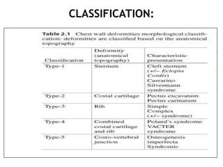
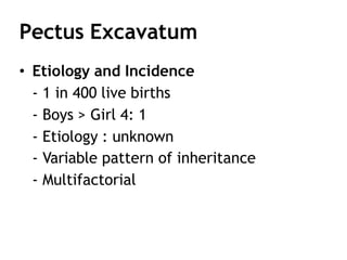
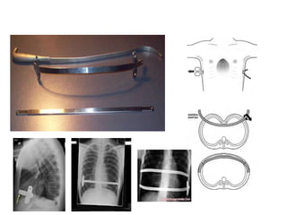
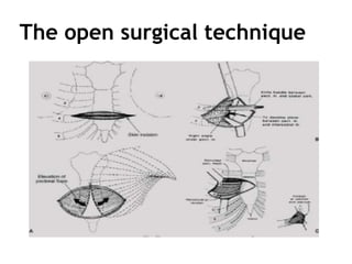
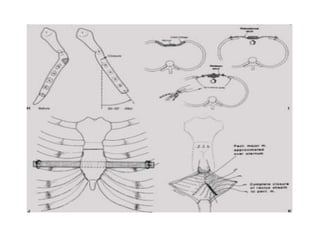
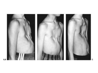
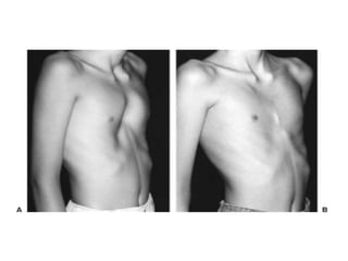
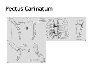
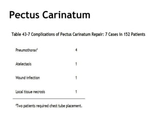
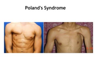
Chest Wall Disorder or
Deformities
Group A
Chest Wall Deformities
Scoliosis
Pectus Excavatum
Ankylosing Spondylitis
Flail chest
Thoracoplasty
Pectus carinatum
Poland Syndrome
Chest wall tumors
CLASSIFICATION:
Scoliosis
• Scoliosis is defined as a
lateral curvature of the
spine and is associated
with rotation of the spine
and viscera adjacent to it.
• It may be (but is usually
not) accompanied by
kyphosis.
• Thoracic scoliosis to the right- most common
presentation.
• More prevalent- Girls.
• Higher & Increase angle of curvature- worse
prognosis.
Cobb’s angle
• Lines are drawn parallel
to the upper border of
the upper vertebral body
and to the lower border
of the lowest vertebra of
the structural curve.
• Perpendiculars are then
erected from these lines
to cross each other, the
angle between the
perpendiculars being the
angle of curvature or
Cobb’s angle
Pathology
• Total lung volume is reduced and the lungs
differ in size, with distortion of lobar shapes
due to the deformity.
• Alveolar size may vary, alveolar numbers per
acinus may correspond to that developmental
stage.
• Vascular medial hypertrophy- Cor pulmonale
Physiology
• Lung volumes are reduced, with a restrictive
pattern.
• VC, TLC, MVV, functional residual capacity
(FRC) and peak expiratory flow (PEF)- reduced.
• The higher the curve in the dorsal spine, the
more severe its effect on function.
• FEV1/FVC ratio is normal unless obstructive
disease such as asthma supervenes.
• Dlco is reduced
• Lung compliance in scoliosis is commonly low-
inability to take a deep breath.
• Exercise tolerance is limited.
• Patients with severe scoliosis (with an angle
≥100°), severe desaturation may occur during
sleep with episodes of central or obstructive
apnoea and hypopnoea.
• In severe disease- increasing hypoxia- Pulmonary
artery hypertension.
• Co2 retention- diminished ventilatory responses.
Clinical features
• Mild scoliosis to have respiratory symptoms-
unusual.
• Daytime somnolence may indicate the presence
of nocturnal desaturation.
• Exertional dyspnoea- due to the restriction.
• PHT and RVH often occurs in the fourth decade.
• Death most commonly occurs as a result of
respiratory failure or cardiac disease.
• Respiratory tract infection.
Treatment- Surgery
Surgical intervention- may ameliorate the angle of
curvature and halt the progression of scoliosis &
halt the progression of scoliosis.
• Surgical fusion of spine- role in children with
congenital spinal deformities or in children with
KS secondary to neurological defects.
• Non-fusion approach includes implantation of
growth-friendly rods such as expandable spinal
rods, titanium rib implants, or remotely
distractible magnetically controlled rods.
Medical treatment
• Long-term domiciliary oxygen therapy-
respiratory failure,
• Assisted ventilation- PPV (nasal mask or full-face
mask),
• Life-threatening hypoventilation include tank
respirators or cuirass shells,
• Less satisfactory for the patient, but equally
effective, is nocturnal ventilation via a permanent
tracheostomy,
• Diuretic therapy- RVH,
Medical treatment
• Bronchodilator therapy,
• Smoking cessation,
• Maintenance of body weight within a desirable
range, engaging in frequent physical activity to
improve exercise capacity,
• Immunization against influenza and
pneumococci,
• Prompt treatment of respiratory infections,
• Psychological support for those with diminished
self-esteem.
Pectus Excavatum
• Funnel chest or trichterbrust
• Posterior depression of the sternum and
costal cartilages
• 1st & 2nd ribs & manubrium : normal
position
• Lower costal cartilages and the body of the
sternum are depressed
• Asymmetry of the depression is frequently
present Rt. > Lt.
Pectus Excavatum
• Present at birth or within first year of life
in the majority : 86%
• Rarely resolves with increasing age
• May worsen during rapid adolescent
growth
• Scoliosis: 26%
• Asthma : 5.2%
• CHD : 1.5%
Pectus Excavatum
• Etiology and Incidence
- 1 in 400 live births
- Boys > Girl 4: 1
- Etiology : unknown
- Variable pattern of inheritance
- Multifactorial
Pectus Excavatum
• Symptoms
- Well tolerated in infancy and childhood
- Older children :
 pain in the area of the deformed cartilages
 precordial pain after sustained exercise
 palpitations
 transient atrial arrhythmias
 may have mitral valve prolapse
Pectus Excavatum
• Surgical Repair
Haller and associates : tripod fixation
- Subperichondrial resection
- Posterior sternal osteotomy
- Most cephalad normal cartilages are then
divided obliquely in a posterolateral
- Sternum elevated, the sternal ends of
the cartilage rest on the costal ends
Pectus Excavatum
• Nuss and procedure
• Minimally Invasive Repair of Pectus
Excavatum (MIRPE)
Before and After Surgery
Non surgical treatment.
The open surgical technique
Complication : Nuss procedure
Early
• Pneumothorax
• Pericarditis
• Pneumonia
• Hemothorax
• Transient extremity
paralysis
• Superficial wound
infection
• Bar infection
Late
• Bar displacement
• Hemothorax
• Overcorrection deformity
• Pectus carinatum
deformity
Flail chest
• double fractures of three or more adjacent ribs or
the combination of sternal and rib fractures are
required to produce a flail segment of the rib
cage and lead to respiratory failure.
Causes:
• trauma related to automobile accidents or falls
• aggressive cardiopulmonary resuscitation
• pathological rib fractures (i.e., multiple myeloma,
other metastases).
• Disruption of rib cage integrity in flail chest
renders motion of the flail segment entirely
dependent on pleural pressure changes during
breathing.
• The most common anatomical location for flail
chest is the lateral rib cage.
• VC and FRC can be reduced to as much as 50% of
predicted.
• With surgical stabilization , VC usually returns to
normal range in about 3 months.
Inspiration
• Pleural pressure becomes
sub-atmospheric, which is
inflationary to the lung and
deflationary to the rib
cage.
• Consequently, the flail
segment moves inward
rather than outward during
inspiration.
Expiration
• Pleural pressure becomes
more positive and the flail
segment moves outward.
• This paradoxical motion of
the flail segment is
augmented by conditions
that load the respiratory
system thereby amplifying
pleural pressure swings.
Pathogenesis of respiratory failure
Treatment
Objective of treatment:
a) Pain control:
• enhance cough efficiency
• use of oral or intravenous narcotics, intercostal nerve
blocks, or epidural anesthesia.
b) Minimize atelectasis:
• frequent tracheal bronchial toilet
c) Mechanical ventilation:
• low impedance modes- CPAP- associated with less chest
wall distortion during inspiration.
• Reduce morbidity and length of hospitalization.
c) Surgical fixation of the flail chest
Thoracoplasty
Pre-antituberculous chemotherapy era,
thoracoplasty was the standard surgical
approach to control PTB.
• Rib removal,
• Rib fracture,
• Phrenic nerve resection,
• Lung compression by filling the pleural space
with foreign material (i.e., ping pong balls)
• Thoracoplasty is indicated for treatment of
bronchopleural fistulae that have failed to
close following decortication or
• For treatment of persistent empyema in which
decortication is not feasible or has failed to
eradicate the infection
Treatment
• Supplemental oxygen,
• Antibiotics for respiratory infections,
• Noninvasive ventilatory support.
Pectus carinatum or pigeon chest
• sternum is prominent, forming an anterior ridge
like the keel of a ship with the ribs falling away
steeply on either side.
• associated with other congenital anomalies,
especially cardiac lesions and coarctation of the
aorta.
• Less frequent than pectus excavatum
• M:F 4:1
• Cause: unknown
• Positive family history in 26%
Pectus Carinatum
• CHD : 18%
• FH of chest wall deformity : 26 %
• Boys > Girls = 78 : 22 %
• Mild form at birth but often progresses
during early childhood
• The chondromanubrial deformity, often
noted at birth
Pectus Carinatum
Pectus Carinatum
Poland’s syndrome
• Hypoplasia of the sternal head of
the pectoralis major and minor
muscles
• Normal underlying ribs to complete
absence anterior portions of the 2nd –
5th ribs and costal cartilages
• The etiology is unknown.
Poland's Syndrome
• Hypoplasia of the sternal head of the
pectoralis major and minor muscles
• Normal underlying ribs to complete
absence anterior portions of the 2nd – 5th
ribs and costal cartilages
Poland's Syndrome
Poland's Syndrome
Poland's Syndrome
• Surgical Repair
- Assessment of the extent of involvement
limited to the sternal component of the
pectoralis major and minor muscles,
• little functional deficit
• repair is not necessary except to
facilitate breast augmentation in
women
Poland's Syndrome
• Surgical Repair
- Assessment of the extent of
involvement underlying costal cartilages
are depressed or absent
• repair must be considered to minimize
the concavity
• to eliminate the paradoxic motion of
the chest wall if ribs are absent
• in girls to provide an optimal base for
breast reconstruction
Poland's Syndrome
• Surgical Repair
Ravitch :
– Correction of posteriorly displaced costal
cartilages by unilateral resection of the
cartilages
– A wedge osteotomy of the sternum, allowing
rotation of the sternum; and fixation with
Rehbein struts and Steinmann pins
– The sternum is then displaced anteriorly and
supported with a retrosternal strut
Thank you

chest wall disorder presentation.pptx

  • 1.
    Chest Wall Disorderor Deformities Group A
  • 2.
    Chest Wall Deformities Scoliosis PectusExcavatum Ankylosing Spondylitis Flail chest Thoracoplasty Pectus carinatum Poland Syndrome Chest wall tumors
  • 3.
  • 4.
    Scoliosis • Scoliosis isdefined as a lateral curvature of the spine and is associated with rotation of the spine and viscera adjacent to it. • It may be (but is usually not) accompanied by kyphosis.
  • 5.
    • Thoracic scoliosisto the right- most common presentation. • More prevalent- Girls. • Higher & Increase angle of curvature- worse prognosis.
  • 6.
    Cobb’s angle • Linesare drawn parallel to the upper border of the upper vertebral body and to the lower border of the lowest vertebra of the structural curve. • Perpendiculars are then erected from these lines to cross each other, the angle between the perpendiculars being the angle of curvature or Cobb’s angle
  • 7.
    Pathology • Total lungvolume is reduced and the lungs differ in size, with distortion of lobar shapes due to the deformity. • Alveolar size may vary, alveolar numbers per acinus may correspond to that developmental stage. • Vascular medial hypertrophy- Cor pulmonale
  • 8.
    Physiology • Lung volumesare reduced, with a restrictive pattern. • VC, TLC, MVV, functional residual capacity (FRC) and peak expiratory flow (PEF)- reduced. • The higher the curve in the dorsal spine, the more severe its effect on function. • FEV1/FVC ratio is normal unless obstructive disease such as asthma supervenes. • Dlco is reduced
  • 9.
    • Lung compliancein scoliosis is commonly low- inability to take a deep breath. • Exercise tolerance is limited. • Patients with severe scoliosis (with an angle ≥100°), severe desaturation may occur during sleep with episodes of central or obstructive apnoea and hypopnoea. • In severe disease- increasing hypoxia- Pulmonary artery hypertension. • Co2 retention- diminished ventilatory responses.
  • 10.
    Clinical features • Mildscoliosis to have respiratory symptoms- unusual. • Daytime somnolence may indicate the presence of nocturnal desaturation. • Exertional dyspnoea- due to the restriction. • PHT and RVH often occurs in the fourth decade. • Death most commonly occurs as a result of respiratory failure or cardiac disease. • Respiratory tract infection.
  • 11.
    Treatment- Surgery Surgical intervention-may ameliorate the angle of curvature and halt the progression of scoliosis & halt the progression of scoliosis. • Surgical fusion of spine- role in children with congenital spinal deformities or in children with KS secondary to neurological defects. • Non-fusion approach includes implantation of growth-friendly rods such as expandable spinal rods, titanium rib implants, or remotely distractible magnetically controlled rods.
  • 13.
    Medical treatment • Long-termdomiciliary oxygen therapy- respiratory failure, • Assisted ventilation- PPV (nasal mask or full-face mask), • Life-threatening hypoventilation include tank respirators or cuirass shells, • Less satisfactory for the patient, but equally effective, is nocturnal ventilation via a permanent tracheostomy, • Diuretic therapy- RVH,
  • 14.
    Medical treatment • Bronchodilatortherapy, • Smoking cessation, • Maintenance of body weight within a desirable range, engaging in frequent physical activity to improve exercise capacity, • Immunization against influenza and pneumococci, • Prompt treatment of respiratory infections, • Psychological support for those with diminished self-esteem.
  • 16.
    Pectus Excavatum • Funnelchest or trichterbrust • Posterior depression of the sternum and costal cartilages • 1st & 2nd ribs & manubrium : normal position • Lower costal cartilages and the body of the sternum are depressed • Asymmetry of the depression is frequently present Rt. > Lt.
  • 18.
    Pectus Excavatum • Presentat birth or within first year of life in the majority : 86% • Rarely resolves with increasing age • May worsen during rapid adolescent growth • Scoliosis: 26% • Asthma : 5.2% • CHD : 1.5%
  • 19.
    Pectus Excavatum • Etiologyand Incidence - 1 in 400 live births - Boys > Girl 4: 1 - Etiology : unknown - Variable pattern of inheritance - Multifactorial
  • 20.
    Pectus Excavatum • Symptoms -Well tolerated in infancy and childhood - Older children :  pain in the area of the deformed cartilages  precordial pain after sustained exercise  palpitations  transient atrial arrhythmias  may have mitral valve prolapse
  • 21.
    Pectus Excavatum • SurgicalRepair Haller and associates : tripod fixation - Subperichondrial resection - Posterior sternal osteotomy - Most cephalad normal cartilages are then divided obliquely in a posterolateral - Sternum elevated, the sternal ends of the cartilage rest on the costal ends
  • 22.
    Pectus Excavatum • Nussand procedure • Minimally Invasive Repair of Pectus Excavatum (MIRPE)
  • 27.
  • 28.
  • 29.
  • 38.
    Complication : Nussprocedure Early • Pneumothorax • Pericarditis • Pneumonia • Hemothorax • Transient extremity paralysis • Superficial wound infection • Bar infection Late • Bar displacement • Hemothorax • Overcorrection deformity • Pectus carinatum deformity
  • 39.
    Flail chest • doublefractures of three or more adjacent ribs or the combination of sternal and rib fractures are required to produce a flail segment of the rib cage and lead to respiratory failure. Causes: • trauma related to automobile accidents or falls • aggressive cardiopulmonary resuscitation • pathological rib fractures (i.e., multiple myeloma, other metastases).
  • 40.
    • Disruption ofrib cage integrity in flail chest renders motion of the flail segment entirely dependent on pleural pressure changes during breathing. • The most common anatomical location for flail chest is the lateral rib cage. • VC and FRC can be reduced to as much as 50% of predicted. • With surgical stabilization , VC usually returns to normal range in about 3 months.
  • 41.
    Inspiration • Pleural pressurebecomes sub-atmospheric, which is inflationary to the lung and deflationary to the rib cage. • Consequently, the flail segment moves inward rather than outward during inspiration.
  • 42.
    Expiration • Pleural pressurebecomes more positive and the flail segment moves outward. • This paradoxical motion of the flail segment is augmented by conditions that load the respiratory system thereby amplifying pleural pressure swings.
  • 43.
  • 44.
    Treatment Objective of treatment: a)Pain control: • enhance cough efficiency • use of oral or intravenous narcotics, intercostal nerve blocks, or epidural anesthesia. b) Minimize atelectasis: • frequent tracheal bronchial toilet c) Mechanical ventilation: • low impedance modes- CPAP- associated with less chest wall distortion during inspiration. • Reduce morbidity and length of hospitalization. c) Surgical fixation of the flail chest
  • 45.
    Thoracoplasty Pre-antituberculous chemotherapy era, thoracoplastywas the standard surgical approach to control PTB. • Rib removal, • Rib fracture, • Phrenic nerve resection, • Lung compression by filling the pleural space with foreign material (i.e., ping pong balls)
  • 47.
    • Thoracoplasty isindicated for treatment of bronchopleural fistulae that have failed to close following decortication or • For treatment of persistent empyema in which decortication is not feasible or has failed to eradicate the infection
  • 48.
    Treatment • Supplemental oxygen, •Antibiotics for respiratory infections, • Noninvasive ventilatory support.
  • 49.
    Pectus carinatum orpigeon chest • sternum is prominent, forming an anterior ridge like the keel of a ship with the ribs falling away steeply on either side. • associated with other congenital anomalies, especially cardiac lesions and coarctation of the aorta. • Less frequent than pectus excavatum • M:F 4:1 • Cause: unknown • Positive family history in 26%
  • 50.
    Pectus Carinatum • CHD: 18% • FH of chest wall deformity : 26 % • Boys > Girls = 78 : 22 % • Mild form at birth but often progresses during early childhood • The chondromanubrial deformity, often noted at birth
  • 51.
  • 52.
  • 53.
    Poland’s syndrome • Hypoplasiaof the sternal head of the pectoralis major and minor muscles • Normal underlying ribs to complete absence anterior portions of the 2nd – 5th ribs and costal cartilages • The etiology is unknown.
  • 54.
    Poland's Syndrome • Hypoplasiaof the sternal head of the pectoralis major and minor muscles • Normal underlying ribs to complete absence anterior portions of the 2nd – 5th ribs and costal cartilages
  • 55.
  • 56.
  • 58.
    Poland's Syndrome • SurgicalRepair - Assessment of the extent of involvement limited to the sternal component of the pectoralis major and minor muscles, • little functional deficit • repair is not necessary except to facilitate breast augmentation in women
  • 59.
    Poland's Syndrome • SurgicalRepair - Assessment of the extent of involvement underlying costal cartilages are depressed or absent • repair must be considered to minimize the concavity • to eliminate the paradoxic motion of the chest wall if ribs are absent • in girls to provide an optimal base for breast reconstruction
  • 60.
    Poland's Syndrome • SurgicalRepair Ravitch : – Correction of posteriorly displaced costal cartilages by unilateral resection of the cartilages – A wedge osteotomy of the sternum, allowing rotation of the sternum; and fixation with Rehbein struts and Steinmann pins – The sternum is then displaced anteriorly and supported with a retrosternal strut
  • 62.