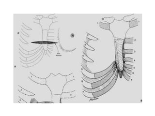
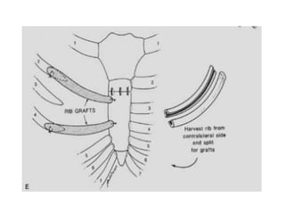
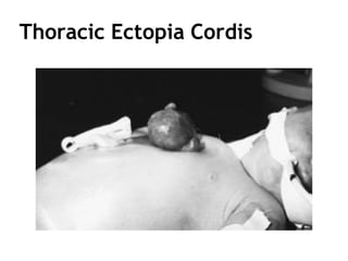
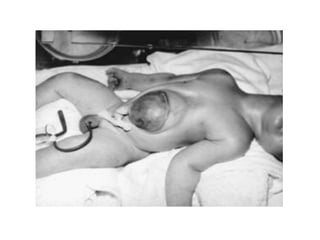
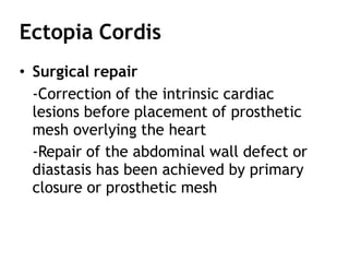
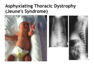
Chest Wall Disorder or
Deformities
Group A
CLASSIFICATION:
Out line
• Pectus Excavatum
• Pectus Carinatum
• Poland's Syndrome
• Sternal Defects
• Cleft Sternum
• Ectopia Cordis
• Thoracic Deformities in Diffuse Skeletal
Disorders
Pectus Excavatum
• Funnel chest or trichterbrust
• Posterior depression of the sternum and
costal cartilages
• 1st & 2nd ribs & manubrium : normal
position
• Lower costal cartilages and the body of the
sternum are depressed
• Asymmetry of the depression is frequently
present Rt. > Lt.
Pectus Excavatum
• Present at birth or within first year of life
in the majority : 86%
• Rarely resolves with increasing age
• May worsen during rapid adolescent
growth
• Scoliosis: 26%
• Asthma : 5.2%
• CHD : 1.5%
Pectus Excavatum
• Etiology and Incidence
- 1 in 400 live births
- Boys > Girl 4: 1
- Etiology : unknown
- Variable pattern of inheritance
- Multifactorial
Pectus Excavatum
• Symptoms
- Well tolerated in infancy and childhood
- Older children :
 pain in the area of the deformed cartilages
 precordial pain after sustained exercise
 palpitations
 transient atrial arrhythmias
 may have mitral valve prolapse
Pectus Excavatum
• Surgical Repair
Haller and associates : tripod fixation
- Subperichondrial resection
- Posterior sternal osteotomy
- Most cephalad normal cartilages are then
divided obliquely in a posterolateral
- Sternum elevated, the sternal ends of
the cartilage rest on the costal ends
Pectus Excavatum
• Nuss and procedure
• Minimally Invasive Repair of Pectus
Excavatum (MIRPE)
The open surgical technique
Complication : Nuss procedure
Early
• Pneumothorax
• Pericarditis
• Pneumonia
• Hemothorax
• Transient extremity
paralysis
• Superficial wound
infection
• Bar infection
Late
• Bar displacement
• Hemothorax
• Overcorrection deformity
• Pectus carinatum
deformity
Pectus Carinatum
• CHD : 18%
• FH of chest wall deformity : 26 %
• Boys > Girls = 78 : 22 %
• Mild form at birth but often progresses
during early childhood
• The chondromanubrial deformity, often
noted at birth
SURGICAL REPAIR
Pectus Carinatum
Pectus Carinatum
Poland's Syndrome
• Hypoplasia of the sternal head of the
pectoralis major and minor muscles
• Normal underlying ribs to complete
absence anterior portions of the 2nd – 5th
ribs and costal cartilages
Poland's Syndrome
Poland's Syndrome
• Surgical Repair
- Assessment of the extent of involvement
limited to the sternal component of the
pectoralis major and minor muscles,
• little functional deficit
• repair is not necessary except to
facilitate breast augmentation in
women
Poland's Syndrome
• Surgical Repair
- Assessment of the extent of
involvement underlying costal cartilages
are depressed or absent
• repair must be considered to minimize
the concavity
• to eliminate the paradoxic motion of
the chest wall if ribs are absent
• in girls to provide an optimal base for
breast reconstruction
Poland's Syndrome
• Surgical Repair
Ravitch :
– Correction of posteriorly displaced costal
cartilages by unilateral resection of the
cartilages
– A wedge osteotomy of the sternum, allowing
rotation of the sternum; and fixation with
Rehbein struts and Steinmann pins
– The sternum is then displaced anteriorly and
supported with a retrosternal strut
Cleft Sternum
• Complete or partial separation of the sternum
but a normally positioned intrathoracic heart
• Results from nonfusion of the sternal bars
• Normal skin coverage is present
• Intact pericardium and a normal diaphragm
• Omphaloceles do not occur in these children
• Dramatic increase in the deformity occurs
with crying or Valsalva's maneuver
Ectopia Cordis
• Thoracic Ectopia Cordis
• Thoracoabdominal Ectopia Cordis
(Cantrell's Pentalogy)
Ectopia Cordis
Etiology
• Disruption of amnion and chorionic layer
or yolk sac
• Chromosomal abnormalities have been
reported
Thoracic Ectopia Cordis
• Severely deficient in the midline somatic
tissues that normally cover the heart
• Many attempts at primary closure fail
because of the inability to mobilize
adequate tissues for coverage
• An abdominal defect is often present as
well
Thoracoabdominal Ectopia Cordis
• Heart is covered by an omphalocele-like membrane or
thin skin, which is often pigmented.
• The five essential features :
(a) a cleft lower sternum
(b)a half moon–shaped anterior diaphragmatic defect
resulting from lack of development of the septum
transversum
(c)absence of the parietal pericardium at the
diaphragmatic defect
(d) omphalocele
(e) in most patients, an intrinsic cardiac anomaly
Thoracoabdominal Ectopia Cordis
Ectopia Cordis
• Surgical repair
• Successful repair and long-term survival are
more frequently achieved in thoracoabdominal
ectopia cordis than in thoracic ectopia cordis
• Return heart to thoracic cavity resulted in
death
• Several cases have been successfully managed
by local application of topical astringents, thus
allowing secondary epithelialization to occur
Ectopia Cordis
• Surgical repair
-Correction of the intrinsic cardiac
lesions before placement of prosthetic
mesh overlying the heart
-Repair of the abdominal wall defect or
diastasis has been achieved by primary
closure or prosthetic mesh
Thoracic Deformities in Diffuse
Skeletal Disorders
• Asphyxiating Thoracic Dystrophy
(Jeune's Syndrome)
• Spondylothoracic Dysplasia
(Jarcho–Levin Syndrome)
Asphyxiating Thoracic Dystrophy
(Jeune's Syndrome)
• Narrow, rigid chest & multiple cartilage
anomalies
• Bell-shaped thorax and protuberant abdomen
• Patient died of respiratory insufficiency early
• Form of osteochondrodystrophy, which has
variable degrees of skeletal involvement
• Inherited in an autosomal recessive
• Not associated with chromosomal
abnormalities
Asphyxiating Thoracic Dystrophy
(Jeune's Syndrome)
Asphyxiating Thoracic Dystrophy
(Jeune's Syndrome)
• Short, stubby extremities with relatively short,
wide bones
• Clavicles fixed and elevated position
• Pelvis is small and hypoplastic, with square iliac
bones
• Involves variable degree of pulmonary
impairment
• Later reports can survive for longer periods of
time
Spondylothoracic Dysplasia (Jarcho–
Levin Syndrome)
Spondylothoracic Dysplasia (Jarcho–
Levin Syndrome)
• Autosomal recessive deformity
• Multiple vertebral and rib malformations
• Multiple alternating hemivertebrae in most or all
of the thoracic and lumbar spine
• Vertebral ossification centers rarely cross the
midline
• Bone formation is normal
• Multiple posterior fusions of the ribs and
remarkable shortening of the thoracic spine
• Crab-like appearance of the ribs on the chest
radiograph
Spondylothoracic Dysplasia (Jarcho–
Levin Syndrome)
• Thoracic deformity is secondary to the spine
anomaly
• Results in close posterior approximation of origin
of ribs
• Most infants with the entity die before 15 months
• No surgical efforts have been proposed or
attempted
• One third of patients with this syndrome have
associated malformations
– congenital heart disease and renal anomalies
Thank you

88.pptx

  • 1.
    Chest Wall Disorderor Deformities Group A
  • 2.
  • 4.
    Out line • PectusExcavatum • Pectus Carinatum • Poland's Syndrome • Sternal Defects • Cleft Sternum • Ectopia Cordis • Thoracic Deformities in Diffuse Skeletal Disorders
  • 5.
    Pectus Excavatum • Funnelchest or trichterbrust • Posterior depression of the sternum and costal cartilages • 1st & 2nd ribs & manubrium : normal position • Lower costal cartilages and the body of the sternum are depressed • Asymmetry of the depression is frequently present Rt. > Lt.
  • 7.
    Pectus Excavatum • Presentat birth or within first year of life in the majority : 86% • Rarely resolves with increasing age • May worsen during rapid adolescent growth • Scoliosis: 26% • Asthma : 5.2% • CHD : 1.5%
  • 8.
    Pectus Excavatum • Etiologyand Incidence - 1 in 400 live births - Boys > Girl 4: 1 - Etiology : unknown - Variable pattern of inheritance - Multifactorial
  • 9.
    Pectus Excavatum • Symptoms -Well tolerated in infancy and childhood - Older children :  pain in the area of the deformed cartilages  precordial pain after sustained exercise  palpitations  transient atrial arrhythmias  may have mitral valve prolapse
  • 10.
    Pectus Excavatum • SurgicalRepair Haller and associates : tripod fixation - Subperichondrial resection - Posterior sternal osteotomy - Most cephalad normal cartilages are then divided obliquely in a posterolateral - Sternum elevated, the sternal ends of the cartilage rest on the costal ends
  • 11.
    Pectus Excavatum • Nussand procedure • Minimally Invasive Repair of Pectus Excavatum (MIRPE)
  • 12.
  • 23.
    Complication : Nussprocedure Early • Pneumothorax • Pericarditis • Pneumonia • Hemothorax • Transient extremity paralysis • Superficial wound infection • Bar infection Late • Bar displacement • Hemothorax • Overcorrection deformity • Pectus carinatum deformity
  • 24.
    Pectus Carinatum • CHD: 18% • FH of chest wall deformity : 26 % • Boys > Girls = 78 : 22 % • Mild form at birth but often progresses during early childhood • The chondromanubrial deformity, often noted at birth
  • 25.
  • 26.
  • 27.
  • 28.
    Poland's Syndrome • Hypoplasiaof the sternal head of the pectoralis major and minor muscles • Normal underlying ribs to complete absence anterior portions of the 2nd – 5th ribs and costal cartilages
  • 29.
  • 31.
    Poland's Syndrome • SurgicalRepair - Assessment of the extent of involvement limited to the sternal component of the pectoralis major and minor muscles, • little functional deficit • repair is not necessary except to facilitate breast augmentation in women
  • 32.
    Poland's Syndrome • SurgicalRepair - Assessment of the extent of involvement underlying costal cartilages are depressed or absent • repair must be considered to minimize the concavity • to eliminate the paradoxic motion of the chest wall if ribs are absent • in girls to provide an optimal base for breast reconstruction
  • 33.
    Poland's Syndrome • SurgicalRepair Ravitch : – Correction of posteriorly displaced costal cartilages by unilateral resection of the cartilages – A wedge osteotomy of the sternum, allowing rotation of the sternum; and fixation with Rehbein struts and Steinmann pins – The sternum is then displaced anteriorly and supported with a retrosternal strut
  • 37.
    Cleft Sternum • Completeor partial separation of the sternum but a normally positioned intrathoracic heart • Results from nonfusion of the sternal bars • Normal skin coverage is present • Intact pericardium and a normal diaphragm • Omphaloceles do not occur in these children • Dramatic increase in the deformity occurs with crying or Valsalva's maneuver
  • 40.
    Ectopia Cordis • ThoracicEctopia Cordis • Thoracoabdominal Ectopia Cordis (Cantrell's Pentalogy)
  • 41.
    Ectopia Cordis Etiology • Disruptionof amnion and chorionic layer or yolk sac • Chromosomal abnormalities have been reported
  • 42.
  • 43.
    Thoracic Ectopia Cordis •Severely deficient in the midline somatic tissues that normally cover the heart • Many attempts at primary closure fail because of the inability to mobilize adequate tissues for coverage • An abdominal defect is often present as well
  • 45.
    Thoracoabdominal Ectopia Cordis •Heart is covered by an omphalocele-like membrane or thin skin, which is often pigmented. • The five essential features : (a) a cleft lower sternum (b)a half moon–shaped anterior diaphragmatic defect resulting from lack of development of the septum transversum (c)absence of the parietal pericardium at the diaphragmatic defect (d) omphalocele (e) in most patients, an intrinsic cardiac anomaly
  • 46.
  • 48.
    Ectopia Cordis • Surgicalrepair • Successful repair and long-term survival are more frequently achieved in thoracoabdominal ectopia cordis than in thoracic ectopia cordis • Return heart to thoracic cavity resulted in death • Several cases have been successfully managed by local application of topical astringents, thus allowing secondary epithelialization to occur
  • 49.
    Ectopia Cordis • Surgicalrepair -Correction of the intrinsic cardiac lesions before placement of prosthetic mesh overlying the heart -Repair of the abdominal wall defect or diastasis has been achieved by primary closure or prosthetic mesh
  • 50.
    Thoracic Deformities inDiffuse Skeletal Disorders • Asphyxiating Thoracic Dystrophy (Jeune's Syndrome) • Spondylothoracic Dysplasia (Jarcho–Levin Syndrome)
  • 51.
    Asphyxiating Thoracic Dystrophy (Jeune'sSyndrome) • Narrow, rigid chest & multiple cartilage anomalies • Bell-shaped thorax and protuberant abdomen • Patient died of respiratory insufficiency early • Form of osteochondrodystrophy, which has variable degrees of skeletal involvement • Inherited in an autosomal recessive • Not associated with chromosomal abnormalities
  • 52.
  • 53.
    Asphyxiating Thoracic Dystrophy (Jeune'sSyndrome) • Short, stubby extremities with relatively short, wide bones • Clavicles fixed and elevated position • Pelvis is small and hypoplastic, with square iliac bones • Involves variable degree of pulmonary impairment • Later reports can survive for longer periods of time
  • 54.
  • 55.
    Spondylothoracic Dysplasia (Jarcho– LevinSyndrome) • Autosomal recessive deformity • Multiple vertebral and rib malformations • Multiple alternating hemivertebrae in most or all of the thoracic and lumbar spine • Vertebral ossification centers rarely cross the midline • Bone formation is normal • Multiple posterior fusions of the ribs and remarkable shortening of the thoracic spine • Crab-like appearance of the ribs on the chest radiograph
  • 56.
    Spondylothoracic Dysplasia (Jarcho– LevinSyndrome) • Thoracic deformity is secondary to the spine anomaly • Results in close posterior approximation of origin of ribs • Most infants with the entity die before 15 months • No surgical efforts have been proposed or attempted • One third of patients with this syndrome have associated malformations – congenital heart disease and renal anomalies
  • 57.