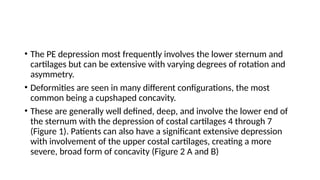
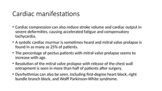
Pectus excavatum
• Pectus chest deformities are among the most common major
congenital anomalies
• Men are afflicted 5 times more often than women
• Pectus excavatum (PE) with sternal depression is approximately 6
times more common than pectus carinatum (protrusion)
• Approximately 40% of patients with pectus deformities have family members who
also have deformities, although a direct genetic link has not been identified.
• Disturbances in the growth of the sternum and costal arches, as well as
biomechanical factors, are suspected in the pathogenesis.
• The involved cartilages can be fused, deformed, or rotated.
• Intrinsic abnormality of the costochondral cartilage is suggested by the significant
occurrence of PE among patients with connective tissue disorders, such as
Marfan syndrome.
• There is also a high percentage of scoliosis associated with the disorder.
• Abnormalities of the cartilages removed during surgery have also been reported,
including stress/strain deformation and abnormal collagen content.
• PE can be present at birth but is most frequently recognized during
early childhood.
• During rapid adolescent growth, many patients experience a marked
increase in the severity of the depression until full skeletal maturity is
achieved.
• In severe cases or those in which depressions directly impinge on the
right ventricle, PE is more than a cosmetic deformity.
• Many patients do not undergo repair during childhood and
subsequently experience progressive a worsening of symptoms and
cardiopulmonary function with increasing age
• One theory for the worsening of symptoms that is experienced with age is that the chest wall
is very compliant in infancy but, with aging, a steady decline in compliance is experienced
and it becomes relatively stiffer compared with the lungs. In adults the loss of elasticity and
flexibility of the chest can lead to cardiac symptoms with only moderate exertion.
• A similar phenomena is also seen in patients with scoliosis as they age.
• Publications regarding the surgical management of adult patients with pectus deformities are
few, but have increased significantly during the past few years with greater recognition of the
physiologic and psychologic impact of the disease.
• Despite well-documented reports to the contrary, the longstanding misconception persists
that PE is a cosmetic defect with no physiologic consequences. This misconception has
resulted in patients reaching adulthood with uncorrected defects.
• Some patients will develop cardiopulmonary symptoms for the first time as they age and
others will experience a worsening of symptoms they have endured for years.
• The PE depression most frequently involves the lower sternum and
cartilages but can be extensive with varying degrees of rotation and
asymmetry.
• Deformities are seen in many different configurations, the most
common being a cupshaped concavity.
• These are generally well defined, deep, and involve the lower end of
the sternum with the depression of costal cartilages 4 through 7
(Figure 1). Patients can also have a significant extensive depression
with involvement of the upper costal cartilages, creating a more
severe, broad form of concavity (Figure 2 A and B)
Clinical features
1. Dyspnea with mild exercise
2. Progressive loss of endurance
3. Inability to keep up with peers
4. Chest pain with activity
5. Worsening, progressive fatigue
6. Palpitations
7. Tachycardia
8. Exercise induced wheezing
9. Frequent upper respiratory infections
10. Easy fatigue
11. Fainting/dizziness
12. Chest pain without exercise
13. Exercise intolerance
Despite multiple studies evaluating cardiac and pulmonary function,
there has been no conclusive or consistent demonstration of
physiologic impact correlating with the symptoms of PE
Pulmonary manifestations
• Anatomic abnormalities, including decreased thoracic volume and cardiac
compression, are thought to explain the physiologic effects noted by
patients.
• The increased work of breathing from a partially restricted chest wall with
impaired oxygen delivery to working muscles as the result of decreased
venous return to the right heart may also play a role.
• Sternal compression is suspected to decrease thoracic volume, which can
reduce the SVO2, exercise tolerance, tidal volume, and vital capacity, causing
dyspnea and a decrease in endurance with compensatory tachypnea during
exercise.
Cardiac manifestations
• Cardiac compression can also reduce stroke volume and cardiac output in
severe deformities, causing accelerated fatigue and compensatory
tachycardia.
• A systolic cardiac murmur is sometimes heard and mitral valve prolapse is
found in as many as 25% of patients.
• The percentage of pectus patients with mitral valve prolapse seems to
increase with age.
• Resolution of the mitral valve prolapse with release of the chest wall
entrapment is seen in more than half of patients after surgery.
• Dysrhythmias can also be seen, including first-degree heart block, right
bundle branch block, and Wolff Parkinson-White syndrome.
Psychological manifestations
• psychosocial issues surrounding body image among teenagers can be significant, life-altering, and
occasionally life-threatening.
• Poor body image and impaired psychosocial function is an enormously important concern for surgical
repair. Children and adolescents with potentially visible physical differences may be at risk for body
image and interpersonal difficulties.
• Psychological research about patients with disfigurement confirm disturbance of the body schema and
altercation of the individual’s self-representation.
• Surgical repair of PE can significantly improve the difficulties with body image and limitations on
physical activity that are experienced by patients.
• These results should prompt physicians to consider the physiologic and psychological implications of PE
just as they would any other physical deformity known to have such consequences. In a multicenter
investigation, geographically dispersed children and adults demonstrated a marked effect of the chest
deformity on perceived ability to exercise, which was consistently improved with surgical correction.
• The importance of these concerns to the child and family should not be underestimated by physicians
• Investigation as to the cause of exercise intolerance and perceived limitation of
ability have yielded mixed results, and whether surgical repair corrects these deficits
is controversial.
• Malek et al22 and Malek and Marelich41 performed a comprehensive metaanalysis
on publications reporting postoperative cardiopulmonary results among PE patients.
• The critical finding of the study was that surgical repair improved cardiovascular
function by more than 1⁄2 standard deviation. The patient’s personal testimony has
been the most convincing evidence for repair.
• The majority of patients with symptoms reported significant improvement after
repair. These findings, along with current ongoing investigations, further the
argument that surgical repair of PE is indicated for symptomatic patients regardless
of physiologic testing and is more than primarily cosmetic.
Evaluation
• The presentation of a patient with PE warrants a thorough workup to
access the significance of the defect. This workup does not necessarily
include every possible physiologic test, but can include those
necessary to determine whether the patient should be referred to a
surgeon for a discussion of repair options.
Radiographic Evaluation
• Imaging for anatomic assessment and documentation of the chest dimensions is important
and remains a mainstay of the anatomic evaluation of PE.
• A noncontrast computerized tomographic (CT) scan is useful to access the deformity of the
bony and cartilaginous skeleton in a 3-dimensional view, which clearly reveals any cardiac
compression (Figure 3a) or cardiac displacement (Figure 3B).
• Magnetic resonance imaging can be used instead of CT scan to reduce radiation exposure
but bony detail is better seen by a CT scan.
• An index of severity(HALLER INDEX) can be calculated by measuring the inner width of the
chest (at the lowest level of the pectus defect) and dividing it by the distance between the
posterior surface of the sternum (at the lowest part of the defect) and the anterior surface
of the spine.
• The severity index for healthy people is 2.52. In general, an index of 3.1 is considered
severe.
Cardiac
• Documentation of any dysrhythmias should be done with a 12-lead
electrocardiogram.
• It may be useful to perform an echocardiogram because the
depressed sternum may compress the right atrium and right ventricle,
interfering with diastolic filling of these structures.
• Mitral valve prolapse is commonly seen among patients with PE and
should also be documented if it is present.
• Evaluation of the aortic root and valve is also critical in patients with
suspected or confirmed Marfan syndrome
Pulmonary Functions
• Both static and exercise pulmonary function tests may be useful in
eliciting a physiologic effect of the PE defect on the patient.
• Static pulmonary function tests are the least sensitive but may show a
significant decrease in forced vital capacity and maximal ventilatory
volumes, which indicate restrictive airway disease.
• Patients may also have evidence of mild obstructive airway disease.
• In general, most patients with significant PE will still have a normal or
low-normal static pulmonary function test
Cardiopulmonary Exercise Testing
• Cardiopulmonary exercise testing can also be used to show significant
physiologic effects of the PE.
• The amount of oxygen that can be delivered to the tissues with
exercise can be examined. A decrease in the O2 pulse (mL/beat) and
amount of oxygen that could be delivered (Vo2 L/min and Ve L/min)
may be significantly below predicted values in patients with PE.
• Low-normal and below-normal values indicate that surgical correction
could benefit the patient.Figures 4 and 5 show the cardiopulmonary
exercise testing Vo2/heart rate and Vo2/ CO2 finding from a
symptomatic PE patient before and after an operation.
• The primary care physician needs to have a good understanding of the disease and realize the
importance of the deformity. Surgical correction of PE can be performed safely with minimal risk.
Referral for consideration of surgical treatment for severe cases should be made. Operation for
correction of PE is readily accomplished in teenagers, who are close to the age of skeletal maturity.
• The timing for surgery is problematic in the younger child. Currently, most surgeons will wait for
patients to reach the adolescence or early teenage years to perform repair. Repair during the later
stages of teenage growth allow the patient to complete their growth and have a lower chance of
recurrence. Younger children with significant cardiopulmonary compromise may also be candidates
for repair depending on the severity of their symptoms; however, repair at too early of an age can
result in improper growth of the chest wall and other complications, including recurrences.
• Adult repair is also feasible and has been reported in patients as old as 78 years. In general, referral
to a surgeon for consultation should occur when a patient demonstrates any of the criteria listed in
Table 2. Most patients will be considered for surgical correction if they demonstrate at least 2 of the
listed criteria. Others may need to be followed closely to watch for progression or worsening of the
defect and symptoms as they age.

Pectus excavatum presentation 15.3.23.pptx

  • 1.
  • 2.
    • Pectus chestdeformities are among the most common major congenital anomalies • Men are afflicted 5 times more often than women • Pectus excavatum (PE) with sternal depression is approximately 6 times more common than pectus carinatum (protrusion)
  • 3.
    • Approximately 40%of patients with pectus deformities have family members who also have deformities, although a direct genetic link has not been identified. • Disturbances in the growth of the sternum and costal arches, as well as biomechanical factors, are suspected in the pathogenesis. • The involved cartilages can be fused, deformed, or rotated. • Intrinsic abnormality of the costochondral cartilage is suggested by the significant occurrence of PE among patients with connective tissue disorders, such as Marfan syndrome. • There is also a high percentage of scoliosis associated with the disorder. • Abnormalities of the cartilages removed during surgery have also been reported, including stress/strain deformation and abnormal collagen content.
  • 4.
    • PE canbe present at birth but is most frequently recognized during early childhood. • During rapid adolescent growth, many patients experience a marked increase in the severity of the depression until full skeletal maturity is achieved. • In severe cases or those in which depressions directly impinge on the right ventricle, PE is more than a cosmetic deformity. • Many patients do not undergo repair during childhood and subsequently experience progressive a worsening of symptoms and cardiopulmonary function with increasing age
  • 5.
    • One theoryfor the worsening of symptoms that is experienced with age is that the chest wall is very compliant in infancy but, with aging, a steady decline in compliance is experienced and it becomes relatively stiffer compared with the lungs. In adults the loss of elasticity and flexibility of the chest can lead to cardiac symptoms with only moderate exertion. • A similar phenomena is also seen in patients with scoliosis as they age. • Publications regarding the surgical management of adult patients with pectus deformities are few, but have increased significantly during the past few years with greater recognition of the physiologic and psychologic impact of the disease. • Despite well-documented reports to the contrary, the longstanding misconception persists that PE is a cosmetic defect with no physiologic consequences. This misconception has resulted in patients reaching adulthood with uncorrected defects. • Some patients will develop cardiopulmonary symptoms for the first time as they age and others will experience a worsening of symptoms they have endured for years.
  • 6.
    • The PEdepression most frequently involves the lower sternum and cartilages but can be extensive with varying degrees of rotation and asymmetry. • Deformities are seen in many different configurations, the most common being a cupshaped concavity. • These are generally well defined, deep, and involve the lower end of the sternum with the depression of costal cartilages 4 through 7 (Figure 1). Patients can also have a significant extensive depression with involvement of the upper costal cartilages, creating a more severe, broad form of concavity (Figure 2 A and B)
  • 8.
    Clinical features 1. Dyspneawith mild exercise 2. Progressive loss of endurance 3. Inability to keep up with peers 4. Chest pain with activity 5. Worsening, progressive fatigue 6. Palpitations 7. Tachycardia 8. Exercise induced wheezing 9. Frequent upper respiratory infections 10. Easy fatigue 11. Fainting/dizziness 12. Chest pain without exercise 13. Exercise intolerance
  • 9.
    Despite multiple studiesevaluating cardiac and pulmonary function, there has been no conclusive or consistent demonstration of physiologic impact correlating with the symptoms of PE
  • 10.
    Pulmonary manifestations • Anatomicabnormalities, including decreased thoracic volume and cardiac compression, are thought to explain the physiologic effects noted by patients. • The increased work of breathing from a partially restricted chest wall with impaired oxygen delivery to working muscles as the result of decreased venous return to the right heart may also play a role. • Sternal compression is suspected to decrease thoracic volume, which can reduce the SVO2, exercise tolerance, tidal volume, and vital capacity, causing dyspnea and a decrease in endurance with compensatory tachypnea during exercise.
  • 11.
    Cardiac manifestations • Cardiaccompression can also reduce stroke volume and cardiac output in severe deformities, causing accelerated fatigue and compensatory tachycardia. • A systolic cardiac murmur is sometimes heard and mitral valve prolapse is found in as many as 25% of patients. • The percentage of pectus patients with mitral valve prolapse seems to increase with age. • Resolution of the mitral valve prolapse with release of the chest wall entrapment is seen in more than half of patients after surgery. • Dysrhythmias can also be seen, including first-degree heart block, right bundle branch block, and Wolff Parkinson-White syndrome.
  • 12.
    Psychological manifestations • psychosocialissues surrounding body image among teenagers can be significant, life-altering, and occasionally life-threatening. • Poor body image and impaired psychosocial function is an enormously important concern for surgical repair. Children and adolescents with potentially visible physical differences may be at risk for body image and interpersonal difficulties. • Psychological research about patients with disfigurement confirm disturbance of the body schema and altercation of the individual’s self-representation. • Surgical repair of PE can significantly improve the difficulties with body image and limitations on physical activity that are experienced by patients. • These results should prompt physicians to consider the physiologic and psychological implications of PE just as they would any other physical deformity known to have such consequences. In a multicenter investigation, geographically dispersed children and adults demonstrated a marked effect of the chest deformity on perceived ability to exercise, which was consistently improved with surgical correction. • The importance of these concerns to the child and family should not be underestimated by physicians
  • 13.
    • Investigation asto the cause of exercise intolerance and perceived limitation of ability have yielded mixed results, and whether surgical repair corrects these deficits is controversial. • Malek et al22 and Malek and Marelich41 performed a comprehensive metaanalysis on publications reporting postoperative cardiopulmonary results among PE patients. • The critical finding of the study was that surgical repair improved cardiovascular function by more than 1⁄2 standard deviation. The patient’s personal testimony has been the most convincing evidence for repair. • The majority of patients with symptoms reported significant improvement after repair. These findings, along with current ongoing investigations, further the argument that surgical repair of PE is indicated for symptomatic patients regardless of physiologic testing and is more than primarily cosmetic.
  • 14.
    Evaluation • The presentationof a patient with PE warrants a thorough workup to access the significance of the defect. This workup does not necessarily include every possible physiologic test, but can include those necessary to determine whether the patient should be referred to a surgeon for a discussion of repair options.
  • 15.
    Radiographic Evaluation • Imagingfor anatomic assessment and documentation of the chest dimensions is important and remains a mainstay of the anatomic evaluation of PE. • A noncontrast computerized tomographic (CT) scan is useful to access the deformity of the bony and cartilaginous skeleton in a 3-dimensional view, which clearly reveals any cardiac compression (Figure 3a) or cardiac displacement (Figure 3B). • Magnetic resonance imaging can be used instead of CT scan to reduce radiation exposure but bony detail is better seen by a CT scan. • An index of severity(HALLER INDEX) can be calculated by measuring the inner width of the chest (at the lowest level of the pectus defect) and dividing it by the distance between the posterior surface of the sternum (at the lowest part of the defect) and the anterior surface of the spine. • The severity index for healthy people is 2.52. In general, an index of 3.1 is considered severe.
  • 16.
    Cardiac • Documentation ofany dysrhythmias should be done with a 12-lead electrocardiogram. • It may be useful to perform an echocardiogram because the depressed sternum may compress the right atrium and right ventricle, interfering with diastolic filling of these structures. • Mitral valve prolapse is commonly seen among patients with PE and should also be documented if it is present. • Evaluation of the aortic root and valve is also critical in patients with suspected or confirmed Marfan syndrome
  • 17.
    Pulmonary Functions • Bothstatic and exercise pulmonary function tests may be useful in eliciting a physiologic effect of the PE defect on the patient. • Static pulmonary function tests are the least sensitive but may show a significant decrease in forced vital capacity and maximal ventilatory volumes, which indicate restrictive airway disease. • Patients may also have evidence of mild obstructive airway disease. • In general, most patients with significant PE will still have a normal or low-normal static pulmonary function test
  • 18.
    Cardiopulmonary Exercise Testing •Cardiopulmonary exercise testing can also be used to show significant physiologic effects of the PE. • The amount of oxygen that can be delivered to the tissues with exercise can be examined. A decrease in the O2 pulse (mL/beat) and amount of oxygen that could be delivered (Vo2 L/min and Ve L/min) may be significantly below predicted values in patients with PE. • Low-normal and below-normal values indicate that surgical correction could benefit the patient.Figures 4 and 5 show the cardiopulmonary exercise testing Vo2/heart rate and Vo2/ CO2 finding from a symptomatic PE patient before and after an operation.
  • 19.
    • The primarycare physician needs to have a good understanding of the disease and realize the importance of the deformity. Surgical correction of PE can be performed safely with minimal risk. Referral for consideration of surgical treatment for severe cases should be made. Operation for correction of PE is readily accomplished in teenagers, who are close to the age of skeletal maturity. • The timing for surgery is problematic in the younger child. Currently, most surgeons will wait for patients to reach the adolescence or early teenage years to perform repair. Repair during the later stages of teenage growth allow the patient to complete their growth and have a lower chance of recurrence. Younger children with significant cardiopulmonary compromise may also be candidates for repair depending on the severity of their symptoms; however, repair at too early of an age can result in improper growth of the chest wall and other complications, including recurrences. • Adult repair is also feasible and has been reported in patients as old as 78 years. In general, referral to a surgeon for consultation should occur when a patient demonstrates any of the criteria listed in Table 2. Most patients will be considered for surgical correction if they demonstrate at least 2 of the listed criteria. Others may need to be followed closely to watch for progression or worsening of the defect and symptoms as they age.