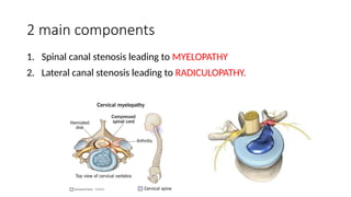
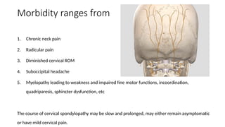
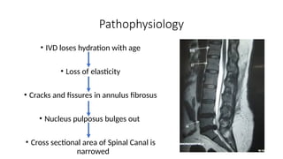
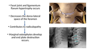
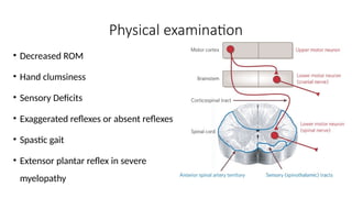
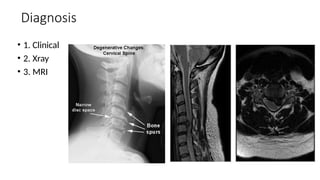
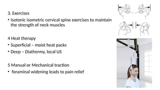
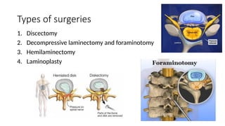
Cervical Spondylosis
Dr Aakash Nandu
M.S. Orthopaedics
Smit Hospital, Anand
Cervical Spondylosis
Definition
It is chronic degenerative disorder of cervical spine which affects
vertebrae, inter vertebral discs and ligaments of cervical spine.
Anatomy
Cervical spine consists of 7 vertebrae beginning from the base of the skull.
There are 8 pairs of nerve roots exiting posteriorly from spinal cord
through foramen in between the vertebrae.
Introduction
• Commonly seen in individuals after the age of 40 years
• Believed to be a part of normal ageing process of vertebral
column.
• Includes degenerative changes in facet joints, longitudinal
ligaments and ligamentum flavum.
2 main components
1. Spinal canal stenosis leading to MYELOPATHY
2. Lateral canal stenosis leading to RADICULOPATHY.
Morbidity ranges from
1. Chronic neck pain
2. Radicular pain
3. Diminished cervical ROM
4. Suboccipital headache
5. Myelopathy leading to weakness and impaired fine motor functions, incoordination,
quadriparesis, sphincter dysfunction, etc
The course of cervical spondylopathy may be slow and prolonged, may either remain asymptomatic
or have mild cervical pain.
Pathophysiology
• IVD loses hydration with age
• Loss of elasticity
• Cracks and fissures in annulus fibrosus
• Nucleus pulposus bulges out
• Cross sectional area of Spinal Canal is
narrowed
• Facet joint and ligamentum
flavum hypertrophy occurs
• Decreases the dorso-lateral
space of the foramen
• Contributes to radiculopathy
• Marginal osteophytes develop
and end plate destruction
occurs.
Clinical features
1. Cervical Pain
• Chronic suboccipital headache
• May radiate to shoulder, scapula or arm
• Pain increases with certain movements
2. Radiculopathy
• Compression of cervical roots leads to radicular pain or muscle weakness
• Mostly resolves with conservative management, rarely surgery is required.
3. CSM (Cervical Spondylotic Myelopathy)
• Most serious consequence of CS
• Occurs due to narrowed vertebral canal
• Quadriparesis, sphinchter involvement occurs
• Complete reversal is rare once CSM occurs
Etiology
1. Age (>40)
2. Trauma
3. Work activity (carrying load on head)
4. Genetics
Physical examination
• Decreased ROM
• Hand clumsiness
• Sensory Deficits
• Exaggerated reflexes or absent reflexes
• Spastic gait
• Extensor plantar reflex in severe
myelopathy
• Spurling sign – exacerbation of radicular pain by
lateral flexion towards affected side (due to
foraminal narrowing)
• Lhermitte sign – generalized electric shock like
sensation due to neck flexion
• Hoffman sign – reflex contraction of thumb and
index finger in response to nipping of middle
finger ( sign of UMN lesion )
Diagnosis
• 1. Clinical
• 2. Xray
• 3. MRI
Treatment
1. Immobilisation
• Reduces irritation of nerves
• Daytime soft cervical collar
• More rigid orthosis (Philadelphia collar, minerva
jacket)
2. Moulded Pillows
• For better alignment of spine during sleep
3. Exercises
• Isotonic isometric cervical spine exercises to maintain
the strength of neck muscles
4 Heat therapy
• Superficial – moist heat packs
• Deep – Diathermy, local US
5 Manual or Mechanical traction
• foraminal widening leads to pain relief
6 Occupational therapy
• Ergonomic adjustment of workplace,
postural correction and vocational therapy.
7 Pharmacological
• NSAIDS
• Opioids
• Pregabalin, Nortryptiline
Surgery
• Indications
1. Progressive neurological deficit
2. Intractable pain
3. Documented compression of nerve root or spinal cord
Types of surgeries
1. Discectomy
2. Decompressive laminectomy and foraminotomy
3. Hemilaminectomy
4. Laminoplasty
Cervical Spondylosis (Nutshell) with pictures

Cervical Spondylosis (Nutshell) with pictures

  • 1.
    Cervical Spondylosis Dr AakashNandu M.S. Orthopaedics Smit Hospital, Anand
  • 2.
    Cervical Spondylosis Definition It ischronic degenerative disorder of cervical spine which affects vertebrae, inter vertebral discs and ligaments of cervical spine. Anatomy Cervical spine consists of 7 vertebrae beginning from the base of the skull. There are 8 pairs of nerve roots exiting posteriorly from spinal cord through foramen in between the vertebrae.
  • 5.
    Introduction • Commonly seenin individuals after the age of 40 years • Believed to be a part of normal ageing process of vertebral column. • Includes degenerative changes in facet joints, longitudinal ligaments and ligamentum flavum.
  • 6.
    2 main components 1.Spinal canal stenosis leading to MYELOPATHY 2. Lateral canal stenosis leading to RADICULOPATHY.
  • 7.
    Morbidity ranges from 1.Chronic neck pain 2. Radicular pain 3. Diminished cervical ROM 4. Suboccipital headache 5. Myelopathy leading to weakness and impaired fine motor functions, incoordination, quadriparesis, sphincter dysfunction, etc The course of cervical spondylopathy may be slow and prolonged, may either remain asymptomatic or have mild cervical pain.
  • 8.
    Pathophysiology • IVD loseshydration with age • Loss of elasticity • Cracks and fissures in annulus fibrosus • Nucleus pulposus bulges out • Cross sectional area of Spinal Canal is narrowed
  • 9.
    • Facet jointand ligamentum flavum hypertrophy occurs • Decreases the dorso-lateral space of the foramen • Contributes to radiculopathy • Marginal osteophytes develop and end plate destruction occurs.
  • 10.
    Clinical features 1. CervicalPain • Chronic suboccipital headache • May radiate to shoulder, scapula or arm • Pain increases with certain movements 2. Radiculopathy • Compression of cervical roots leads to radicular pain or muscle weakness • Mostly resolves with conservative management, rarely surgery is required.
  • 11.
    3. CSM (CervicalSpondylotic Myelopathy) • Most serious consequence of CS • Occurs due to narrowed vertebral canal • Quadriparesis, sphinchter involvement occurs • Complete reversal is rare once CSM occurs
  • 12.
    Etiology 1. Age (>40) 2.Trauma 3. Work activity (carrying load on head) 4. Genetics
  • 13.
    Physical examination • DecreasedROM • Hand clumsiness • Sensory Deficits • Exaggerated reflexes or absent reflexes • Spastic gait • Extensor plantar reflex in severe myelopathy
  • 14.
    • Spurling sign– exacerbation of radicular pain by lateral flexion towards affected side (due to foraminal narrowing) • Lhermitte sign – generalized electric shock like sensation due to neck flexion • Hoffman sign – reflex contraction of thumb and index finger in response to nipping of middle finger ( sign of UMN lesion )
  • 15.
  • 16.
    Treatment 1. Immobilisation • Reducesirritation of nerves • Daytime soft cervical collar • More rigid orthosis (Philadelphia collar, minerva jacket) 2. Moulded Pillows • For better alignment of spine during sleep
  • 17.
    3. Exercises • Isotonicisometric cervical spine exercises to maintain the strength of neck muscles 4 Heat therapy • Superficial – moist heat packs • Deep – Diathermy, local US 5 Manual or Mechanical traction • foraminal widening leads to pain relief
  • 18.
    6 Occupational therapy •Ergonomic adjustment of workplace, postural correction and vocational therapy. 7 Pharmacological • NSAIDS • Opioids • Pregabalin, Nortryptiline
  • 19.
    Surgery • Indications 1. Progressiveneurological deficit 2. Intractable pain 3. Documented compression of nerve root or spinal cord
  • 20.
    Types of surgeries 1.Discectomy 2. Decompressive laminectomy and foraminotomy 3. Hemilaminectomy 4. Laminoplasty