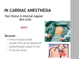
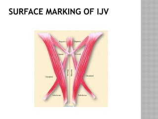
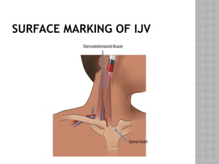
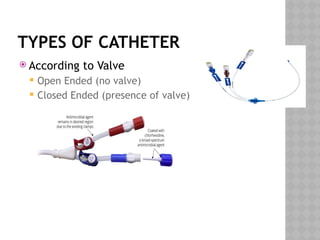
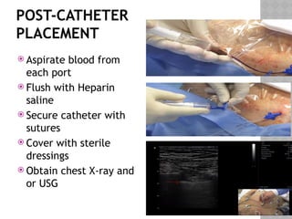
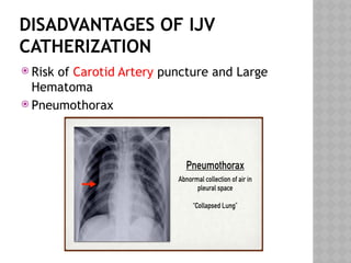
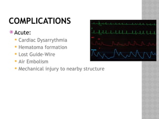
CENTRAL VENOUS
CATHETERIZATION
DR. MD. SHARIF HASAN
MBBS, DA
Specialist, Cardiac Anaesthesia
SQUARE HOSPITALS LTD.
INDICATIONS
 Central Venous Pressure Monitoring
 Hemodynamic Monitoring
 Volume Resuscitation
INDICATIONS
 Administration of
Drugs likely to
induce Phlebitis (eg-
Potassium,
Amiodarone, 3%
NaCl)
 Administration of
Multiple Ionotrops
INDICATIONS
 Lack of Peripheral Access
 Repeated Blood Sampling
 Parenteral Feeding
CHOISE OF VEINS
 Internal Jugular Vein
 Subclavian Vein
 External Jugular Vein
 Chephalic Vein
 Femoral Vein
Subclavian
IN CARDIAC ANESTHESIA
Our Choice is Internal Jugular
Vein (IJV)
WHY?
Because-
 Free of Surgical Site
 Actual CVP can be Measured
 Anatomically closure to RA
 It has no valves
D.H. LAWRENCE QUOTE:
“THE EYES DON'T SEE WHAT THE
MIND DOESN'T KNOW.”
SURFACE MARKING OF IJV
Sternocleidomastoid-Clavicle
Triangle
SURFACE MARKING OF IJV
INSTRUMENTS FOR
CATHETERIZATION
INSTRUMENTS FOR
CATHETERIZATION
INSTRUMENTS FOR
CATHETERIZATION
TYPES OF CATHETER
 According to Duration
 Non-Tunneled (Short duration)
 Tunneled
 Peripherally inserted
 Implantable ports
 According to Lumen
 Single Lumen
 Multiple Lumen (Double, Three
lumen, Four Lumen)
TYPES OF CATHETER
 According to Valve
 Open Ended (no valve)
 Closed Ended (presence of valve)
TECHNIQUE OF INSERTION
Blind Technique USG Guided
Technique
BLIND TECHNIQUE
 Right side preferred – lower
pleural dome and thoracic duct
on left
 Trendelenburg position – 10-15
degrees
 Head rotated approximately 15
degree to left
 At cricoid level while palpating
the carotid pulse, introducer
needle into the apex of the
sternocleidomastoid-clavicular
triangle at a 30-40 degree angle
to the skin. Aim the needle
caudally towards the patient’s
ipsilateral nipple.
SELDINGER
TECHNIQUE
 Wire is threaded
through the needle
 Needle is removed
 Skin and vessel are
dilated with the
Dilator
 Catheter is placed
over the wire
 Wire is removed
 Catheter is secured
in place
POST-CATHETER
PLACEMENT
 Aspirate blood from
each port
 Flush with Heparin
saline
 Secure catheter with
sutures
 Cover with sterile
dressings
 Obtain chest X-ray and
or USG
USG GUIDED TECHNIQUE
DISADVANTAGES OF IJV
CATHERIZATION
 Risk of Carotid Artery puncture and Large
Hematoma
 Pneumothorax
COMPLICATIONS
 Acute:
 Cardiac Dysarrythmia
 Hematoma formation
 Lost Guide-Wire
 Air Embolism
 Mechanical injury to nearby structure
COMPLICATIONS
 Chronic
 Infection
 Non-function or Blockage
 Thromboembolism
 Catheter fragmentation
WHAT WE NEED ???
USG
Machine
CENTRAL VENOUS CATHETERIZATION FOR CARDIAC

CENTRAL VENOUS CATHETERIZATION FOR CARDIAC