Recommended
PDF
51a9f15c-8424-4bcf-8e0f-7265ae2a5856.pdf
PDF
51a9f15c-8424-4bcf-8e0f-7265ae2a5856.pdf
PPTX
Cell Cycle and Cell Division.pptx
PDF
Cell-Cycle-and-Cell-Divisssssssssssssion.pdsf
PPTX
The Cell Cycle: Phases and Regulation Explained
PPTX
Lec1 cell cycle and mitosis
PPTX
PPTX
PDF
Lecture on stages of Mitosis-cell di.pdf
PPTX
PDF
Class 11th Ncert Cbse Neet BIOLOGY CELL CYCLE AND CELL DIVISION.pdf
PPT
Cell cycle,cell cycle checkpoint and control
PPT
The cell cycle , the different phases and the cell division
PPTX
CELL CYCLE, MITOSIS & MEIOSIS SMG
PPT
-CELL_ STRUCTURE_ AND_ FUNCTION_PPTX.ppt
PPTX
Cell cycle-1.pptx pharmacology, pharmacy students
PPTX
PPTX
DOCX
Concept Of Cell cycle phases.docx
PDF
10 – cell cycle and cell division
PPTX
PPTX
Cell division (Mitosis and Meiosis)
PPTX
PPT ABOUT CELL CYCLES SS - Angelina.pptx
PPTX
Cel cycle and its regulation
PPTX
Mitosis division by Arifa Bee M.Sc. I year.pptx
PPT
PPT
Structure , function and growth of prokaryote and eukaryote cells
DOCX
PPTX
12TH ENGLISH BIHAR BOARD GRAMMAR FOR 2026
PPTX
PLATHELMINTHES.pptx TAXONOMY FOR CLASS UG PG
More Related Content
PDF
51a9f15c-8424-4bcf-8e0f-7265ae2a5856.pdf
PDF
51a9f15c-8424-4bcf-8e0f-7265ae2a5856.pdf
PPTX
Cell Cycle and Cell Division.pptx
PDF
Cell-Cycle-and-Cell-Divisssssssssssssion.pdsf
PPTX
The Cell Cycle: Phases and Regulation Explained
PPTX
Lec1 cell cycle and mitosis
PPTX
PPTX
Similar to cell cycle and cell division.pptx biology
PDF
Lecture on stages of Mitosis-cell di.pdf
PPTX
PDF
Class 11th Ncert Cbse Neet BIOLOGY CELL CYCLE AND CELL DIVISION.pdf
PPT
Cell cycle,cell cycle checkpoint and control
PPT
The cell cycle , the different phases and the cell division
PPTX
CELL CYCLE, MITOSIS & MEIOSIS SMG
PPT
-CELL_ STRUCTURE_ AND_ FUNCTION_PPTX.ppt
PPTX
Cell cycle-1.pptx pharmacology, pharmacy students
PPTX
PPTX
DOCX
Concept Of Cell cycle phases.docx
PDF
10 – cell cycle and cell division
PPTX
PPTX
Cell division (Mitosis and Meiosis)
PPTX
PPT ABOUT CELL CYCLES SS - Angelina.pptx
PPTX
Cel cycle and its regulation
PPTX
Mitosis division by Arifa Bee M.Sc. I year.pptx
PPT
PPT
Structure , function and growth of prokaryote and eukaryote cells
DOCX
More from asfu8409
PPTX
12TH ENGLISH BIHAR BOARD GRAMMAR FOR 2026
PPTX
PLATHELMINTHES.pptx TAXONOMY FOR CLASS UG PG
PPTX
Paid CRASH,Bihar,English 12th,The Earth,Full Chapter Explanation.pptx
PPTX
PSYCHOLOGY CH-9, OBJECTIVE.pptx 12th bseb
PPTX
URDU TO HINDI TABULAR FORM.pptx urdu translator
PPTX
Echinoderm phylum echinodermata for bseb
PPTX
cell the unit of life Biology Class 12th
Recently uploaded
PDF
Why Projects Fail – The Need to “Do the Right Project” and “Do the Project Ri...
PPTX
Health9_Q4 PPT_Week 1_Lesson 1 (Intentional Injuries).pptx
PPTX
industrial arts grade 6 tle_q4_w3_melc.pptx
PPTX
Chapter 1: Introduction to Economics - Macroeconomics and Microeconomics.pptx
PDF
Judgement Regarding Land Acquisition Act not Applicable to Encroachers.pdf
PPTX
ECP Demo ppt.pptx Visualizing and Identifying solid figures using concrete an...
PPTX
A brief introduction to Minor vegetable crops.pptx
PDF
Unit Plan and Unit Test-pdf-Dr. Rajashekhar Shirvalkar, Principal, SMRS B.Ed ...
PPTX
World cancer day 2026 4febuary united by unique
PPTX
Chapter 1 - Introduction to Business Research.pptx
PPTX
Q4_PPT MUSIC & ARTS 7 week 1-2, matatag curriculum
PDF
TỔNG HỢP 156 ĐỀ CHÍNH THỨC KỲ THI CHỌN HỌC SINH GIỎI TIẾNG ANH LỚP 12 CÁC TỈN...
PDF
Using T-Test to Analyze Research Data.pdf
PDF
MP High Court Stenographer (Grade 3) Previous Year's Question Paper's Compute...
PPTX
Complete Overview of Revamp in Odoo 18 Debug
PPTX
Lesson_1 Acceleration.pptx_Science 8-4th quarter
PPTX
GRADE 8 - QUARTER 4 TYPES AND PARTS OF LETTER.pptx
PPTX
Chapter 1: Introduction to Strategic Management.pptx
PDF
operation with integers and finding common ground worksheet solutions/7th cla...
PDF
Bones by Sadu Kassam (play) story ppt pdf
cell cycle and cell division.pptx biology 1. 2. Cell cycle
Cell and cell
division
Interphase
Phases of cell
cycle
G1 phase
S phase
G2 phase
G0 phase
Cell cycle
checkpoints:
Interphase
G1 /S Checkpoint
G2/M Checkpoint
Key takeaways
1
2
3
4
5
3. Key takeaways
Mitosis phase
Stages of
karyokinesis
Prophase
Metaphase
Anaphase
Telophase
Cytokinesis
Cell furrow
formation
Cell plate
formation
R
egulation of cell cycle
Significance of
mitosis
7
8
9
10
6
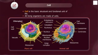
4. 5. ⚫ Cell is the basic structural and functional unit of
life.
⚫ All living organisms are made of cells.
Cell
Cell
wall
Vacuole
Endoplasmic
reticulum
Cytoplasm
Nucleolus
Nucleus
Mitochondria
Nuclear
membrane
Golgi
complex
Cell
membrane
Ribosomes
Plant cell Animal cell
Lysosomes
Centrioles
Ribosomes
Chloroplast
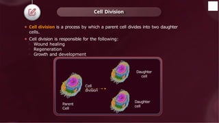
6. ⚫ Cell division is a process by which a parent cell divides into two daughter
cells.
⚫ Cell division is responsible for the following:
o Wound healing
o Regeneration
o Growth and development
Cell Division
Parent
Cell
Daughter
cell
Daughter
cell
Cell
division
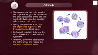
7. ⚫ The sequence of events by which a
cell duplicates its genome, synthesises
the other constituents of the cell and
eventually divides into two daughter
cells is termed cell cycle.
⚫ During the growth of a cell, the
cell organelles duplicate, and
DNA replication takes place.
⚫ Cell growth results in disturbing the
ratio between the nucleus and the
cytoplasm.
⚫ Therefore, it becomes essential for
the cell to divide and restore the
nucleo- cytoplasmic ratio.
Cell Cycle
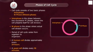
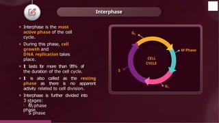
8. ⚫ Cell cycle consists of two basic phases
o Interphase
o M Phase (Mitosis phase)
⚫ Interphase is the phase between
two successive M phases, where the
cell prepares itself for cell division.
⚫ M phase is the phase where actual
cell division occurs.
⚫ Period of cell cycle varies from
organism to
organism.
⚫ A human cell divides approximately
every
24 hours.
⚫ A yeast cell divides every 90
minutes.
Phases of Cell Cycle
Interphase
M Phase
9. Interphase
CELL
CYCLE
M Phase
G1
G2
o G2
phase
© 2022, Aakash BYJU'S. All rights reserved
S
⚫ Interphase is the most
active phase of the cell
cycle.
⚫ During this phase, cell
growth and
DNA replication takes
place.
⚫ I
t lasts for more than 95% of
the duration of the cell cycle.
⚫ I
t is also called as the resting
phase as there is no apparent
activity related to cell division.
⚫ Interphase is further divided into
3 stages:
o G1 phase
o S phase
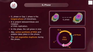
10. ⚫ G1 phase or Gap 1 phase is the
longest phase of interphase.
⚫ It is present betweenmitosis and
initiation
of DNA replication.
⚫ In this phase, the cell grows in size.
⚫ Also, active synthesis of RNA and
proteins takes place in this phase.
⚫ The cell organelles duplicate during
this phase.
G1 Phase
CELL
CYCLE
M Phase
G1
G2
S
G1 phase
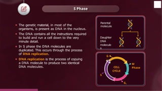
11. S Phase
⚫ The genetic material, in most of the
organisms, is present as DNA in the nucleus.
⚫ The DNA contains all the instructions required
to build and run a cell down to the very
minute detail.
⚫ In S phase the DNA molecules are
duplicated. This occurs through the process
of DNA replication.
⚫ DNA replication is the process of copying
a DNA molecule to produce two identical
DNA molecules.
Parental
molecule
Daughter
DNA
molecule
s
CELL
CYCLE
M
Phase
G1
G
2
© 2022, Aakash BYJU'S. All rights reserved
S
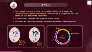
12. ⚫ Even though the DNA content gets doubled during the S phase, the
chromosome number remains the same since the 2 copies of the DNA
strand are still attached to each other.
⚫ In animal cells, centrioles also duplicate in this phase.
⚫ The centrioles help in distributing the duplicated genetic material equally.
S Phase
Before S
phase (G1
Phase)
After S
Phase
M Phase
CELL
CYCLE
G1
G2
S
© 2022, Aakash BYJU'S. All rights reserved
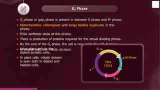
13. ⚫ G2 phase or gap2 phase is present in between S phase and M phase.
⚫ Mitochondria, chloroplast and Golgi bodies duplicate in this
phase.
⚫ DNA synthesis stops at this phase.
⚫ There is production of proteins required for the actual dividing phase.
⚫ By the end of the G2 phase, the cell is now ready for cell division.
⚫ In animal cells, the mitotic division
takes place only in the
diploid somatic cells.
⚫ In plant cells, mitotic division
is seen both in diploid and
haploid cells.
G2 Phase
CELL
CYCLE
M Phase
G1
G2
S
© 2022, Aakash BYJU'S. All rights reserved
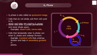
14. ⚫ G0 phase is also called as quiescent stage.
⚫ Cells that do not divide, exit from cell cycle
in G1
phase and enter into inactive G0 phase.
⚫ Some cells enter G0 phase permanently
and never divide again.
o Examples: Heart cells, nerve cells.
⚫ Cells that temporarily enter G0 phase can
enter G1 phase and undergo division.
o Example: Cambial cells that undergo
division and help in secondary growth
in
plants.
G0 Phase
CELL
CYCLE
M Phase
G1
G2
S
G0
Phase
© 2022, Aakash BYJU'S. All rights reserved
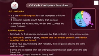
15. Cell Cycle Checkpoints: Interphase
G1/S checkpoint
⚫ It is the main checkpoint for a cell to progress or halt cell
cycle.
⚫ It checks for nutrients, growth factors, DNA damage
⚫ If conditions are not favourable, the cell exits G1 phase and
enters G0 phase.
G2/M checkpoint
⚫ Cell checks for DNA damage and ensures that DNA replication is done without errors.
⚫ G2 checkpoint, before M phase, ensures that cell division proceeds and healthy
daughter cells are formed.
⚫ If errors have occurred during DNA replication, then cell pauses allowing the cell to
undergo repair.
⚫ If errors are not rectified, then cell undergoes programmed cell death, where the cell’s
lysosomes
release their hydrolytic enzymes to destroy itself.
G2
© 2022, Aakash BYJU'S. All rights reserved
M
S
DNA
synt
hesi
s
G1
G0
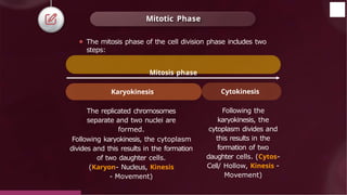
16. Mitotic Phase
Following the
karyokinesis, the
cytoplasm divides and
this results in the
formation of two
daughter cells. (Cytos-
Cell/ Hollow, Kinesis -
Movement)
The replicated chromosomes
separate and two nuclei are
formed.
Following karyokinesis, the cytoplasm
divides and this results in the formation
of two daughter cells.
(Karyon- Nucleus, Kinesis
- Movement)
⚫ The mitosis phase of the cell division phase includes two
steps:
Mitosis phase
Karyokinesis Cytokinesis
© 2022, Aakash BYJU'S. All rights reserved
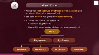
17. Mitosis Phase
⚫ Mitosis was first observed by Strasburger in plant cell and
by Walter Flemming in animal cell.
⚫ The term mitosis was given by Walter Flemming.
⚫ A type of cell division that produces:
○ Two similar daughter cells
○ Having the same number of chromosomes as parent cell.
Mitosis
Prophase Metaphase Anaphase Telophase
Karyokinesis Cytokinesis
© 2022, Aakash BYJU'S. All rights reserved
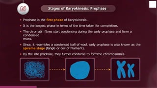
18. ⚫ Prophase is the first phase of karyokinesis.
⚫ It is the longest phase in terms of the time taken for completion.
⚫ The chromatin fibres start condensing during the early prophase and form a
condensed
mass.
⚫ Since, it resembles a condensed ball of wool, early prophase is also known as the
spireme stage (tangle or coil of filament).
⚫ By the late prophase, they further condense to formthe chromosomes.
Stages of Karyokinesis: Prophase
© 2022, Aakash BYJU'S. All rights reserved
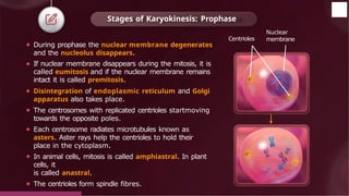
19. ⚫ During prophase the nuclear membrane degenerates
and the nucleolus disappears.
⚫ If nuclear membrane disappears during the mitosis, it is
called eumitosis and if the nuclear membrane remains
intact it is called premitosis.
⚫ Disintegration of endoplasmic reticulum and Golgi
apparatus also takes place.
⚫ The centrosomes with replicated centrioles startmoving
towards the opposite poles.
⚫ Each centrosome radiates microtubules known as
asters. Aster rays help the centrioles to hold their
place in the cytoplasm.
⚫ In animal cells, mitosis is called amphiastral. In plant
cells, it
is called anastral.
⚫ The centrioles form spindle fibres.
Stages of Karyokinesis: Prophase
Nuclear
membrane
© 2022, Aakash BYJU'S. All rights reserved
Centrioles
20. ⚫ The complete degradation of the nuclear membrane
marks
the start of metaphase.
⚫ The chromosomes come to lie at the equatorial
plate (equidistant fromthe two poles). This process is
known as congression.
⚫ Congression occurs with the assembly of the mitotic
spindle that mediates the microtubule-chromosome
interactions required for the movement of
chromosomes.
⚫ The centromere is surrounded by a small disc
shaped structure called kinetochore. The
kinetochore formthe site of attachment of
microtubules.
⚫ Chromosomes are observed to be the thickest and
Stages of Karyokinesis: Metaphase
© 2022, Aakash BYJU'S. All rights reserved
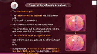
21. ⚫ The centromere splits.
⚫ The sister chromatids separate into two identical
and
independent chromosomes.
⚫ Each chromatid now has its own centromere.
⚫ The spindle fibres pull the chromatids along with the
centromere towards their respective poles.
⚫ The chromatids move to opposite poles.
⚫ Half of them reach one pole and the other half reach
the
other.
⚫ Duringmigration, the centromere of chromosomes
face towards the poles. The chromatids or arms of
chromosomes trail behind.
Stages of Karyokinesis: Anaphase
© 2022, Aakash BYJU'S. All rights reserved
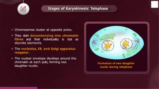
22. ⚫ Chromosomes cluster at opposite poles.
⚫ They start decondensing into chromatin
fibres and their individuality is lost as
discrete elements.
⚫ The nucleolus, ER, and Golgi apparatus
reappear.
⚫ The nuclear envelope develops around the
chromatin at each pole, forming two
daughter nuclei.
Formation of two daughter
nuclei during telophase
Stages of Karyokinesis: Telophase
© 2022, Aakash BYJU'S. All rights reserved
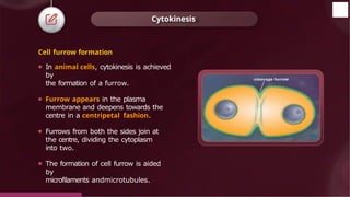
23. Cell furrow formation
⚫ In animal cells, cytokinesis is achieved
by
the formation of a furrow.
⚫ Furrow appears in the plasma
membrane and deepens towards the
centre in a centripetal fashion.
⚫ Furrows from both the sides join at
the centre, dividing the cytoplasm
into two.
⚫ The formation of cell furrow is aided
by
microfilaments andmicrotubules.
Cytokinesis
© 2022, Aakash BYJU'S. All rights reserved
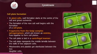
24. Cell plate formation
⚫ In plant cells, wall formation starts at the centre of the
cell and grows outwards.
⚫ The formation of the new cell wall begins with the
formation
of a cell plate.
⚫ Fragments from the Golgi complex
(phragmoplast), which are known as vesicles,
fuse together to form cell plates.
⚫ The cell plate is laid in a centrifugal manner.
⚫ The cell plate represents the middle lamella between
the walls of two adjacent cells.
⚫ Mitochondria and plastids get distributed between the
two
daughter cells.
Cytokinesis
© 2022, Aakash BYJU'S. All rights reserved
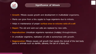
25. Significance of Mitosis
© 2022, Aakash BYJU'S. All rights reserved
⚫ Growth: Mitosis causes growth and development in multicellular organisms.
⚫ Plants can grow from a tiny zygote to huge organisms due to mitosis.
⚫ Helps in maintenance of proper surface area to volume ratio of a cell
⚫ Repair: The old and worn-out cells are replaced by new cells.
⚫ Reproduction: Unicellular organisms reproduce (multiply) throughmitosis.
⚫ In unicellular organisms, replication of cells is synonymous with growth.
⚫ Regeneration: Mitosis causes cell growth that causes the revival of the lost body
parts in animals such as starfish, planaria, the tail of a lizard, etc.
26. Regulation of Cell Cycle
© 2022, Aakash BYJU'S. All rights reserved
G2
M
S
DNA
synt
hesi
s
G1
G0
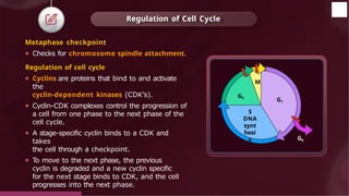
Metaphase checkpoint
⚫ Checks for chromosome spindle attachment.
Regulation of cell cycle
⚫ Cyclins are proteins that bind to and activate
the
cyclin-dependent kinases (CDK’s).
⚫ Cyclin-CDK complexes control the progression of
a cell from one phase to the next phase of the
cell cycle.
⚫ A stage-specific cyclin binds to a CDK and
takes
the cell through a checkpoint.
⚫ To move to the next phase, the previous
cyclin is degraded and a new cyclin specific
for the next stage binds to CDK, and the cell
progresses into the next phase.
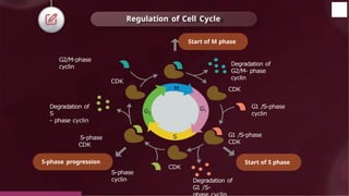
27. Regulation of Cell Cycle
Degradation of
G2/M- phase
cyclin
CDK
G1 /S-phase
cyclin
G1 /S-phase
CDK
CDK
CDK
S-phase
CDK
G2/M-phase
cyclin
S-phase
cyclin
Degradation of
S
- phase cyclin
Degradation of
G1 /S-
S
G1
M
G2
Start of M phase
Start of S phase
S-phase progression
© 2022, Aakash BYJU'S. All rights reserved
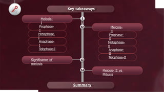
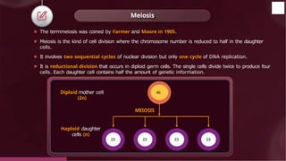
28. Meiosis
⚫ The termmeiosis was coined by Farmer and Moore in 1905.
⚫ Meiosis is the kind of cell division where the chromosome number is reduced to half in the daughter
cells.
⚫ It involves two sequential cycles of nuclear division but only one cycle of DNA replication.
⚫ It is reductional division that occurs in diploid germ cells. The single cells divide twice to produce four
cells. Each daughter cell contains half the amount of genetic information.
Diploid mother cell
(2n)
Haploid daughter
cells (n)
MEIOSIS
46
23 23 23 23
© 2022, Aakash BYJU'S. All rights reserved
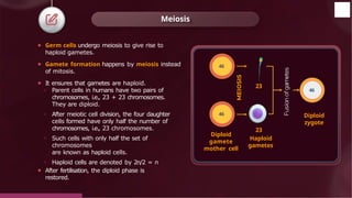
29. Meiosis
⚫ Germ cells undergo meiosis to give rise to
haploid gametes.
⚫ Gamete formation happens by meiosis instead
of mitosis.
⚫ It ensures that gametes are haploid.
o Parent cells in humans have two pairs of
chromosomes, i.e., 23 + 23 chromosomes.
They are diploid.
o After meiotic cell division, the four daughter
cells formed have only half the number of
chromosomes, i.e., 23 chromosomes.
o Such cells with only half the set of
chromosomes
are known as haploid cells.
o Haploid cells are denoted by 2n/2 = n
⚫ After fertilisation, the diploid phase is
restored.
Diploid
gamete
mother cell
23
23
Haploid
gametes
46
Diploid
zygote
46
46
© 2022, Aakash BYJU'S. All rights reserved
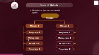
30. Steps of Meiosis
Meiosis I Meiosis II
Prophase I
Metaphase
I
Anaphase I
Telophase I
Prophase II
Metaphase II
Anaphase II
Telophase II
⚫ Meiosis involves two sequential
cycles:
Meiosis
© 2022, Aakash BYJU'S. All rights reserved
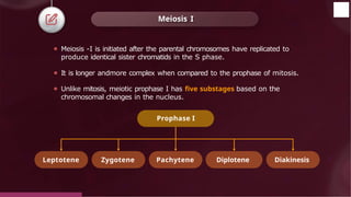
31. Meiosis I
⚫ Meiosis -I is initiated after the parental chromosomes have replicated to
produce identical sister chromatids in the S phase.
⚫ It is longer andmore complex when compared to the prophase of mitosis.
⚫ Unlike mitosis, meiotic prophase I has five substages based on the
chromosomal changes in the nucleus.
Prophase I
Leptotene Zygotene Pachytene Diplotene Diakinesis
© 2022, Aakash BYJU'S. All rights reserved
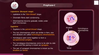
32. Prophase-I
Leptotene (Bouquet stage)
⚫ Leptotene is the ‘thin thread’ stage.
⚫ Chromatin fibres start condensing.
⚫ Chromosomes become gradually visible under
light microscope.
Zygotene
⚫ Zygotene is the paired thread stage.
⚫ The two chromosomes which are similar in form, size
and structure are called homologous chromosomes.
⚫ Homologous pairs come together to form a
synaptonemal complex.
⚫ The homologous chromosomes come to lie side by side
in pairs and this pairing is known as synapsis.
⚫ The pair of synapsed chromosomes is known as the
bivalent or tetrad.
Bivalent
Tetrad
Homologous
chromosomes
© 2022, Aakash BYJU'S. All rights reserved
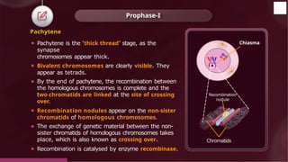
33. Pachytene
⚫ Pachytene is the ‘thick thread’ stage, as the
synapse
chromosomes appear thick.
⚫ Bivalent chromosomes are clearly visible. They
appear as tetrads.
⚫ By the end of pachytene, the recombination between
the homologous chromosomes is complete and the
two chromatids are linked at the site of crossing
over.
⚫ Recombination nodules appear on the non-sister
chromatids of homologous chromosomes.
⚫ The exchange of genetic material between the non-
sister chromatids of homologous chromosomes takes
place, which is also known as crossing over.
⚫ Recombination is catalysed by enzyme recombinase.
Prophase-I
Chiasma
Recombination
nodule
Chromatids
© 2022, Aakash BYJU'S. All rights reserved
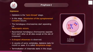
34. Diplotene
⚫ Diplotene is the ‘twin thread’ stage.
⚫ In this stage, dissolution of the synaptonemal
complex occurs.
⚫ The homologous chromosomes start separating
i.e.
desynapsis occurs.
⚫ Recombined homologous chromosomes separate
from each other at all sites except at the site of
crossover.
⚫ X-shaped chiasmata is observed.
⚫ In oocytes of some vertebrates, diplotene lasts for
month or years. It is called dictyotene stage.
⚫ Terminalisation of chiasmata starts in this stage.
Prophase-I
X-shaped
chiasmata
© 2022, Aakash BYJU'S. All rights reserved
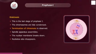
35. Diakinesis
⚫ This is the last stage of prophase I.
⚫ The chromosomes are fully condensed.
⚫ Termination of chiasmata is observed.
⚫ Spindle apparatus assembles.
⚫ The nuclear membrane breaks down.
⚫ Nucleolus also disappears.
Prophase-I
© 2022, Aakash BYJU'S. All rights reserved
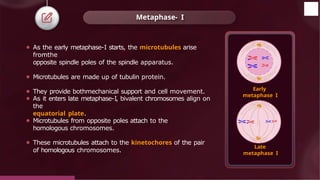
36. ⚫ As the early metaphase-I starts, the microtubules arise
fromthe
opposite spindle poles of the spindle apparatus.
⚫ Microtubules are made up of tubulin protein.
⚫ They provide bothmechanical support and cell movement.
⚫ As it enters late metaphase-I, bivalent chromosomes align on
the
equatorial plate.
⚫ Microtubules from opposite poles attach to the
homologous chromosomes.
⚫ These microtubules attach to the kinetochores of the pair
of homologous chromosomes.
Metaphase- I
Early
metaphase I
Late
metaphase I
© 2022, Aakash BYJU'S. All rights reserved
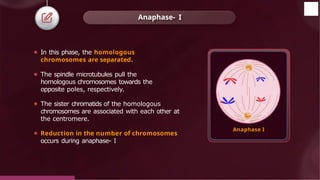
37. ⚫ In this phase, the homologous
chromosomes are separated.
⚫ The spindle microtubules pull the
homologous chromosomes towards the
opposite poles, respectively.
⚫ The sister chromatids of the homologous
chromosomes are associated with each other at
the centromere.
⚫ Reduction in the number of chromosomes
occurs during anaphase- I
Anaphase- I
Anaphase I
© 2022, Aakash BYJU'S. All rights reserved
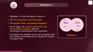
38. ⚫ Telophase- I is the final step of meiosis -I.
⚫ The chromosomes reach the poles.
⚫ The spindle fibres completely disappear.
⚫ At this stage, the nuclear membrane and
the nucleolus reappear after the
homologous chromosomes have separated.
⚫ It produces two daughter nuclei each containing half
the number of chromosomes but double the amount
of nuclear DNA.
Telophase- I
Telophase I
© 2022, Aakash BYJU'S. All rights reserved
39. ⚫ Telophase I is followed by cytokinesis.
⚫ Cytokinesis is the process where the cytoplasm is divided equally into daughter cells.
⚫ The daughter cells formed at the end of meiosis have bivalent chromosomes, and this
chromosome is also known as a dyad (one pair of chromosomes fromthe tetrad).
Interkinesis
⚫ It is a short-lived stage betweenmeiosis I andmeiosis II.
⚫ During this phase, the chromosomes are elongated but do not form chromatin fibres.
⚫ This stage has no DNA replication.
⚫ The RNA and protein required duringmeiosis -II are synthesized during this phase.
Cytokinesis
© 2022, Aakash BYJU'S. All rights reserved
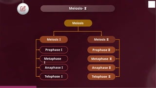
40. Meiosis- II
Meiosis
Meiosis I Meiosis II
Prophase I
Metaphase
I
Anaphase I
Telophase I
Prophase II
Metaphase II
Anaphase II
Telophase II
© 2022, Aakash BYJU'S. All rights reserved
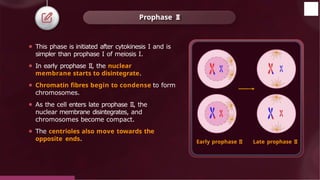
41. ⚫ This phase is initiated after cytokinesis I and is
simpler than prophase I of meiosis I.
⚫ In early prophase II, the nuclear
membrane starts to disintegrate.
⚫ Chromatin fibres begin to condense to form
chromosomes.
⚫ As the cell enters late prophase II, the
nuclear membrane disintegrates, and
chromosomes become compact.
⚫ The centrioles also move towards the
opposite ends.
Prophase II
Early prophase II
© 2022, Aakash BYJU'S. All rights reserved
Late prophase II
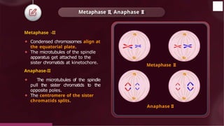
42. Metaphase -II
⚫ Condensed chromosomes align at
the equatorial plate.
⚫ The microtubules of the spindle
apparatus get attached to the
sister chromatids at kinetochore.
Anaphase-II
⚫ The microtubules of the spindle
pull the sister chromatids to the
opposite poles.
⚫ The centromere of the sister
chromatids splits.
Metaphase II, Anaphase II
Metaphase II
Anaphase II
© 2022, Aakash BYJU'S. All rights reserved
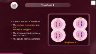
43. ⚫ It marks the end of meiosis II.
⚫ The nuclear membrane and
the
nucleolus reappear.
⚫ The chromosomes decondense
into chromatin.
⚫ The spindle fibers degenerate.
Telophase II
Telophase II
© 2022, Aakash BYJU'S. All rights reserved
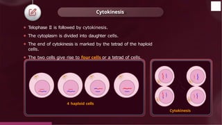
44. ⚫ Telophase II is followed by cytokinesis.
⚫ The cytoplasm is divided into daughter cells.
⚫ The end of cytokinesis is marked by the tetrad of the haploid
cells.
⚫ The two cells give rise to four cells or a tetrad of cells.
Cytokinesis
Cytokinesis
4 haploid cells
© 2022, Aakash BYJU'S. All rights reserved
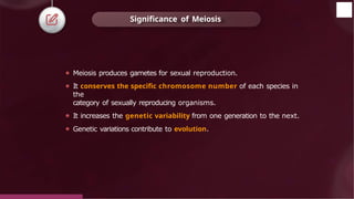
45. ⚫ Meiosis produces gametes for sexual reproduction.
⚫ It conserves the specific chromosome number of each species in
the
category of sexually reproducing organisms.
⚫ It increases the genetic variability from one generation to the next.
⚫ Genetic variations contribute to evolution.
Significance of Meiosis
© 2022, Aakash BYJU'S. All rights reserved
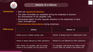
46. ⚫ Both are equatorial divisions.
⚫ The sister chromatids are separated during the anaphase to become
the chromosomes of the daughter cells.
⚫ Microtubules attach fromthe opposite directions to the centromere of each
sister chromatid pair.
⚫ Chromosomes decondense during telophase.
Meiosis- II vs Mitosis
Similarities:
Mitosis Meiosis -II
Mitosis occurs in diploid somatic cells. Meiosis -II always occurs in haploid germ cells.
Mitosis is always followed by DNA replication. Meiosis-II is not followed by DNA replication.
After mitosis, the daughter cells are exactly
similar to one another and the parent cell.
The daughter cells formed are neither similar
to each other nor similar to the parent cell.
© 2022, Aakash BYJU'S. All rights reserved
Differences:
47. © 2022, Aakash BYJU'S. All rights
Summary
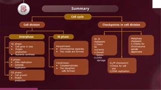
Cell cycle
Cell division
Interphase M phase
G1 phase:
● Cell grow in size
● Protein
production
● Nutrients synthesis
S phase:
● DNA replication
● Centriole
duplication
G2 phase:
● Cell growth
● Protein
production
Karyokinesis:
● Chromosomes separate
● Two nuclei are formed
Cytokinesis:
● Cytoplasmdivides
● Two daughter
cells formed
Checkpoints in cell division
G1 /S
checkpoint:
i) Check
for
nutrients
ii) Growth
factors
iii)DNA
damage
G2/M checkpoint:
i)Check for cell
size
ii)DNA replication
Metaphase
checkpoint:
i) Checks for
chromosome
spindle
attachment.
48. © 2022, Aakash BYJU'S. All rights
Summary
Karyokinesis
Karyon = Nucleus; Kinesis =
Movement
It is the division of the nucleus.
Prophase
- Condensation of chromatin
fibres
- Nuclear membrane
degenerates
Anaphase
- Centromere splits and chromatids
separate.
- Chromatids move to opposite poles.
Metaphase
- Chromosomes are attached to spindle
fibres.
- Chromosomes are arranged in
the equatorial plane.
Telophase
- Chromosome reach the poles
- Disappearance of spindle fibres
- Decondensation of
chromosomes
Cytokinesis
Cytos = Cell, Kinesis =
Movement
It is the division of the
cytoplasm.
Cell furrow formation
Observed in animals
Cell plate formation
Observed in plants
Mitosis
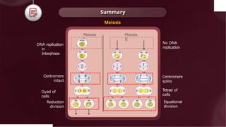
49. © 2022, Aakash BYJU'S. All rights
Summary
Reduction
division
Equational
division
Centromere
intact
DNA replication
in
Interphase
No DNA
replication
Meiosis
I
Meiosis
II
Centromere
splits
Dyad of
cells
Tetrad of
cells
Meiosis