More Related Content

PPTX
Cell the unit of life ncert cell wall to golgi complex 
PPTX
EUKARYOTIC CELLS PPT BT I.pptx 
PPTX
2. structure and function of organelles 
PPTX
Cell membrane and endoplasmic reticulum 
PPTX
cellstructurefunctions-240227121448-bddcc97f.pptx 
PDF
Biology 189 the_cell_spring_2012.ppt 
PPT
2 Plant Cell physiology and their role tt 
PPTX
Cell structure slideshare.pptx Unlocking the Secrets of Cells: Structure, Fun... Similar to cell the unit of life Biology Class 12th

PDF

PPTX
Cellular level of organization 
PPTX
Ultra structure of Plant Cell by Salman Saeed Lecturer Botany UCMS Khanewal 
PPTX
Cell :Structure & Functions for Medical and Health allied Students 
PPT
STRUCTURE OF ORGANELLES AND FUNCTIONS.ppt 
PPTX
Cell Structure and Function.pptx 
PDF

PPT

PPTX
Cell: The Code of Life (Exploring Anatomy and Physiology of Cell) 
PPTX
Endoplasmic Reticulum and Golgi Apparatus.pptx 
PPTX
cell organelle and Its composition function 
PPTX
Cell the fundamental unit of life 
PPTX

PPTX
Cell - The Unit of Life organelles about cell class 11 standard.pptx 
PPTX
The cell its organells and their functions 
PPTX
human cell anatomy and function 
PPT

PPTX

PPTX
CELLULAR COMPONENT.ldkdkddldldldldldldlddl 
PPT
Module II_2_Pro,eukaryote&cellstructure,ecm.ppt Recently uploaded

PDF
Scalable-MADDPG-Based Cooperative Target Invasion for a Multi-USV System.pdf 
PDF
DHA/HAAD/MOH/DOH OPTOMETRY MCQ PYQ. .pdf 
PPTX
Semester 6 unit 2 Atopic dermatitis.pptx 
PPTX
ICH Harmonization A Global Pathway to Unified Drug Regulation.pptx 
PPTX
Limpitlaw "Licensing: From Mindset to Milestones" 
PDF
Current Electricity for first year physiotherapy 
PPTX
Details of Epithelial and Connective Tissue.pptx 
PDF
Projecte de la porta de la classe de primer A: Mar i cel. 
PDF
Projecte de la porta d'i5B: Els animals marins 
PPTX
Basics in Phytochemistry, Extraction, Isolation methods, Characterisation etc. 
PDF
IMANI Africa files RTI request seeking full disclosure on 2026 SIM registrati... 
PPTX
ATTENTION -PART 2.pptx Shilpa Hotakar for I semester BSc students 
PPTX
Searching in PubMed andCochrane_Practical Presentation.pptx 
PPTX
10-12-2025 Francois Staring How can Researchers and Initial Teacher Educators... 
PPTX
How to Manage Reception Report in Odoo 18 Inventory 
PPTX
PURPOSIVE SAMPLING IN EDUCATIONAL RESEARCH RACHITHRA RK.pptx 
PDF
NAVIGATE PHARMACY CAREER OPPORTUNITIES.pdf 
PPTX
The Cell & Cell Cycle-detailed structure and function of organelles.pptx 
PPTX
Pig- piggy bank in Big Data Analytics.ppt.pptx 
PPTX
How to Configure Push & Pull Rule in Odoo 18 Inventory cell the unit of life Biology Class 12th
- 2.
Eukaryotic Cells
Characteristic features:
● Presence of true nucleus enclosed by a nuclear envelope
● Presence of membrane bound organelles
● Genetic material is organised into chromosomes
● Has a variety of complex locomotory and cytoskeletal structures
● These cells occur in protists, fungi, plants and animals
Nucleus
Centrioles
Lysosome
RER
SER
Peroxisome
Cytoskeleton
Mitochondria
Golgi apparatus
Plasma
membrane
Nucleus
Ribosomes
Animal cell
Nucleus
Cytosol
Endoplasmic
Reticulum
Ribosomes
Golgi apparatus
Mitochondria
Vacuole
Cell wall
© 2022, A k s igh
Cell
membrane
Chlor
oplast
Plant cell
- 3.
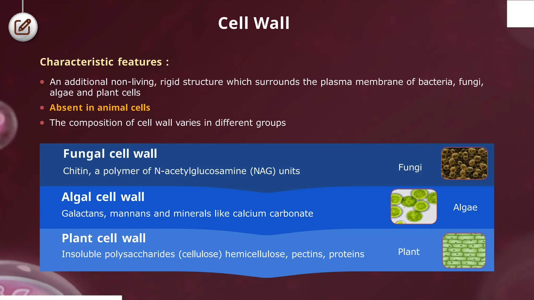
Cell Wall
Plant cellwall
Insoluble polysaccharides (cellulose) hemicellulose, pectins, proteins
Algal cell wall
Galactans, mannans and minerals like calcium carbonate
Fungal cell wall
Chitin, a polymer of N-acetylglucosamine (NAG) units Fungi
© 2022, A k s igh
Algae
Plant
Characteristic features :
● An additional non-living, rigid structure which surrounds the plasma membrane of bacteria, fungi,
algae and plant cells
● Absent in animal cells
● The composition of cell wall varies in different groups
- 4.
Cell Wall
Characteristic features:
● The cell wall of plants consists of two regions : primary
wall and secondary wall.
● Primary wall:
● It is found in young plant
cells.
● It is a thin single layer which is elastic in nature
and capable of expanding in a growing cell such
as, meristematic and parenchymatous cells.
● Secondary
wall :
● It is found in mature cells.
● It has more layers than primary wall, which brings
about thickening of the cell wall such as, lignified
and suberised cell wall.
PPllaanntt
cceellll
Secondary
cell wall
layers
S1
S2
S3
© 2022, A k s igh
Primary cell wall
- 5.
Cell Wall
Characteristic features:
● Middle lamella : Hold adjacent cells together by a thin,
sticky, amorphous layer of cementing material
● Made up of calcium and magnesium pectate
● Plasmodesmata : Intercellular cytoplasmic connections
Endoplasmic reticulum plays a role in origin of plasmodesmata
Plant cell Plant cell
Plant cell Plant cell
Middle lamella
Plasmodesmata
Functions :
● It maintains shape of the cells.
● It protects the cell from mechanical injury.
© 2022, A k s igh
● It wards off the attacks of pathogens like viruses, bacteria, fungi, etc.
● It allows the materials to pass in and out of the cell.
● It helps in cell-to-cell interaction and provides barrier to undesirable macromolecules.
- 6.
© 2022, AakashBYJU'S. All rights reserved
Cell Wall
Middle lamella (white)
Primary wall (blue)
Secondary wall (olive)
Pit
Torus
Bordered pits Simple pits
Pits
Pits :
● At certain places secondary wall is not laid
down. Such unthickened areas are called
pits
● Adjacent cells are generally opposite to each
other and form pit pairs
● Pits are of two types :
o Simple pit : Uniform pit cavity in diameter
o Bordered pit: Flask-shaped pit
cavity as in tracheid
● Presence of number of plasmodesmata or
cytoplasmic strands are in pit through which the
cytoplasm of one cell is in contact with other
o Lined by plasma membrane and contains a
fine tubule called desmotubule
- 7.
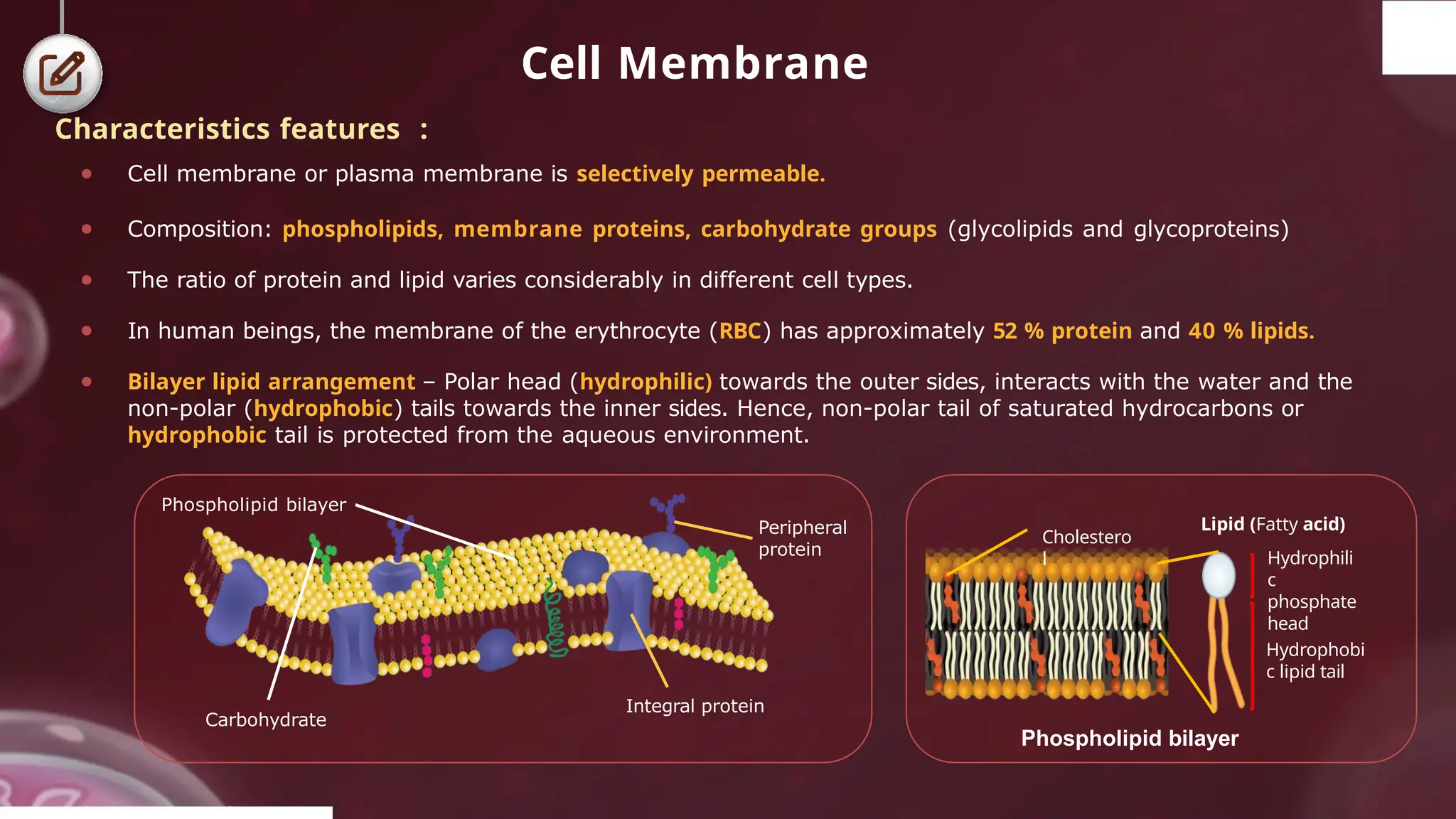
Cell Membrane
Characteristics features:
● Cell membrane or plasma membrane is selectively permeable.
● Composition: phospholipids, membrane proteins, carbohydrate groups (glycolipids and glycoproteins)
● The ratio of protein and lipid varies considerably in different cell types.
● In human beings, the membrane of the erythrocyte (RBC) has approximately 52 % protein and 40 % lipids.
● Bilayer lipid arrangement – Polar head (hydrophilic) towards the outer sides, interacts with the water and the
non-polar (hydrophobic) tails towards the inner sides. Hence, non-polar tail of saturated hydrocarbons or
hydrophobic tail is protected from the aqueous environment.
Integral protein
Carbohydrate
Lipid (Fatty acid)
Hydrophili
c
phosphate
head
Hydrophobi
c lipid tail
© 2022, A k s igh
Cholestero
l
Phospholipid bilayer
Peripheral
protein
Phospholipid bilayer
- 8.
Cell Membrane
Integral proteins
Theseproteins cannot be removed easily, and their removal requires crude methods of
treatment like detergents. Thus, the membrane has been described as protein
icebergs floating in sea of phospholipids.
Peripheral proteins
● Lie on the membrane surface
● Partially or totally buried in
membrane
● Tunnel proteins, which run
through the lipid bilayer are
known as trans membrane
proteins
Peripheral
membrane
protein
Integral
membrane
proteins
© 2022, A k s igh
Characteristics features :
● In cell membrane, two types of membrane proteins are present, depending on the ease of extraction:
peripheral and integral
Membrane proteins
- 9.
Cell Membrane
Characteristics features:
● Structure: Fluid mosaic model proposed by Singer and Nicolson (1972)
● Fluidity: Quasi-fluid nature of lipid allows lateral movement of proteins within the bilayer
● Cell growth, formation of intercellular junctions, secretion, endocytosis, cell division, etc.
● Transport of the molecules
Movement
Lateral
● Seen in lipids and proteins
● Occurs within the same
monolayer
● More common
Functions :
© 2022, A k s igh
Flip-flop
● Seen in lipids (more common) but not
in proteins due to their large size
● Slower than lateral movement
● Movement from one monolayer to the
other
- 10.
Cell Membrane
● Bysimple diffusion
● No energy utilised
Membrane transport
Passive transport
● Movement of neutral solutes along the
concentration gradient (Higher to
lower concentration)
Active transport
● Movement of ions or molecules against the
concentration gradient (Lower to higher
concentration)
© 2022, A k s igh
● Transporters such as Na+/K+ pump in animal
cells
● Energy dependant (ATP is utilised)
- 11.
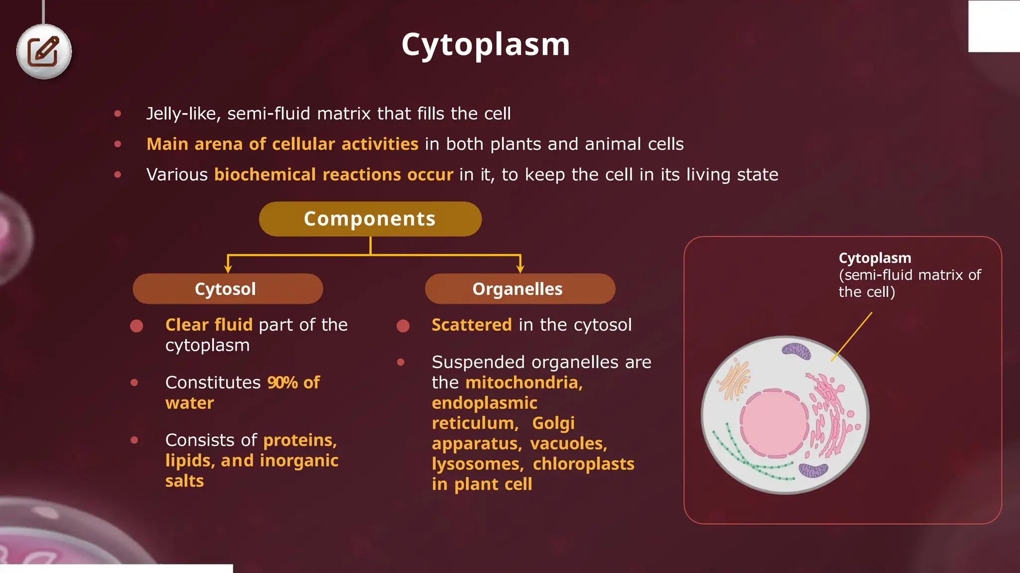
Cytoplasm
● Jelly-like, semi-fluidmatrix that fills the cell
● Main arena of cellular activities in both plants and animal cells
● Various biochemical reactions occur in it, to keep the cell in its living state
Components
Cytosol
● Clear fluid part of the
cytoplasm
● Constitutes 90% of
water
● Consists of proteins,
lipids, and inorganic
salts
Organelles
● Scattered in the cytosol
● Suspended organelles are
the mitochondria,
endoplasmic
reticulum, Golgi
apparatus, vacuoles,
lysosomes, chloroplasts
in plant cell
Cytoplasm
(semi-fluid matrix of
the cell)
© 2022, A k s igh
- 12.
Endomembrane System
● Membranouscell organelles which function in a coordinated manner
● Involved in the packaging and transport of materials
● Absent in prokaryotic cells and RBCs of mammals
Endomembrane system
Golgi
complex
Lysosomes
Endoplasmic
reticulum
Vacuoles
© 2022, A k s igh
- 13.
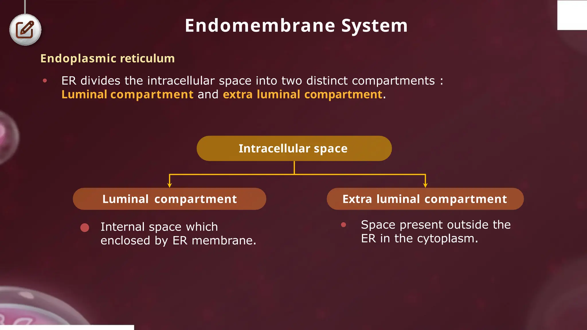
Endomembrane System
Endoplasmic reticulum
●A network of reticulum of tiny tubular structures scattered in the cytoplasm.
Endomembrane system
(composed of three kind of structures)
Tubules Vesicles
Cisternae
● Abundant in the
pancreatic cells and
these are the only
ER structures found
in spermatocytes
● Involved in lipid and
sterol synthesis
● Actively involved in
protein synthesis; e.g.,
cells of pancreas and
brain. Associated with
large subunit (60 S) Tubules
Cisternal space
Cisternae
© 2022, A k s igh
- 14.
Endomembrane System
Endoplasmic reticulum
●ER divides the intracellular space into two distinct compartments :
Luminal compartment and extra luminal compartment.
● Internal space which
enclosed by ER membrane.
Intracellular space
Luminal compartment Extra luminal compartment
● Space present outside the
ER in the cytoplasm.
© 2022, A k s igh
- 15.
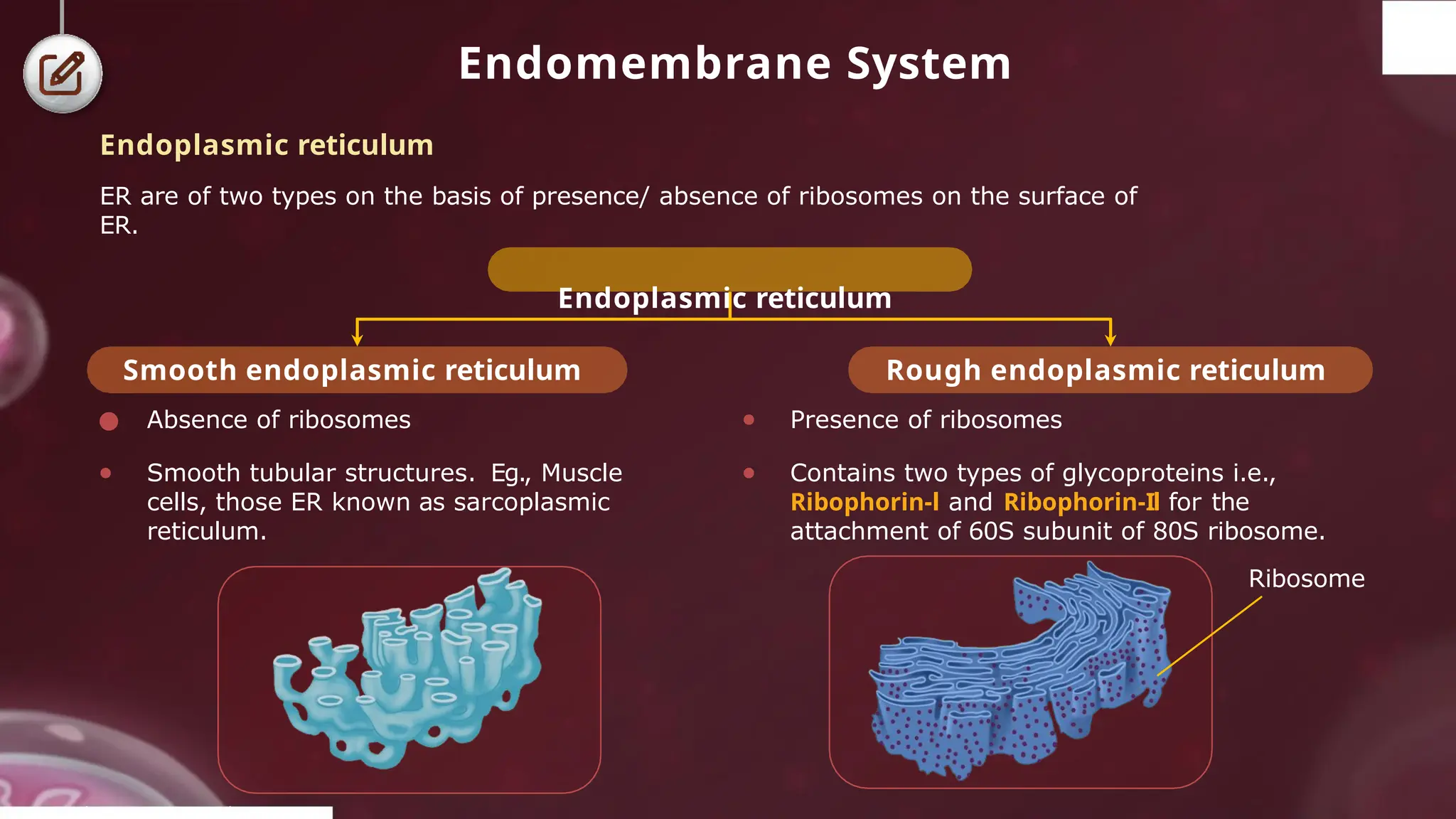
Endomembrane System
Smooth endoplasmicreticulum
● Absence of ribosomes
● Smooth tubular structures. Eg., Muscle
cells, those ER known as sarcoplasmic
reticulum.
Rough endoplasmic reticulum
● Presence of ribosomes
● Contains two types of glycoproteins i.e.,
Ribophorin-l and Ribophorin-Il for the
attachment of 60S subunit of 80S ribosome.
Ribosome
© 2022, A k s igh
Endoplasmic reticulum
ER are of two types on the basis of presence/ absence of ribosomes on the surface of
ER.
Endoplasmic reticulum
- 16.
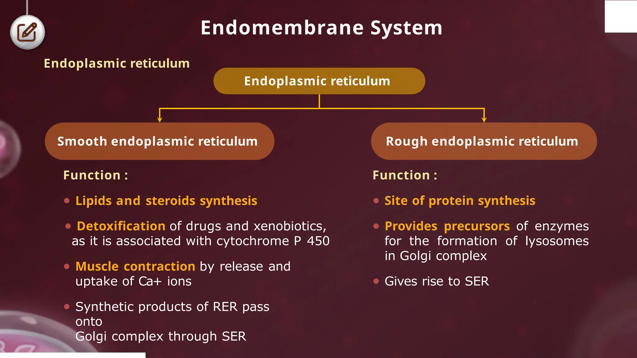
Endomembrane System
Function :
●Lipids and steroids synthesis
● Detoxification of drugs and xenobiotics,
as it is associated with cytochrome P 450
● Muscle contraction by release and
uptake of Ca+ ions
● Synthetic products of RER pass
onto
Golgi complex through SER
Rough endoplasmic reticulum
Smooth endoplasmic reticulum
Function :
● Site of protein synthesis
● Provides precursors of enzymes
for the formation of lysosomes
in Golgi complex
● Gives rise to SER
© 2022, A k s igh
Endoplasmic reticulum
Endoplasmic reticulum
- 17.
Endomembrane System
Cisternae
● Flattenedsac-like
structures stacked
on one another
Tubules
● Small, flat,
interconnecting
structures
Vesicles
● Small rounded sacs
present at the edges
of cisternae in
clusters
Golgian Vacuoles
● Large, spherical
vacuoles produced at
maturing face
Vesicle
Cisternae
Tubules
© 2022, A k s igh
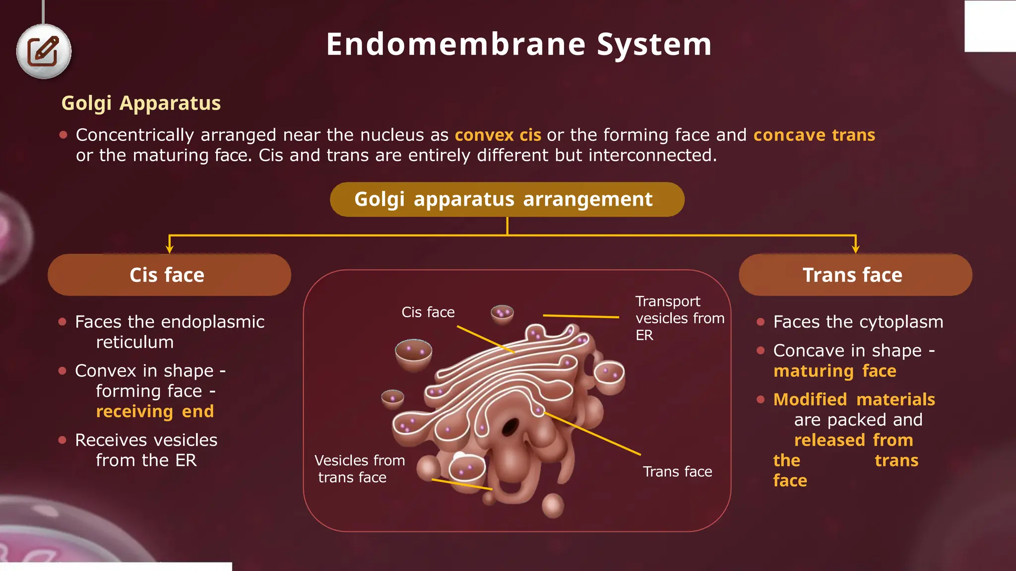
Golgi Apparatus
● First observed by Camillo Golgi in 1898
● Densely stained reticular structures; present near the nucleus of the cell
● Present in eukaryotic cells, except in mature sieve tubes of plants,
mature RBCs of mammals, sperm cells of bryophytes and
pteridophytes, etc
● In plants, it is called dictyosomes as Golgi apparatus is made up of
unconnected units
Golgi apparatus
- 18.
Endomembrane System
⚫ Facesthe endoplasmic
reticulum
⚫ Convex in shape -
forming face -
receiving end
⚫ Receives vesicles
from the ER
⚫ Faces the cytoplasm
⚫ Concave in shape -
maturing face
⚫ Modified materials
are packed and
released from
the trans
face
Trans face
Cis face
Cis face
Transport
vesicles from
ER
Trans face
© 2022, A k s igh
Vesicles from
trans face
Golgi Apparatus
⚫ Concentrically arranged near the nucleus as convex cis or the forming face and concave trans
or the maturing face. Cis and trans are entirely different but interconnected.
Golgi apparatus arrangement
- 19.
Endomembrane System
Golgi Apparatus:Functions
To process, package and transport the materials for secretions
© 2022, A k s igh
Site of formation of glycoproteins and glycolipids
1
2
3 Root cap cells are rich in Golgi bodies which secrete mucilage for
the lubrication of root tip
4
5
Acrosome of the sperm is modified Golgi apparatus
Formation of plasma membrane during cytokinesis
- 20.
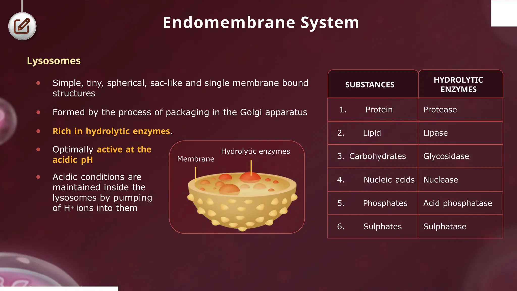
Endomembrane System
● Simple,tiny, spherical, sac-like and single membrane bound
structures
● Formed by the process of packaging in the Golgi apparatus
● Rich in hydrolytic enzymes.
Membrane
Hydrolytic enzymes
SUBSTANCES
HYDROLYTIC
ENZYMES
© 2022, A k s igh
1. Protein Protease
2. Lipid Lipase
3. Carbohydrates Glycosidase
4. Nucleic acids Nuclease
5. Phosphates Acid phosphatase
6. Sulphates Sulphatase
● Optimally active at the
acidic pH
● Acidic conditions are
maintained inside the
lysosomes by pumping
of H+ ions into them
Lysosomes
- 21.
Endomembrane System
Lysosomes
Lysosomes polymorphism
Residual
Primary
Newlyformed
Secondary /
Heterolysosomes
Primary lysosome
+ Phagosome
Undigested
materials
Autophagic/Vacuolar
lysosomes
⚫ Formed by union of
many primary
lysosomes around old
or dead organelles
⚫ Surrounds and digest
them by autolysis or
autodigestion
The disappearance of larval organs during metamorphosis (e.g., tail in frog) is due to
autolysis.
Hence, lysosomes are known as “suicide bags”
© 2022, A k s igh
- 22.
Endomembrane System
Vacuoles
● Vacuolesare large membrane-bound space.
They are prominently found in the cytoplasm.
● It contains water, sap, excretory products.
These are also called sap vacuoles.
● Its membrane is called tonoplast.
● Tonoplast facilitates the transport of ions and
other materials against concentration
gradients into the vacuole.
● Thus, ions concentration is significantly higher
in the vacuole than in the cytoplasm.
● In plant cells, the vacuoles can occupy upto
90 % of the volume of the cell.
Chloroplast
Cell wall
Cell membrane
SER
RER
Nucleus
Vacuole
Mitochondria
Tonoplast
(membrane
of vacuole)
Golgi body
Cytoplasm
Plant cell
© 2022, A k s igh
- 23.
Endomembrane System
Food vacuole
Gasvacuole/
pseudo vacuoles
Contractile
vacuole
● Membrane less vacuoles
found in prokaryotes
● Provides buoyancy
● In Amoeba, it helps
in excretion
● Helps in
osmoregulation
● In many cells, as in protists,
food vacuoles are formed
by engulfing the food
particles
Food Food
vacuole
particle
Vacuoles
Types of vacuoles
© 2022, A k s igh
- 24.
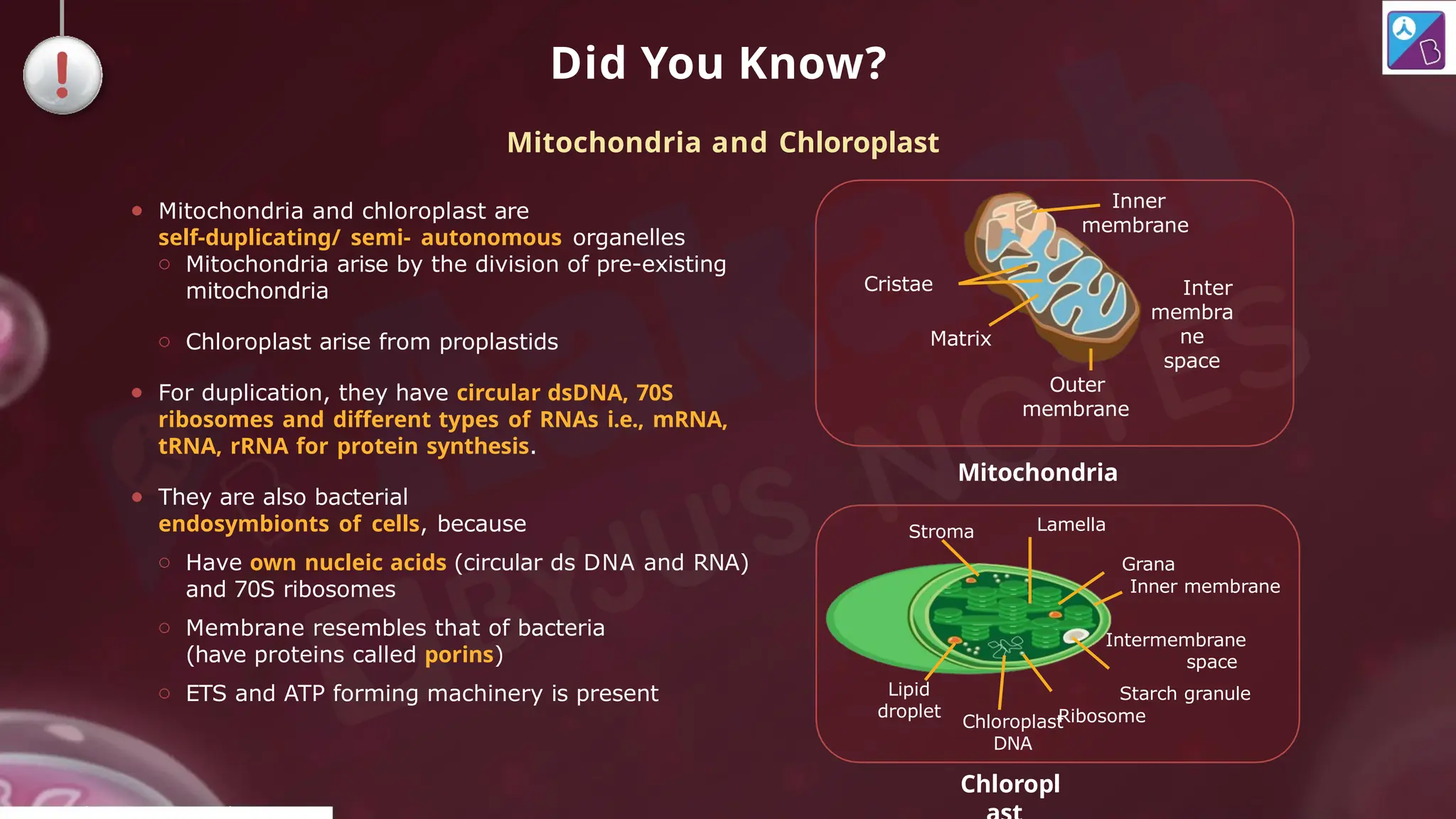
Did You Know?
●Mitochondria and chloroplast are
self-duplicating/ semi- autonomous organelles
o Mitochondria arise by the division of pre-existing
mitochondria
o Chloroplast arise from proplastids
● For duplication, they have circular dsDNA, 70S
ribosomes and different types of RNAs i.e., mRNA,
tRNA, rRNA for protein synthesis.
● They are also bacterial
endosymbionts of cells, because
o Have own nucleic acids (circular ds DNA and RNA)
and 70S ribosomes
o Membrane resembles that of bacteria
(have proteins called porins)
o ETS and ATP forming machinery is present
Mitochondria and Chloroplast
Inter
membra
ne
space
Cristae
Matrix
Outer
membrane
Inner
membrane
Lipid
droplet
Grana
Inner membrane
Intermembrane
space
Starch granule
Ribosome
Lamella
Stroma
Mitochondria
Chloroplast
DNA
Chloropl
© 2022, A k s igh
- 25.
Mitochondria
Inner membrane
● Numberof infoldings
called the cristae
● Has 80% protein and
20% lipids and is rich
in cardiolipins
● Contains ATP
synthase/F0-F1
Outer membrane
● Smooth
● Chemically
composed of 40%
lipid and 60%
proteins
● Contains transport
proteins
Inter
membrane
space
Cristae
© 2022, A k s igh
Matrix
Outer
membrane
Inner
membrane
● Sausage-shaped double membraned organelles.
●Since they are not visible easily, they are stained by a vital stain Janus Green.
Number : Depends on the amount of work done by the cell and its energy
requirement
Structure : Double membrane
Structure
- 26.
Mitochondria
⚫ It isbetween the two
mitochondrial membranes.
⚫ It is also called peri-
mitochondrial space.
Inner compartment or
matrix
Outer compartment or
intermembrane space
⚫ It is inside the inner
membrane.
⚫ The cristae are formed from
particle infolding of inner
membrane towards the matrix
which increases the surface
area for enzyme action.
© 2022, A k s igh
Has two distinct chambers filled with aqueous fluid
Chambers
- 27.
Mitochondria
Functions :
● Mitochondriaare main sites of aerobic
respiration and ATP synthesis, therefore
“Powerhouse of the cell”.
● They bring about the oxidation of
carbohydrates, proteins and ß-oxidation
of fats.
Inter membrane
space
Cristae
© 2022, A k s igh
Matrix
Outer
membrane
● Matrix contains single circular dsDNA molecule (with high G = C content), a few RNA
molecules, 70S ribosomes and enzymes for TCA (Tricarboxylic acid) cycle.
● Mitochondria divide by fission.
● The cristae and inner surface of the inner membrane are studied with numerous
spherical or knob like protuberances called elementary particles or Particles
of Fernandez and Moran or F, particles or oxysomes.
● Each oxysome is differentiated into base, stalk and headpiece. The head piece
contains enzyme ATP synthetase which brings about oxidative phosphorylation
coupled with release of ATP.
Inner
membrane