Lower urinary tract
•Download as PPTX, PDF•
1 like•656 views

Micturition reflex and bladder dysfunctions
![Introduction
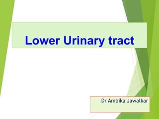
Renal pelvis Ureter
Ureter – muscular tube. Length - 30 cm
2 ureters [i.e. one from each kidney] enter
urinary bladder on posterior side above
bladder neck.
Urinary bladder-
i. Upper – fundus;
ii. Lower – bladder neck.
Functional valve of ureter – due to oblique
passage thro wall of urinary bladder](data:image/gif;base64,R0lGODlhAQABAIAAAAAAAP///yH5BAEAAAAALAAAAAABAAEAAAIBRAA7)
Recommended
More Related Content
What's hot
What's hot (20)
Similar to Lower urinary tract
Similar to Lower urinary tract (20)
More from Ambika Jawalkar
More from Ambika Jawalkar (18)
Recently uploaded
Recently uploaded (20)
Lower urinary tract
- 1. Lower Urinary tract Dr Ambika Jawalkar
- 2. Introduction Renal pelvis Ureter Ureter – muscular tube. Length - 30 cm 2 ureters [i.e. one from each kidney] enter urinary bladder on posterior side above bladder neck. Urinary bladder- i. Upper – fundus; ii. Lower – bladder neck. Functional valve of ureter – due to oblique passage thro wall of urinary bladder
- 4. Filling of urinary bladder – Peristaltic contractions of muscular wall of ureters push urine in spurts. Gravity helps. Applied – Prevention of reflux of urine – due to functional valve. If absent leads to Cystitis glomerulonephritis Chronic renal failure.
- 5. Structure of urethra Male – Length - 15 -20 cm [extend through penis] Female - Length – 4 cm 2 orifices – Internal urethral orifice – formed by detrusor muscle fibres in bladder neck External urethral orifice – formed by skeletal muscle fibres Trigone of urinary bladder – Triangular portion between openings of ureters & int urethral orifice Microscopic – Transitional epithelium which has ability to distend
- 6. Innervation of bladder [sympathetic, parasympathetic & somatic] Sympathetic – Lateral horn cells of L1, L2, L3 spinal cord segments sympathetic ganglion. Postganlionic thro Hypogastric nerve body, bladder neck & urethra Body - b adrenergic receptors. On stim relaxation of detrusor muscle. Trigone + Int Ure Sphi - a adrenergic receptors. On stim contraction of muscle. Sympathetic stim storage of urine. [Relaxation of body + contraction of Int US].
- 8. Parasympathetic innervation Carry both afferent [sensory] & efferent [motor] Origin – S1,S2,S3 spinal cord segments. pelvic nerves & relay in ganglia in bladder wall. Fundus of bladder – post ganglionic fibers Sensory – sense of fullness, pain to sacral segments afferent limb of spinal reflex of urination [note – input also to higher center] Parasympathetic stim. urination. [contraction of body + relaxation of IUS]
- 10. Somatic Innervation Origin – S2,S3,S4 spinal segments. Pudendal nerve ext. urethral sphicter. Note – EUS is striated , voluntary muscle.
- 11. MICTURITION Defn – it is a process of emptying urinary bladder. It is an automatic spinal reflex which can be modified by higher centers in the brain stem & cerebral cortex i.e. can be initiated voluntarily & can be stopped voluntarily because of cerebral cortical control of EUS. 1. Stimulation of stretch receptors in UB – due to filling of UB 2. Afferent – Parasympathetic sensory thro PELVIC NERVE [S1,S2,S3] 3. Center - [S1,S2,S3] spinal segments. 4. Efferent - Parasympathetic motor thro PELVIC NERVE [S1,S2,S3] 5. Effector – detrusor + IUS 6. Response – Detrusor – contraction; IUS - relaxation
- 12. Control of micturition by Higher centers Higher centers – at brain stem & cerebral cortex. Proprioceptive like feeling of fullness & desire to void urine thro fasciculus Gracilis cerebral cortex efferent fibres thro pyramidal tract spinal cord. Spinal micturition reflex starts – 5th week of IUL. Continued upto 3 yrs of age. Voluntary control attained slowly by training the child
- 13. I] Function of brainstem center [at unconscious level] i. Function of brainstem inhibitory center at mid brain [at unconscious level] a) 1st desire to void urine = at 150 -250 ml. IVV [intra vesical volume]. Can be inhibited by brainstem inhibitory center. b) Urge to void at 500 -600ml IVV associated with discomfort & pain. ii. Function of brainstem fascilitatory center at pons [at unconscious level]. II]Function of CC [at conscious level]. Due to voluntary control of EUS + perineal muscles. a) Inhibit spinal centers when conditions are unfavorable +
- 15. Continence – is urine retaining power of UB for some time due to tone of smooth muscle of IUS. Incontinence – lack of voluntary control over urination. Normal in children below 2 yrs. In adults – abnormal. Due to a) Injury to spinal cord b) Injury to spinal nerves innervating UB c) States of unconsciousness.
- 16. Urinary retention is failure to void urine – Due to Obstruction of urethra. e.g. enlarged prostrate Lack of sensation to urinate
- 17. Abnormalities of bladder function 1. Atonic Bladder or Tabetic Bladder 2. Autonomous Bladder 3. Automatic Bladder 4. Spastic Neurogenic Bladder 5. Nocturnal enuresis
- 18. Atonic Bladder or Tabetic Bladder Damage to afferent limb of reflex arc – i. e.g. tabes dorsalis [neurosyphilis], ii. Injury to nerve Reflex contraction of UB absent. Urinary Bladder is – a) Thin , b) Hypotonic & c) Distended No periodic emptying of UB full UB overflow incontinence
- 19. Autonomous Bladder Damage to both afferent & efferent nerves e.g. tumours of cauda equina. Urinary Bladder is i. Flaccid & ii. Distended dribbling of urine with shrunken & hypertrophied UB
- 20. Automatic Bladder In transection of spinal cord Urinary Bladder is i. Hypotonic, ii. Distended, Overflow incontinence Note – if catheterization is done during the stage of spinal shock, micturition reflex reappears but no voluntary control.
- 21. Spastic Neurogenic Bladder Lesion in Brainstem OR spinal cord. a) Inhibitory influence = Absent ; b) Fascilitatory signals = Present. Urinary Bladder is i. Hyperactive; Uncontrollable micturition
- 22. Nocturnal enuresis Poorly developed nervous control of bladder. i. In children - may be normal. ii. In Adults – diseases of sacral spinal segments.
- 23. Cystometry Detrusor is smooth muscle & has plasticity property i.e. urine gets filled without much increase in Intra vesical pressure [IVP]. Cystometry – study of relationship IVP to IVV. Procedure – 1. Double lumen catheter into UB & empty it. 2. Fill with 50ml of water increments & measure IVP 3. Cystometrogram - Graph plotted with IVV in X axis & IVP in Y axis.
- 24. Cystometrogram - Graph plotted with IVV [in ml] at X axis & IVP [in cm of water] at Y axis.
- 25. CYSTOMETROGRAM [ 3 phases] 1st phase [Ia] - Slight in pressure [IVP] 2nd phase[Ib] - Flat segment due to manifestation of Law of Laplace. i.e. In a hollow organ P= 2T/R. To maintain low pressure [P], Tension [T] of wall of UB reduces Due to smooth muscle plasticity property by it’s relaxation 3rd phase[II] - Sharp rise in IVP when IVV exceeds 400 ml