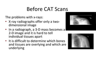
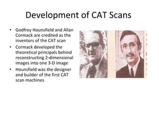
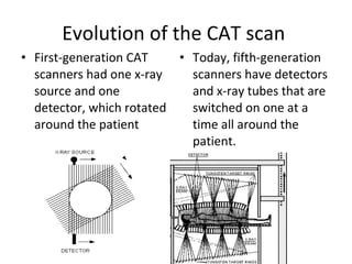
Computerized Axial   Tomography   (CAT) SCANS   By Abby Jeffers  And  James Kloss
Before CAT Scans  The problems with x-rays: X-ray radiographs offer only a two-dimensional image In a radiograph, a 3-D mass becomes a 2-D image and it is hard to tell individual tissues apart It is difficult to determine which bones and tissues are overlying and which are underlying
Development of CAT Scans  Godfrey Hounsfield and Allan Cormack are credited as the inventors of the CAT scan Cormack developed the theoretical principals behind reconstructing 2-dimensional images into one 3-D image Hounsfield was the designer and builder of the first CAT scan machines
Evolution of the CAT scan  First-generation CAT scanners had one x-ray source and one detector, which rotated around the patient Today, fifth-generation scanners have detectors and x-ray tubes that are switched on one at a time all around the patient.
What do they scan for in the body CAT scans internal organs, bones, soft tissue, and blood vessels.  CAT scans provide better clarity than X-rays and reveals more important details than X-rays too.
How do they work In many ways CAT scans work very much like any other X-ray examination devices.  In conventional X-rays small burst of radiation are aimed a the body and shot through the body.  The result if a image that is recorded on photographic film  or a special image recording plate.
How do they work 2  Now CAT scans are very similar to X-rays because in a CAT scan there are numerous X-rays that rotate around A At the same time the X-rays  are shooting beams of radiation that is being received by X-ray detectors on the opposite side of the patient.  At the same time the examination table that you are laying on moves in and out of the machine.  The movement of the table creates a spiral path for the X-ray beam to follow.
How do they work 3 The CAT scan shoot X-ray beams at every angle for 360 ° around the body.  A special computer process this large amount of data and produces a 2-D cross section of the human body.  This technique is called Helical or Spiral CAT
Benefits of CAT scan It is painless, noninvasive, and very accurate. CAT scans can provide information on bone, soft tissue, and blood vessels all at the same time.  Fast and simple  Provides real time imaging  No radiation remains in patients body after CAT scan.
Risks of CAT scans  Slight chance of cancer from excessive exposure to radiation.  Pregnant women should not use CAT scans unless needed to because of the potential risk to the baby. Children should not have too many CAT scans because they are more sensitive to radiation.
What they look like CAT scans are typically a large box like machine with a hole or a short tunnel in the center.  There is a examination table that slides into and out of the tunnel.  The computer workstation the process  information from the scan is located in a separate room.
Works Cited  http://www.asnt.org/publications/materialseval/basics/may00basics/may00basics.htm http://www.aps.org/publications/apsnews/200411/history.cfm http://www.radiologyinfo.org/en/info.cfm?pg=bodyct#part_one http://www.mayfieldclinic.com/pe-ct.htm

CAT scan Final

  • 1.
    Computerized Axial Tomography (CAT) SCANS By Abby Jeffers And James Kloss
  • 2.
    Before CAT Scans The problems with x-rays: X-ray radiographs offer only a two-dimensional image In a radiograph, a 3-D mass becomes a 2-D image and it is hard to tell individual tissues apart It is difficult to determine which bones and tissues are overlying and which are underlying
  • 3.
    Development of CATScans Godfrey Hounsfield and Allan Cormack are credited as the inventors of the CAT scan Cormack developed the theoretical principals behind reconstructing 2-dimensional images into one 3-D image Hounsfield was the designer and builder of the first CAT scan machines
  • 4.
    Evolution of theCAT scan First-generation CAT scanners had one x-ray source and one detector, which rotated around the patient Today, fifth-generation scanners have detectors and x-ray tubes that are switched on one at a time all around the patient.
  • 5.
    What do theyscan for in the body CAT scans internal organs, bones, soft tissue, and blood vessels. CAT scans provide better clarity than X-rays and reveals more important details than X-rays too.
  • 6.
    How do theywork In many ways CAT scans work very much like any other X-ray examination devices. In conventional X-rays small burst of radiation are aimed a the body and shot through the body. The result if a image that is recorded on photographic film or a special image recording plate.
  • 7.
    How do theywork 2 Now CAT scans are very similar to X-rays because in a CAT scan there are numerous X-rays that rotate around A At the same time the X-rays are shooting beams of radiation that is being received by X-ray detectors on the opposite side of the patient. At the same time the examination table that you are laying on moves in and out of the machine. The movement of the table creates a spiral path for the X-ray beam to follow.
  • 8.
    How do theywork 3 The CAT scan shoot X-ray beams at every angle for 360 ° around the body. A special computer process this large amount of data and produces a 2-D cross section of the human body. This technique is called Helical or Spiral CAT
  • 9.
    Benefits of CATscan It is painless, noninvasive, and very accurate. CAT scans can provide information on bone, soft tissue, and blood vessels all at the same time. Fast and simple Provides real time imaging No radiation remains in patients body after CAT scan.
  • 10.
    Risks of CATscans Slight chance of cancer from excessive exposure to radiation. Pregnant women should not use CAT scans unless needed to because of the potential risk to the baby. Children should not have too many CAT scans because they are more sensitive to radiation.
  • 11.
    What they looklike CAT scans are typically a large box like machine with a hole or a short tunnel in the center. There is a examination table that slides into and out of the tunnel. The computer workstation the process information from the scan is located in a separate room.
  • 12.
    Works Cited http://www.asnt.org/publications/materialseval/basics/may00basics/may00basics.htm http://www.aps.org/publications/apsnews/200411/history.cfm http://www.radiologyinfo.org/en/info.cfm?pg=bodyct#part_one http://www.mayfieldclinic.com/pe-ct.htm