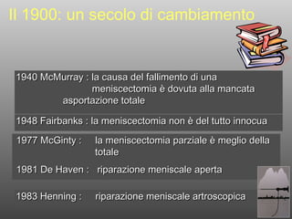
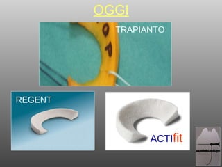
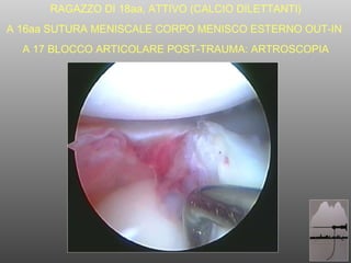
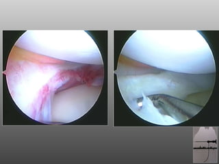
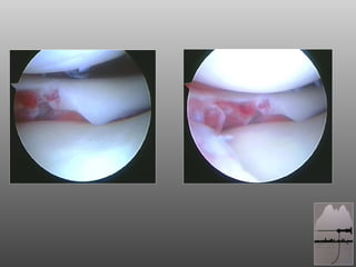
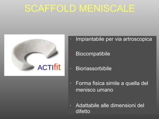
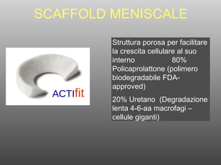
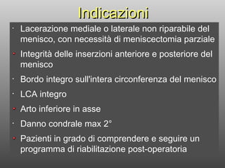
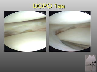
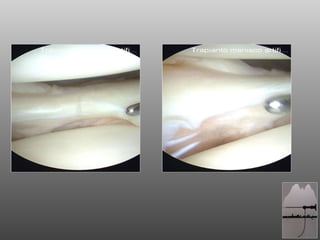
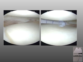
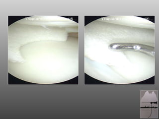
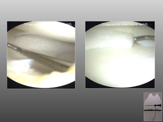
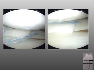
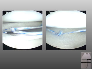
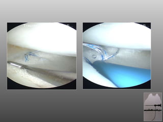
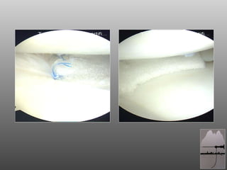
Pinning laterale bloccato del radio distale: tecnica di LegnagoAlberto Mantovani
CORSO ANNUALE S.E.R.T.O.T. 2013, Piacenza
Fratture di polso
TRATTAMENTO CHIRURGICO MINI INVASIVO
Pinning laterale bloccato del radio distale: Tecnica di Legnago
Artrolisi del gomito a cielo aperto nella rigidità post traumaticaAlberto Mantovani
ARTROLISI A CIELO APERTO NELLA RIGIDITA’ POST TRAUMATICA DEL GOMITO: ASPETTI CONSOLIDATI E INNOVAZIONI.
SCOPO DEL LAVORO: si vuole confermare con questo lavoro il ruolo dell’artrolisi a cielo aperto nelle rigidità post traumatiche del gomito secondo le indicazioni consolidate in letteratura. Ma, in assenza di ossificazioni eterotopiche o malunion omero-ulnari, si mette in evidenza anche la possibilità di eseguire la resezione del solo fascio posteriore del legamento collaterale mediale per recuperare la flessione del gomito e di trattare con protesi laterale di rivestimento l’artrosi o la perdità di sostanza ossea del condilo omerale laterale.
MATERIALI E METODI: sono stati esaminati 8 pazienti operati di artrolisi a cielo aperto del gomito per rigidità post traumatica con follow up da 1 a 5 anni. Si sono registrati prima e dopo l’intervento il grado di rigidità secondo Morrey, il dolore con scala visuale analogica, il grado di artrosi secondo Retting & Hastings e l’instabilità con il table top relocation test. In tutti i casi si è praticata la resezione del fascio posteriore del legamento collaterale mediale e in 4 casi si è applicata una protesi laterale di rivestimento: 2 casi per artrosi e 2 casi per perdita di sostanza ossea.
RISULTATI: il miglioramento è stato molto significativo in tutti i casi operati e non si sono registrate né instabilità del gomito operato né altre complicanze.
CONCLUSIONI: il release del legamento collaterale mediale e l’applicazione della protesi laterale di rivestimento sono due gesti chirurgici innovativi e aggiuntivi nell’intervento di artrolisi del gomito per rigidità post traumatica. Si suggerisce di utilizzare la protesi laterale di rivestimento sia per l’artrosi post traumatica che per i difetti ossei del condilo omerale laterale. La resezione del solo fascio posteriore obliquo del legamento collaterale mediale consente un recupero costante della flessione senza creare instabilità.
Il ritorno al transosseo, nuove potenzialità nella riparazione della cuffia d...baudispallaonline
Il sistema sharc-ft permette di creare un construtto stabile massimizzando la copertura del foot-print e la pressione di contatto tra tendine e trochite.
L'impianto sharc-ft può essere considerato una piattaforma su cui caricare medialmente fino a 4 suture e consente una variabilità e flessibilità di modelli riparativi.
E' in grado di replicare la classica riparazione trans-ossea evitando l'unica possibilità di fallimento che è il cedimento dei tunnel per azione dei fili ad alta resistenza e consente alle suture di lavorare verticalmente e in tutti i range di articolarità.
il dottor Marco Spoliti illustra come curare nel modo migliore con Protesi la fratture di spalla di persone anziane, frattura omero prossimale, protesi inversa
Osteosintesi percutanea del radio distale: Tecnica di Legnago (2013)Alberto Mantovani
SUMMARY
Purpose: We have developed and used a system of percutaneous fixation of unstable distal radius fractures (DRF)
using 4 Kirschner (K) wires. These wires are passed from the lateral side of the radius and connected among themselves
using a clamp. We call this the “Legnago technique” and the objective of this study is to standardize the
method and make it safe and easily reproducible. Methods: 27 patients aged from 45 to 102, 3 men and 24 women,
were treated using this technique. The indications were strictly limited to type A2 and A3 of the AO classification,
excluding the A3.3. These were usually emergency procedures, performed under local anaesthesia and under
image intensifier control. We recommend a small incision at the entry point of each K wire and blunt dissection up
to the bone in order to avoid impalement of vessels, tendons or nerves.We follow a standard sequence of passing four
K wires, starting with a 2 mm K wire from the radial styloid into the medullary canal of the radius. This is inserted
dorsal to the tendons of the first extensor compartment. The K wire was mounted on a Jacob’s chuck handle and
was pre-bent at its leading end to around 30 degrees. This helps to control the direction of the wire within the bone
and, also, helps in achieving the reduction. The subsequent three wires of diameter 1.8 mm are passed using a motorised
drill from the lateral aspect of the lower end of the radius across the fracture site to engage the opposite cortex.
Finally, each of the wires is bent adequately in a convergent direction along the axis of the wrist on the lateral side
and held together with the help of a clamp. Results: Each patient was evaluated according to MayoWrist Score criteria,
with a follow-up ranging from 4-26 months.We noted 17 excellent results, 7 good and 3 satisfactory. Radiological
consolidation of the fracture was achieved in each patient, at an average delay of 40 days. Union occurred
with no change in the radiological parameters achieved by the operation. The complications included three cases of
superficial infection around the K wires and a partial lesion of the superficial radial nerve. The patients regained
complete autonomy in the use of the affected upper limb for activities of daily living within a week from the operation.
None of the patients underwent supervised physiotherapy. Conclusions: The Legnago technique of percutaneous
fixation of the DRF has proved efficacious in the treatment of unstable extra-articular fractures. The particular
arrangement of insertion of the K-wires and their connection using an external fixator clamp allowed early
active mobilisation of the wrist without plaster support. This concurs with recent experimental demonstrations according
to which the biomechanical stability of the percutaneous fixation of the DRF with externally connected
crossing K wires is superimposable to that obtained by volar locked plates. RivChirMano 2012; 3: 339-349
This document analyzes the results of unicompartmental knee prostheses with mobile and fixed plates from the 1950s to the 2000s. It summarizes the history and development of these prostheses over time. Key findings from literature on survival rates of unicondylar prostheses show survival rates comparable to total knee replacements at 10 years and questions remain about survival rates at 15-20 years. The document advocates for unicondylar prostheses based on appropriate patient selection and surgical technique to achieve long prosthesis life expectancy.
The document discusses the anatomical double bundle ACL reconstruction technique. It provides details on the surgical procedure, including patient setup at 90 degrees of flexion, use of the accessory AM portal, placement of femoral and tibial tunnels, and tensioning of the AM and PL bundles separately. It also mentions an ongoing prospective randomized clinical study comparing outcomes of SB versus DB ACL reconstruction that is ongoing, with preliminary results still pending after only 40% of patients reaching the 6 month follow up so far.
This document summarizes the results of meniscus allograft transplantation (MAT) procedures performed by the author and colleagues on 38 patients over 5 years of follow up. Key findings include:
- Good short term outcomes in reducing pain and improving function based on clinical scores.
- Success depends on careful patient selection criteria like age, activity level, and degree of cartilage damage.
- Longer follow up is still needed to determine the biomechanical effects and ability of MAT to prevent cartilage degeneration over time.
- MAT is technically demanding but shows promise as an alternative to treat symptomatic knees with meniscus deficiency.
1. CASO CLINICO: SCAFFOLD MENISCALE A. TRIPODO - G.C. COARI CASA DI CURA “SAN CAMILLO” - VERSILIA RIGHI
2. 1940 McMurray : la causa del fallimento di una meniscectomia è dovuta alla mancata asportazione totale 1948 Fairbanks : la meniscectomia non è del tutto innocua Il 1900: un secolo di cambiamento 1977 McGinty : la meniscectomia parziale è meglio della totale 1981 De Haven : riparazione meniscale aperta 1983 Henning : riparazione meniscale artroscopica
9. SCAFFOLD MENISCALE ACTI fit Struttura porosa per facilitare la crescita cellulare al suo interno 80% Policaprolattone (polimero biodegradabile FDA-approved) 20% Uretano (Degradazione lenta 4-6-aa macrofagi – cellule giganti)