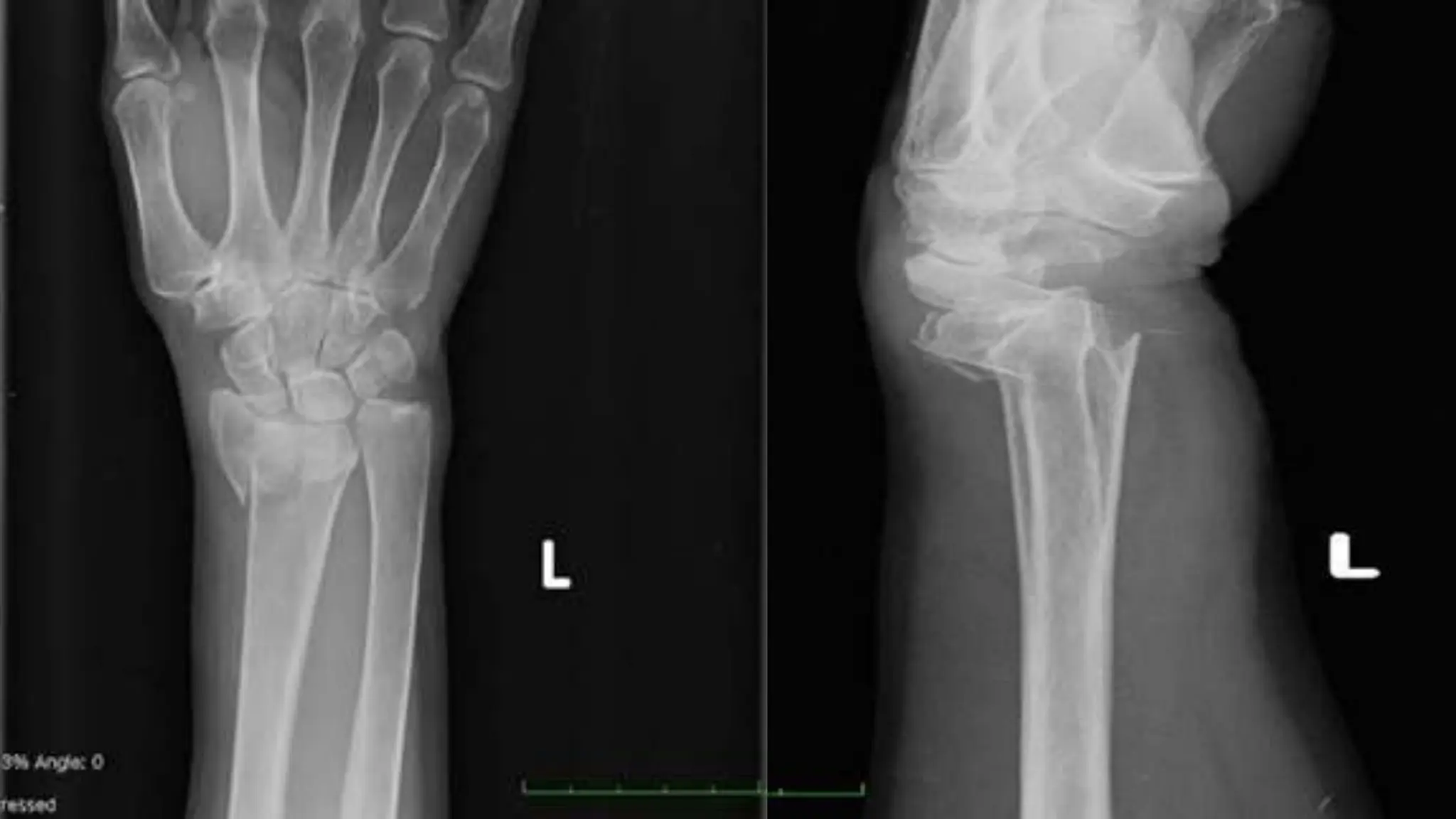
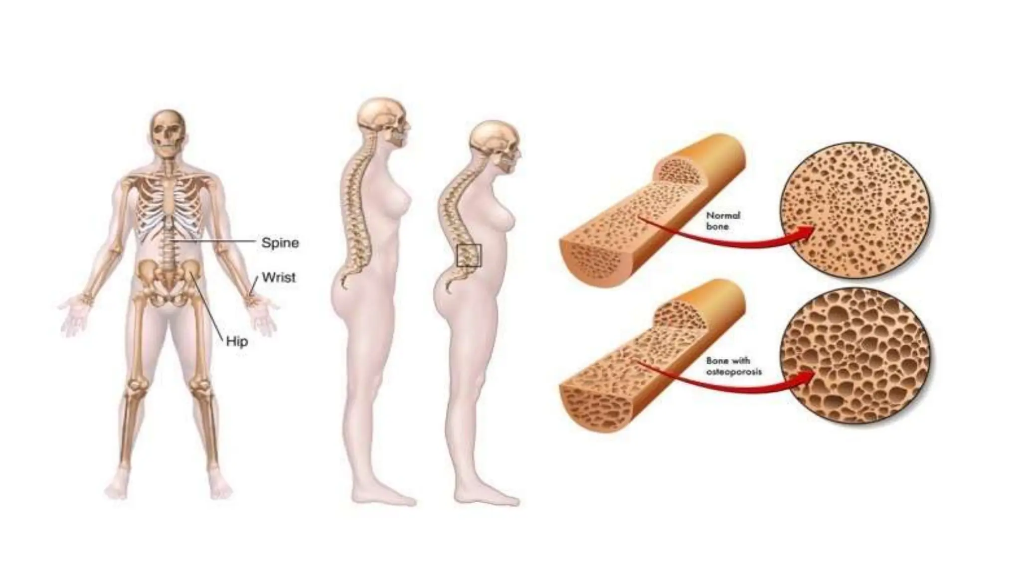
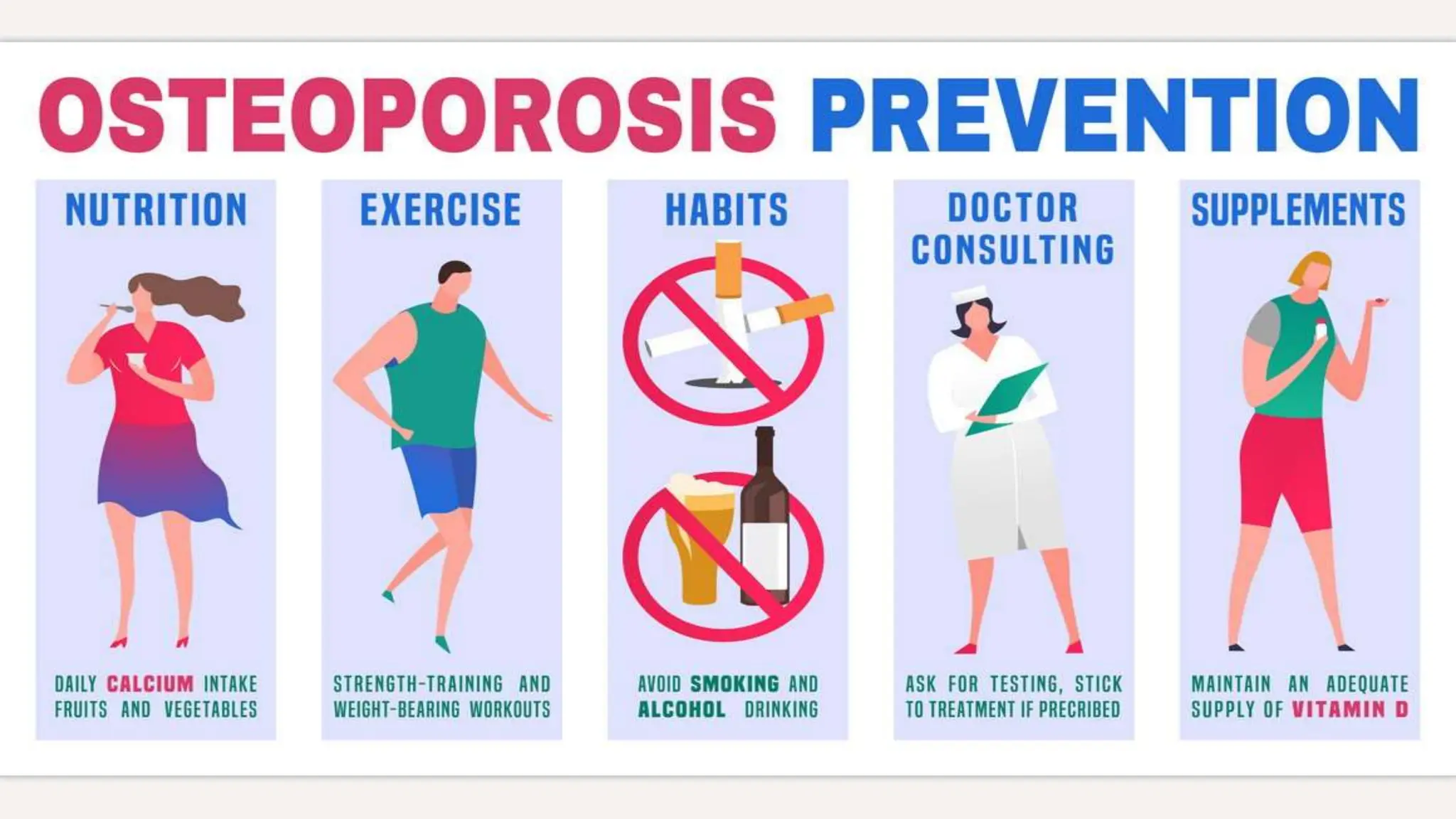
A 58-year-old postmenopausal woman presented with a left wrist fracture following a fall. Her medical history included asthma treated with corticosteroids, gastric ulcer, and a previous wrist fracture. Examination found a displaced, swollen wrist with no neurovascular compromise. X-ray revealed a Colles' fracture. Risk factors for osteoporosis from her history included advanced age, menopause, corticosteroid use, smoking, and lack of exercise. Further workup was needed to confirm the diagnosis and guide management, which may include bisphosphonates, parathyroid hormone, or calcium and vitamin D supplementation to prevent future fractures.
Osteoporosis is a disease where bones become weak and brittle, increasing the risk of fractures, especially in the hips, spine and wrists. It affects over 10 million Americans aged 50 and older, with women at higher risk than men. Risk factors include age, gender, family history and previous fractures. Symptoms may not appear until a fracture occurs. Treatment focuses on lifestyle changes like diet, exercise and medication to slow bone loss and reduce fracture risk. Complications can include pain, loss of mobility and independence from broken bones.
Osteoporosis is a systemic skeletal disease characterized by low bone mass and deterioration of bone microarchitecture, leading to increased bone fragility and risk of fracture. It is diagnosed based on a combination of clinical history, risk factors, physical examination, imaging findings, and bone mineral density (BMD) measurement via dual-energy X-ray absorptiometry. BMD T-scores are used to classify individuals as having normal bone density, osteopenia, or osteoporosis according to World Health Organization criteria. Other imaging techniques like quantitative computed tomography and peripheral BMD measurement can provide additional information.
This document summarizes a presentation on osteoarthritis (OA) phenotypes and risk factors. The presentation discusses evidence that OA may consist of distinct subtypes including generalized vs. joint-specific, secondary vs. primary, painful vs. non-painful, and malaligned vs. neutrally aligned joints. Identifying OA phenotypes is important for developing effective prevention and treatment strategies that may differ between subtypes.
The document defines osteoarthritis as a degenerative joint disease characterized by destruction of articular cartilage and new bone formation at joint surfaces. It most commonly affects weight-bearing joints like the hip and knee. Treatment involves a combination of approaches to relieve pain, restore function, and reduce disability, including weight loss, exercises, braces, and medications like acetaminophen, NSAIDs, or opioids. Conservative treatment succeeds for about 50% of patients before considering surgical options.
OSTEOPOROSIS:A Barebone guide to diagnosis and managementGovindRankawat1
“Progressive systemic skeletal disease characterized by low bone mass and microarchitectural deterioration of bone tissue, leading to enhanced bone fragility and a consequent increase in fracture risk”
True Definition: bone with lower density and higher fracture risk
WHO: utilizes Bone Mineral Density as definition (T score <-2.5)
Osteoporosis is silent because there are no symptoms initially.
The most common are fractures of the spine, hip, and wrist.
Osteoporosis is not an inevitable part of aging, but is a disease that can be prevented and treated, provided it is detected early.
The main goal of treating osteoporosis is to prevent such fractures in the first place.
Bare bone term used for “necked bone with necked eye”
“There is clearly a problem of underdiagnosis and undertreatment of osteoporosis and we want to raise awareness about the risk factors for osteoporosis so that those who need treatment get treatment”.
Learning Objectives
Utilize recent recommendations for osteoporosis prevention and treatment and how to apply them in practice.
Explain controversies surrounding pharmacologic osteoporosis therapy including side effects and the risk/benefit ratio of therapy.
Determine when and how to utilize the current pharmacologic therapies including anabolic versus anti-resorptive approaches and how to transition or discontinue treatment
Osteoporosis only causes symptoms when it is far advanced.
Symptoms include loss of height, deformed spine (“dowager’s hump”), unexplained back pain, and fractures.
It is best to detect problems at an early stage, when treatment is most effective.
The best test for detecting osteoporosis is bone densitometry, done with a technique called “Dual-energy X-ray Absorptiometry” or DXA.
This document discusses osteoporosis and osteomalacia. It provides information on:
- A case of a 68-year-old woman who presents with a wrist fracture following a fall.
- Risk factors for osteoporosis including corticosteroid use, menopause, family history of fractures.
- Diagnostic tests for osteoporosis including DXA scan, FRAX score to evaluate 10-year fracture risk.
- Treatment involves lifestyle changes like exercise, calcium and vitamin D supplementation, as well as pharmacologic therapies like bisphosphonates or teriparatide.
This document provides an overview of osteoporosis including its definition, epidemiology, pathophysiology, causes, clinical features, diagnosis, and treatment. Some key points include:
- Osteoporosis is a skeletal disorder characterized by compromised bone strength and increased risk of fracture. It is defined by the WHO as a bone density 2.5 standard deviations below the mean.
- It is a major global health problem, particularly affecting post-menopausal women and the elderly. Lifetime risk of osteoporotic fractures is 30-50% in females and 15-30% in males.
- Causes include failure to achieve peak bone mass, increased bone resorption, and inadequate bone formation
This document discusses osteoporosis, including what it is, symptoms, diagnosis, risk factors, incidence rates, treatment options, and preventative measures. Some key points:
- Osteoporosis is the thinning and weakening of bones, making them fragile and prone to fractures. It has no symptoms until a fracture occurs.
- Risk factors include genetics, low vitamin D, low calcium intake, lack of physical activity, menopause for women. India has a high incidence, especially in men and those aged 50-60.
- Treatment options include hormone replacement therapy, SERMs, bisphosphonates, and Teriparatide injections to rebuild bone density and reduce fracture risk.
The document discusses osteoporosis and questions whether it should be considered a disease. It summarizes the WHO conference that defined osteoporosis and established T-scores. It raises concerns about the definition and how DEXA scans are used to diagnose based on measurements that may not be accurate or applicable to all groups. Risk factors are highlighted and questions are raised about whether they apply to all populations equally.
This document provides an overview of osteoporosis, including:
- Osteoporosis is a disease characterized by low bone density and increased susceptibility to fractures. It is underdiagnosed and undertreated.
- Vertebral compression fractures are the most common osteoporotic fractures.
- By 2050, the number of osteoporotic fractures occurring worldwide each year is projected to increase from 1.66 million to 6.26 million.
This document provides an overview of osteoporosis, including its definition, demographics, risk factors, diagnosis, treatment options and guidelines for follow-up. It defines osteoporosis as a skeletal disorder characterized by compromised bone strength and increased risk of fracture. Key points include that osteoporosis is diagnosed based on fractures or low bone mineral density (BMD), affects millions of Americans especially postmenopausal women, and treatment focuses on increasing calcium/vitamin D intake and medications to reduce fracture risk.
Osteoporosis in elderly causes and managementGovindRankawat1
“Progressive systemic skeletal disease characterized by low bone mass and microarchitectural deterioration of bone tissue, leading to enhanced bone fragility and a consequent increase in fracture risk”
True Definition: bone with lower density and higher fracture risk
WHO: utilizes Bone Mineral Density as definition (T score <-2.5)
Osteoporosis is silent because there are no symptoms initially.
The most common are fractures of the spine, hip, and wrist.
Osteoporosis is not an inevitable part of aging, but is a disease that can be prevented and treated, provided it is detected early.
The main goal of treating osteoporosis is to prevent such fractures in the first place.
The Battle Sport Traumatology 2023 Castrocaro Terme FC.pdfNicola Taddio
In this presentation the author analyzes the various problems relating to the functional and mechanical instability of the ankle which has suffered a lesion of the lateral ligaments, the complications, failures and short and long term outcomes in order to have a 360 degree vision of the problem , the possible solutions and the correct management to avoid them.
Frequency of Osteoporotic Fractures, Parameters of Bone Mineral Density and T...CrimsonPublishersOPROJ
Frequency of Osteoporotic Fractures, Parameters of Bone Mineral Density and Trabecular Bone Score in Postmenopausal Women by Grygorieva N* in Orthopedic Research Online Journal
Osteoporosis is a systemic skeletal disease characterized by low bone mass and deterioration of bone tissue, leading to increased bone fragility and risk of fractures. It is most commonly seen in elderly women. Bone mineral density testing is used to diagnose osteoporosis, with scores more than 2.5 standard deviations below normal indicating the disease. Treatment options include calcium, vitamin D, exercise, bisphosphonates, calcitonin, parathyroid hormone, and selective estrogen receptor modulators.
This document discusses osteoporosis and bone health. It provides an overview of osteoporosis, defining it as a disease characterized by low bone mass and deterioration of bone structure. It notes that osteoporosis is most common in post-menopausal women and those over age 65. The document discusses the risk of fractures from osteoporosis and their impact, including loss of height, disability, and increased mortality. It also covers osteoporosis risks and treatments.
Osteoporosis is a disease characterized by reduced bone mass and deterioration of bone tissue, leading to an increased risk of bone fractures. It is defined as a bone mineral density (BMD) score of more than 2.5 standard deviations below the average for a young healthy adult. Osteoporosis affects many post-menopausal women and older adults due to age-related bone loss. It has no symptoms until a fracture occurs, with vertebral compression fractures and wrist fractures being common. Diagnosis involves assessing risk factors and performing BMD tests and imaging to evaluate for fractures.
Osteogenesis imperfecta, also known as brittle bone disease, is a genetic disorder characterized by fragile bones that break easily. It is caused by a defect in the genes responsible for producing collagen, which provides structure and strength to bones. Symptoms can range from mild to severe and include bone fractures, weak muscles, small stature, and bone deformities. While there is no cure, treatment focuses on pain management, surgery, and physical therapy.
Osteoporosis is a chronic, progressive skeletal disease characterized by low bone mass, microarchitecture deterioration of bone tissue, bone fragility, and a consequent increase in fracture risk.
This document discusses osteoporosis and provides information on evaluating individuals for the disease. It defines osteoporosis as a skeletal disorder characterized by compromised bone strength and increased fracture risk. Key points include:
- Osteoporosis is most prevalent in postmenopausal women and those over age 70. It can also occur secondary to certain diseases, medications, and risk factors.
- Evaluation for osteoporosis involves assessing bone mineral density via DXA scan and considering risk factors like prior fractures, family history, smoking status, and certain medical conditions.
- Identifying those at risk helps prevent fragility fractures through lifestyle changes, medication, and fall prevention strategies.
Ckd-MBD & osteoporosis the management dilemma Ayman Seddik
This document discusses the management of chronic kidney disease-mineral and bone disorder (CKD-MBD) and osteoporosis in elderly patients. It outlines that CKD-MBD and osteoporosis are common in elderly populations and impact mortality and morbidity. Management is based on the stage of CKD and involves controlling serum phosphorus and calcium levels, using phosphate binders to treat hyperphosphatemia, and treating abnormal PTH levels. Guidelines recommend treating bone disease with bisphosphonates and other osteoporosis medications according to the condition and stage of CKD. The risks and benefits of different treatment options must be considered based on each patient's situation.
Bone densitometry uses x-rays to measure bone mineral density and detect osteoporosis, a disease where bones become porous and fragile. The DEXA machine generates two x-ray energies to measure radiation passing through bones. Bone density tests indicate if a person has low bone density before a fracture, monitor changes in density over time, predict future fracture risk, and help determine treatment. Results compare a patient's density to age-matched peers and young adults to derive z-scores and t-scores. Lower scores indicate increased risk of osteoporosis and fracture.
Osteoporosis is a bone disorder characterized by low bone density and compromised bone strength, predisposing patients to fractures. It occurs when bone resorption by osteoclasts exceeds bone formation by osteoblasts, disrupting the normal bone remodeling process. Key risk factors include increasing age, estrogen deficiency, family history, and lack of physical activity. Diagnosis is made through dual-energy x-ray absorptiometry scans, which measure bone mineral density and compare it to healthy young adults. Treatment focuses on lifestyle changes like calcium supplementation and exercise to prevent fractures and stabilize remaining bone mass.
This document discusses osteoporosis and its prevention and management. It begins with definitions of osteoporosis and descriptions of normal bone versus osteoporotic bone. It then covers the risk factors, clinical presentation, diagnosis through bone mineral density testing, investigations for secondary causes, and imaging findings of osteoporosis. The document concludes with sections on screening and preventing osteoporosis through nutrition, exercise, and fall prevention strategies.
Common Musculoskeletal (orthopedic) disorders in elderlyBhaskarBorgohain4
elderly and geriatric old age people tend to suffer many orthopedic disability due to common functional limitations and mobility issues as a result of pain from osteoarthritis, osteoporotic fractures, low back pain and degenerative spinal disorders like lumbar spondylosis and vitamin D and nutritional deficiencies. early diagnosis , prevention, timely surgical interventions and optimum rehabilitation are paramount to bring elderly to pre-injury state of functional independence.
The document discusses testosterone and men's health. It describes how testosterone levels naturally decrease with age, which can lead to symptoms like decreased energy and vitality. The author details his own experience with low testosterone at age 63 and how replacing his testosterone through treatment resolved many of his symptoms. Based on his clinical practice, the author advocates for evaluating older men's testosterone levels if they present with nonspecific complaints, as low testosterone may be an underlying cause and replacement can effectively treat symptoms.
Osteoporosis is a systemic skeletal disease characterized by low bone mass and deterioration of bone tissue, leading to fragile bones that are prone to fractures. It often presents without symptoms until a fracture occurs. Key risk factors include advanced age, female sex, small body frame, family history, smoking, excessive alcohol use, low calcium intake, physical inactivity, and certain medications. Screening high-risk individuals is important for diagnosis and treatment before fractures manifest.
1. The document provides information on examining the shoulder, elbow, wrist, and hand, including anatomy, inspection, palpation, range of motion tests, and special tests.
2. Common causes of pain in these areas are described, including rotator cuff injuries, arthritis, tendinitis, bursitis, and neurological issues.
3. Examination techniques for each area include inspection for deformities, swelling, atrophy; palpation for temperature, tenderness, crepitus; and range of motion and special tests like impingement signs and drop arm test for shoulders.
F waves are produced when a supramaximal stimulus is applied to a motor nerve, causing antidromic impulses that travel to the spinal cord. Some motor neurons then backfire, producing the F wave. F wave latencies provide information about motor nerve conduction velocity and can help detect neuropathies. The H-reflex is produced when a weak stimulus excites muscle spindle Ia afferents, causing antidromic impulses in the spinal cord that excite alpha motor neurons. F wave and H-reflex latencies are used to evaluate spinal and peripheral nerve conduction.
More Related Content
Similar to Case senario presentation...............
This document discusses osteoporosis, including what it is, symptoms, diagnosis, risk factors, incidence rates, treatment options, and preventative measures. Some key points:
- Osteoporosis is the thinning and weakening of bones, making them fragile and prone to fractures. It has no symptoms until a fracture occurs.
- Risk factors include genetics, low vitamin D, low calcium intake, lack of physical activity, menopause for women. India has a high incidence, especially in men and those aged 50-60.
- Treatment options include hormone replacement therapy, SERMs, bisphosphonates, and Teriparatide injections to rebuild bone density and reduce fracture risk.
The document discusses osteoporosis and questions whether it should be considered a disease. It summarizes the WHO conference that defined osteoporosis and established T-scores. It raises concerns about the definition and how DEXA scans are used to diagnose based on measurements that may not be accurate or applicable to all groups. Risk factors are highlighted and questions are raised about whether they apply to all populations equally.
This document provides an overview of osteoporosis, including:
- Osteoporosis is a disease characterized by low bone density and increased susceptibility to fractures. It is underdiagnosed and undertreated.
- Vertebral compression fractures are the most common osteoporotic fractures.
- By 2050, the number of osteoporotic fractures occurring worldwide each year is projected to increase from 1.66 million to 6.26 million.
This document provides an overview of osteoporosis, including its definition, demographics, risk factors, diagnosis, treatment options and guidelines for follow-up. It defines osteoporosis as a skeletal disorder characterized by compromised bone strength and increased risk of fracture. Key points include that osteoporosis is diagnosed based on fractures or low bone mineral density (BMD), affects millions of Americans especially postmenopausal women, and treatment focuses on increasing calcium/vitamin D intake and medications to reduce fracture risk.
Osteoporosis in elderly causes and managementGovindRankawat1
“Progressive systemic skeletal disease characterized by low bone mass and microarchitectural deterioration of bone tissue, leading to enhanced bone fragility and a consequent increase in fracture risk”
True Definition: bone with lower density and higher fracture risk
WHO: utilizes Bone Mineral Density as definition (T score <-2.5)
Osteoporosis is silent because there are no symptoms initially.
The most common are fractures of the spine, hip, and wrist.
Osteoporosis is not an inevitable part of aging, but is a disease that can be prevented and treated, provided it is detected early.
The main goal of treating osteoporosis is to prevent such fractures in the first place.
The Battle Sport Traumatology 2023 Castrocaro Terme FC.pdfNicola Taddio
In this presentation the author analyzes the various problems relating to the functional and mechanical instability of the ankle which has suffered a lesion of the lateral ligaments, the complications, failures and short and long term outcomes in order to have a 360 degree vision of the problem , the possible solutions and the correct management to avoid them.
Frequency of Osteoporotic Fractures, Parameters of Bone Mineral Density and T...CrimsonPublishersOPROJ
Frequency of Osteoporotic Fractures, Parameters of Bone Mineral Density and Trabecular Bone Score in Postmenopausal Women by Grygorieva N* in Orthopedic Research Online Journal
Osteoporosis is a systemic skeletal disease characterized by low bone mass and deterioration of bone tissue, leading to increased bone fragility and risk of fractures. It is most commonly seen in elderly women. Bone mineral density testing is used to diagnose osteoporosis, with scores more than 2.5 standard deviations below normal indicating the disease. Treatment options include calcium, vitamin D, exercise, bisphosphonates, calcitonin, parathyroid hormone, and selective estrogen receptor modulators.
This document discusses osteoporosis and bone health. It provides an overview of osteoporosis, defining it as a disease characterized by low bone mass and deterioration of bone structure. It notes that osteoporosis is most common in post-menopausal women and those over age 65. The document discusses the risk of fractures from osteoporosis and their impact, including loss of height, disability, and increased mortality. It also covers osteoporosis risks and treatments.
Osteoporosis is a disease characterized by reduced bone mass and deterioration of bone tissue, leading to an increased risk of bone fractures. It is defined as a bone mineral density (BMD) score of more than 2.5 standard deviations below the average for a young healthy adult. Osteoporosis affects many post-menopausal women and older adults due to age-related bone loss. It has no symptoms until a fracture occurs, with vertebral compression fractures and wrist fractures being common. Diagnosis involves assessing risk factors and performing BMD tests and imaging to evaluate for fractures.
Osteogenesis imperfecta, also known as brittle bone disease, is a genetic disorder characterized by fragile bones that break easily. It is caused by a defect in the genes responsible for producing collagen, which provides structure and strength to bones. Symptoms can range from mild to severe and include bone fractures, weak muscles, small stature, and bone deformities. While there is no cure, treatment focuses on pain management, surgery, and physical therapy.
Osteoporosis is a chronic, progressive skeletal disease characterized by low bone mass, microarchitecture deterioration of bone tissue, bone fragility, and a consequent increase in fracture risk.
This document discusses osteoporosis and provides information on evaluating individuals for the disease. It defines osteoporosis as a skeletal disorder characterized by compromised bone strength and increased fracture risk. Key points include:
- Osteoporosis is most prevalent in postmenopausal women and those over age 70. It can also occur secondary to certain diseases, medications, and risk factors.
- Evaluation for osteoporosis involves assessing bone mineral density via DXA scan and considering risk factors like prior fractures, family history, smoking status, and certain medical conditions.
- Identifying those at risk helps prevent fragility fractures through lifestyle changes, medication, and fall prevention strategies.
Ckd-MBD & osteoporosis the management dilemma Ayman Seddik
This document discusses the management of chronic kidney disease-mineral and bone disorder (CKD-MBD) and osteoporosis in elderly patients. It outlines that CKD-MBD and osteoporosis are common in elderly populations and impact mortality and morbidity. Management is based on the stage of CKD and involves controlling serum phosphorus and calcium levels, using phosphate binders to treat hyperphosphatemia, and treating abnormal PTH levels. Guidelines recommend treating bone disease with bisphosphonates and other osteoporosis medications according to the condition and stage of CKD. The risks and benefits of different treatment options must be considered based on each patient's situation.
Bone densitometry uses x-rays to measure bone mineral density and detect osteoporosis, a disease where bones become porous and fragile. The DEXA machine generates two x-ray energies to measure radiation passing through bones. Bone density tests indicate if a person has low bone density before a fracture, monitor changes in density over time, predict future fracture risk, and help determine treatment. Results compare a patient's density to age-matched peers and young adults to derive z-scores and t-scores. Lower scores indicate increased risk of osteoporosis and fracture.
Osteoporosis is a bone disorder characterized by low bone density and compromised bone strength, predisposing patients to fractures. It occurs when bone resorption by osteoclasts exceeds bone formation by osteoblasts, disrupting the normal bone remodeling process. Key risk factors include increasing age, estrogen deficiency, family history, and lack of physical activity. Diagnosis is made through dual-energy x-ray absorptiometry scans, which measure bone mineral density and compare it to healthy young adults. Treatment focuses on lifestyle changes like calcium supplementation and exercise to prevent fractures and stabilize remaining bone mass.
This document discusses osteoporosis and its prevention and management. It begins with definitions of osteoporosis and descriptions of normal bone versus osteoporotic bone. It then covers the risk factors, clinical presentation, diagnosis through bone mineral density testing, investigations for secondary causes, and imaging findings of osteoporosis. The document concludes with sections on screening and preventing osteoporosis through nutrition, exercise, and fall prevention strategies.
Common Musculoskeletal (orthopedic) disorders in elderlyBhaskarBorgohain4
elderly and geriatric old age people tend to suffer many orthopedic disability due to common functional limitations and mobility issues as a result of pain from osteoarthritis, osteoporotic fractures, low back pain and degenerative spinal disorders like lumbar spondylosis and vitamin D and nutritional deficiencies. early diagnosis , prevention, timely surgical interventions and optimum rehabilitation are paramount to bring elderly to pre-injury state of functional independence.
The document discusses testosterone and men's health. It describes how testosterone levels naturally decrease with age, which can lead to symptoms like decreased energy and vitality. The author details his own experience with low testosterone at age 63 and how replacing his testosterone through treatment resolved many of his symptoms. Based on his clinical practice, the author advocates for evaluating older men's testosterone levels if they present with nonspecific complaints, as low testosterone may be an underlying cause and replacement can effectively treat symptoms.
Osteoporosis is a systemic skeletal disease characterized by low bone mass and deterioration of bone tissue, leading to fragile bones that are prone to fractures. It often presents without symptoms until a fracture occurs. Key risk factors include advanced age, female sex, small body frame, family history, smoking, excessive alcohol use, low calcium intake, physical inactivity, and certain medications. Screening high-risk individuals is important for diagnosis and treatment before fractures manifest.
Similar to Case senario presentation............... (20)
1. The document provides information on examining the shoulder, elbow, wrist, and hand, including anatomy, inspection, palpation, range of motion tests, and special tests.
2. Common causes of pain in these areas are described, including rotator cuff injuries, arthritis, tendinitis, bursitis, and neurological issues.
3. Examination techniques for each area include inspection for deformities, swelling, atrophy; palpation for temperature, tenderness, crepitus; and range of motion and special tests like impingement signs and drop arm test for shoulders.
F waves are produced when a supramaximal stimulus is applied to a motor nerve, causing antidromic impulses that travel to the spinal cord. Some motor neurons then backfire, producing the F wave. F wave latencies provide information about motor nerve conduction velocity and can help detect neuropathies. The H-reflex is produced when a weak stimulus excites muscle spindle Ia afferents, causing antidromic impulses in the spinal cord that excite alpha motor neurons. F wave and H-reflex latencies are used to evaluate spinal and peripheral nerve conduction.
The document provides an overview of the radiological anatomy of the spine. It describes the key anatomical structures of the cervical, thoracic, and lumbar regions of the spine, including the vertebral bodies, intervertebral discs, transverse processes, pedicles, lamina, spinous processes, ribs, and sacrum. Diagrams are included to illustrate these structures for each region.
Neuropathic arthropathy, also known as Charcot joint, is a condition characterized by progressive joint destruction and deformity caused by loss of sensation in the joints. It was first described in detail by Jean-Martin Charcot in 1868. The major theories for its pathophysiology are neurotrauma from repetitive micro-injuries without pain sensation, and neurovascular dysregulation leading to hyperemia and bone resorption. Diabetes is the most common cause. Imaging shows bone fragmentation, joint dislocation, and deformity. Treatment involves prolonged casting or bracing to immobilize joints during the acute inflammatory phase, followed by bracing and accommodative footwear. Later stages may require surgery.
power point presentation brucellosis.pptxazzaelnenaey
The document provides information about brucellosis, including its definition as a zoonotic disease caused by Brucella bacteria. It discusses the epidemiology, transmission, clinical presentation, diagnosis, and treatment of the disease. Brucellosis commonly involves the musculoskeletal system, with spondylitis, sacroiliitis, and peripheral arthritis being frequent manifestations. Diagnosis involves serological testing and culture of infected tissues. Treatment requires prolonged antibiotic therapy.
This document discusses rheumatoid arthritis and seronegative spondyloarthritis. It lists common hand deformities in rheumatoid arthritis such as swan neck and boutonniere deformities. It also lists criteria for arthritis including redness, swelling and tenderness. The sacroiliac joint is commonly affected in seronegative spondyloarthritis. Criteria for inflammatory back pain and members of the seronegative spondyloarthritis family are also enumerated.
1) The document describes several lysosomal storage disorders including Tay-Sachs disease, Sandhoff disease, and GM2 Activator deficiency. These disorders are caused by defects in genes encoding hexosaminidase enzymes, resulting in ganglioside accumulation.
2) The phenotypes range from infantile to juvenile to adult-onset, with infantile having the earliest onset of symptoms around 6 months and most severe progression leading to death by age 3-4. Juvenile and adult-onset phenotypes have later onset and slower progression.
3) Initial symptoms vary by phenotype but often involve developmental delay, loss of motor skills, weakness, and neurological decline. Disease progression results in loss of abilities and eventual death due to
Urate crystals can initiate and perpetuate acute inflammation by inducing cells like phagocytes and synovial cells to release inflammatory mediators. This leads to neutrophil infiltration into joints, causing synovitis. Colchicine inhibits this process by interfering with neutrophil migration, metabolism, and phagocytosis. It does this by disrupting microtubules involved in cell structure and movement, reducing neutrophil motility, chemotaxis, and the release of inflammatory factors. Colchicine is used to treat gout attacks and prevent recurrences by dampening the inflammatory response driven by neutrophils.
This document discusses psoriatic arthritis (PsA), including its clinical manifestations, patterns, imaging, diagnosis, and treatment. PsA is a chronic inflammatory arthritis occurring in 7-42% of psoriasis patients, typically presenting as painful, stiff joints. Five patterns are recognized: oligoarticular disease in 70% of cases affecting distal joints; asymmetric DIP involvement in 10% of cases; arthritis mutilans deforming fingers and toes in 5% of cases; symmetric polyarthritis resembling rheumatoid arthritis in 15% of cases; and psoriatic spondyloarthritis in 5% presenting with axial inflammation. Treatment involves NSAIDs, DMARDs like methotrexate, and biological agents
This document discusses the classification of vasculitis. It begins by defining vasculitis as a heterogeneous group of conditions where inflammation of blood vessels occurs independently or secondary to other diseases. It then describes how vasculitis typically presents depending on vessel size, such as limb claudication for large vessel vasculitis or palpable purpura for small vessel vasculitis. Specific types of vasculitis are then outlined according to vessel size and other characteristics, such as the diagnostic criteria for giant cell arteritis and Takayasu arteritis as examples of large vessel vasculitis.
Non steroid anti inflammatory drug.pptxazzaelnenaey
NSAIDs work by inhibiting cyclooxygenase enzymes (COX-1 and COX-2) which are involved in prostaglandin synthesis. This decreases inflammation but also inhibits platelet aggregation and increases risk of gastrointestinal ulcers/bleeds. Common side effects include gastrointestinal issues, renal toxicity, and cardiovascular effects. COX-2 inhibitors have fewer gastrointestinal side effects but increase risk of heart attack and thrombosis. Proper use and monitoring can help reduce risks.
Juvenile Idiopathic Arthritis (JIA) is defined as arthritis of unknown cause that begins before age 16 and lasts over 6 weeks. It is classified based on symptoms into subtypes including systemic onset JIA, oligoarticular JIA, and polyarticular JIA. Treatment involves a stepwise approach starting with NSAIDs and intra-articular steroids and escalating to DMARDs and biologicals. Complications can include chronic anterior uveitis, osteoporosis, and potentially life-threatening macrophage activation syndrome.
This document provides an overview of targeted disease-modifying antirheumatic drugs (DMARDs) for the treatment of rheumatoid arthritis (RA). It discusses the Janus kinase (JAK) signaling pathway and how JAK inhibitors block specific kinases to treat RA. The document reviews the timeline of RA treatments from early interventions like baths and injections to current first-line methotrexate and additions of targeted therapies like JAK inhibitors if treatment goals are not met. It profiles approved and investigational JAK inhibitors for RA including tofacitinib, baricitinib, upadacitinib, filgotinib, and peficitinib.
Statin myopathy refers to muscle-related side effects caused by statin medications used to lower cholesterol. There are two main types: autoimmune statin myopathy and toxic statin myopathy. Autoimmune statin myopathy is rare but serious, involving muscle inflammation and weakness. Toxic statin myopathy is more common and involves temporary muscle pain and fatigue that resolves after stopping the statin. The document discusses the differences between the two conditions and reviews evidence on evaluating and managing statin intolerance. Non-statin lipid-lowering options are also summarized.
Hypertension and it's role of physiotherapy in it.Vishal kr Thakur
This particular slides consist of- what is hypertension,what are it's causes and it's effect on body, risk factors, symptoms,complications, diagnosis and role of physiotherapy in it.
This slide is very helpful for physiotherapy students and also for other medical and healthcare students.
Here is summary of hypertension -
Hypertension, also known as high blood pressure, is a serious medical condition that occurs when blood pressure in the body's arteries is consistently too high. Blood pressure is the force of blood pushing against the walls of blood vessels as the heart pumps it. Hypertension can increase the risk of heart disease, brain disease, kidney disease, and premature death.
Get Covid Testing at Fit to Fly PCR TestNX Healthcare
A Fit-to-Fly PCR Test is a crucial service for travelers needing to meet the entry requirements of various countries or airlines. This test involves a polymerase chain reaction (PCR) test for COVID-19, which is considered the gold standard for detecting active infections. At our travel clinic in Leeds, we offer fast and reliable Fit to Fly PCR testing, providing you with an official certificate verifying your negative COVID-19 status. Our process is designed for convenience and accuracy, with quick turnaround times to ensure you receive your results and certificate in time for your departure. Trust our professional and experienced medical team to help you travel safely and compliantly, giving you peace of mind for your journey.www.nxhealthcare.co.uk
Michigan HealthTech Market Map 2024. Includes 7 categories: Policy Makers, Academic Innovation Centers, Digital Health Providers, Healthcare Providers, Payers / Insurance, Device Companies, Life Science Companies, Innovation Accelerators. Developed by the Michigan-Israel Business Accelerator
Joker Wigs has been a one-stop-shop for hair products for over 26 years. We provide high-quality hair wigs, hair extensions, hair toppers, hair patch, and more for both men and women.
The Importance of Black Women Understanding the Chemicals in Their Personal C...bkling
Certain chemicals, such as phthalates and parabens, can disrupt the body's hormones and have significant effects on health. According to data, hormone-related health issues such as uterine fibroids, infertility, early puberty and more aggressive forms of breast and endometrial cancers disproportionately affect Black women. Our guest speaker, Jasmine A. McDonald, PhD, an Assistant Professor in the Department of Epidemiology at Columbia University in New York City, discusses the scientific reasons why Black women should pay attention to specific chemicals in their personal care products, like hair care, and ways to minimize their exposure.
VEDANTA AIR AMBULANCE SERVICES IN REWA AT A COST-EFFECTIVE PRICE.pdfVedanta A
Air Ambulance Services In Rewa works in close coordination with ground-based emergency services, including local Emergency Medical Services, fire departments, and law enforcement agencies.
More@: https://tinyurl.com/2shrryhx
More@: https://tinyurl.com/5n8h3wp8
Sectional dentures for microstomia patients.pptxSatvikaPrasad
Microstomia, characterized by an abnormally small oral aperture, presents significant challenges in prosthodontic treatment, including limited access for examination, difficulties in impression making, and challenges with prosthesis insertion and removal. To manage these issues, customized impression techniques using sectional trays and elastomeric materials are employed. Prostheses may be designed in segments or with flexible materials to facilitate handling. Minimally invasive procedures and the use of digital technologies can enhance patient comfort. Education and training for patients on prosthesis care and maintenance are crucial for compliance. Regular follow-up and a multidisciplinary approach, involving collaboration with other specialists, ensure comprehensive care and improved quality of life for microstomia patients.
Digital Health in India_Health Informatics Trained Manpower _DrDevTaneja_15.0...DrDevTaneja1
Digital India will need a big trained army of Health Informatics educated & trained manpower in India.
Presently, generalist IT manpower does most of the work in the healthcare industry in India. Academic Health Informatics education is not readily available at school & health university level or IT education institutions in India.
We look into the evolution of health informatics and its applications in the healthcare industry.
HIMMS TIGER resources are available to assist Health Informatics education.
Indian Health universities, IT Education institutions, and the healthcare industry must proactively collaborate to start health informatics courses on a big scale. An advocacy push from various stakeholders is also needed for this goal.
Health informatics has huge employment potential and provides a big business opportunity for the healthcare industry. A big pool of trained health informatics manpower can lead to product & service innovations on a global scale in India.
End-tidal carbon dioxide (ETCO2) is the level of carbon dioxide that is released at the end of an exhaled breath. ETCO2 levels reflect the adequacy with which carbon dioxide (CO2) is carried in the blood back to the lungs and exhaled.
Non-invasive methods for ETCO2 measurement include capnometry and capnography. Capnometry provides a numerical value for ETCO2. In contrast, capnography delivers a more comprehensive measurement that is displayed in both graphical (waveform) and numerical form.
Sidestream devices can monitor both intubated and non-intubated patients, while mainstream devices are most often limited to intubated patients.
TEST BANK FOR Health Assessment in Nursing 7th Edition by Weber Chapters 1 - ...rightmanforbloodline
TEST BANK FOR Health Assessment in Nursing 7th Edition by Weber Chapters 1 - 34.
3. 58 years old white woman presents to accident and emergency
department with left wrist pain, swelling, and displacement following a
fall onto outstretched hand at home.
Menstrual history: menopause 50 years’ old
Past history: asthma since childhood (treated with corticosteroids),
gastric ulcer aged 45, right wrist fracture aged 57.
Family history: stroke in sister aged 65, hip fracture in mother aged 78.
4. Social history: lives alone, 2 children, retired, smokes 5 cigarettes
per day, occasional alcohol, takes no exercise, fully mobile and
able to complete all ADLs (activities of daily living).
On examination: wrist displaced, swollen, no open wound No loss
of sensation or vascular compromise.
X-ray: Colles’ fracture of distal radius present. Colles’ fracture
treated conservatively with cast and analgesia
7. Differential diagnosis for this case? How to differentiate?
What underlying condition contributed to this fracture? Key point
from history and examination?
Age and Sex related differences in bone mass?
What is bone strength?
What is RANK, RANKL and OPG pathway?
Mention pathogenesis of this disease? risk factors?
What sites of fracture are most common in this condition?
Further Investigation to support your diagnosis?
What is the differences between T-score and Z-score?
Management Plan?
8. Pathologic fractures :(minor trauma ) occur through
areas of weakened bone due to primary malignant
lesions, benign lesions, metastasis, or underlying
metabolic abnormalities as fragility fracture which
result from a minor trauma, most commonly occur at
the hip, spine, or wrist, and indicate that osteoporosis.
Traumatic fracture : (major trauma) occurs when
significant or extreme force is applied to a bone.
caused by a fall or car accident as stress fractures
which are tiny cracks in a bone ,commonly, in the
weight-bearing bones of the lower leg and foot.
9. Differential diagnosis for this case? How to differentiate?
What underlying condition contributed to this fracture? Key
point from history and examination?
Age and Sex related differences in bone mass?
What is bone strength?
What is RANK, RANKL and OPG pathway?
Mention pathogenesis of this disease? risk factors?
What sites of fracture are most common in this condition?
Further Investigation to support your diagnosis?
What is the differences between T-score and Z-score?
Management Plan?
10. Key points from history and examination?
o 58 years old woman (advanced age >50 years)
o Post menopausal state
o White ethnicity
o Full onto outstretched hand
o Treated with corticosteroids
o History of gastric ulcer ( may be treated with PPI)
o History of fractures
o Smoking 5 cigarettes per day
o Occasionally alcohol
o No exercise inspite of fully mobile and able to complete ADLs
o Wrist displaced
o No loss of sensation o vascular
compromise
o Colles fracture of distal radius
o Family history of fracture
11. Differential diagnosis for this case? How to differentiate?
What underlying condition contributed to this fracture? Key point
from history and examination?
Age and Sex related differences in bone mass?
What is bone strength?
What is RANK, RANKL and OPG pathway?
Mention pathogenesis of this disease? risk factors?
What sites of fracture are most common in this condition?
Further Investigation to support your diagnosis?
What is the differences between T-score and Z-score?
Management Plan?
12. In young: the body makes new bone faster than it breaks down
old bones, and the bone mass increases.
In mid 30s: reaches the beak bone mass.
After 40: bone remolding continues, but loses slightly more
than gain.
At menopause: when estrogen levels drops, bone loss
increases.
The role of sex hormones: Low estrogen or testosterone as in
late menarche, premature menopause, menopausal state,
testosterone deficiency in males decreases the bone mass.
Bone mass decreases in female more than male
13. Differential diagnosis for this case? How to differentiate?
What underlying condition contributed to this fracture? Key point
from history and examination?
Age and Sex related differences in bone mass?
What is bone strength?
What is RANK, RANKL and OPG pathway?
Mention pathogenesis of this disease? risk factors?
What sites of fracture are most common in this condition?
Further Investigation to support your diagnosis?
What is the differences between T-score and Z-score?
Management Plan?
14. Bone strength ( density and quality of
bone):
It is determined by bone geometry (size
,shape), cortical thickness, porosity and
trabecular bone morphology.
Bone strength is indirectly estimated by
bone mineral density using Dual energy
X-Ray absorptiometry (DXA).
15. Differential diagnosis for this case? How to differentiate?
What underlying condition contributed to this fracture? Key point
from history and examination?
Age and Sex related differences in bone mass?
What is bone strength?
What is RANK, RANKL and OPG pathway?
Mention pathogenesis of this disease? risk factors?
What sites of fracture are most common in this condition?
Further Investigation to support your diagnosis?
What is the differences between T-score and Z-score?
Management Plan?
16. RANK ligand:
o It is a protein on the surface of
osteoblast.
o when RANK ligand is attached with
RANK on osteoclast precursor, this
activate osteoclast which actives bone
resorption OPG (osteoprotegenin )
osteoclastgenesis inhibiting factor
17. Differential diagnosis for this case? How to differentiate?
What underlying condition contributed to this fracture? Key point
from history and examination?
Age and Sex related differences in bone mass?
What is bone strength?
What is RANK, RANKL and OPG pathway?
Mention pathogenesis of this disease? risk factors?
What sites of fracture are most common in this condition?
Further Investigation to support your diagnosis?
What is the differences between T-score and Z-score?
Management Plan?
18. Pathogenesis
Bone mass peaks around the third decade of life and slowly
decrease afterward. Therefore nutrition and physical activity
are important during growth and development .
Physiologically bone remodeling has 4 sequential phases :
19. Activation :
conversion of bone surface
from quiscence to Active form
Reversal :
octeoclast complete the
resorption. produce signals that
initiate bone formation
Resorption :
differentiation of osteoclast
into maturity
Formation:
Mesenchyme cells differentiate
into functional osteoblasts to
make the bone matrix
01
03
02
04
Bone Remodeling :4 phase
20. Under physiologic conditions bone formation and resorption are in fair balance
a change in this balance may lead to osteoporosis.
23. Differential diagnosis for this case? How to differentiate?
What underlying condition contributed to this fracture? Key point
from history and examination?
Age and Sex related differences in bone mass?
What is bone strength?
What is RANK, RANKL and OPG pathway?
Mention pathogenesis of this disease? risk factors?
What sites of fracture are most common in this condition?
Further Investigation to support your diagnosis?
What is the differences between T-score and Z-score?
Management Plan?
24. Fractures caused by osteoporosis
most often occur in the spine.
Spinal fractures — called vertebral
compression fractures and also
affect ribs ,hip and wrist.
25.
26. Differential diagnosis for this case? How to differentiate?
What underlying condition contributed to this fracture? Key point
from history and examination?
Age and Sex related differences in bone mass?
What is bone strength?
What is RANK, RANKL and OPG pathway?
Mention pathogenesis of this disease? risk factors?
What sites of fracture are most common in this condition?
Further Investigation to support your diagnosis?
What is the differences between T-score and Z-score?
Management Plan?
27. o Laboratory markers: to exclude secondary cause
of osteoporosis .
o Plain radiography: indicated if fracture is
already suspected or if patient have lost more
than 1.5 inch.
o Dual energy x ray( DXA) : Gold standard used
to calculate BMD.
o Quantitative computed tomography (QCT).
o Magnetic resonance imaging (MRI).
28.
29. Differential diagnosis for this case? How to differentiate?
What underlying condition contributed to this fracture? Key point
from history and examination?
Age and Sex related differences in bone mass?
What is bone strength?
What is RANK, RANKL and OPG pathway?
Mention pathogenesis of this disease? risk factors?
What sites of fracture are most common in this condition?
Further Investigation to support your diagnosis?
What is the differences between T-score and Z-score?
Management Plan?
30. The T-score is a comparison of a person’s bone density with
that of a healthy 30-year-old of the same sex.
The Z-score is a comparison of a person’s bone density with
that of an average person of the same age and sex.
Lower scores (more negative) mean lower bone density:
A T-score of -2.5 or lower qualifies as osteoporosis.
A T-score of -1.0 to -2.5 signifies osteopenia, meaning
below-normal bone density without full .
Z-scores are not used to formally diagnose osteoporosis. Low
Z-scores can sometimes be a clue to look for a cause of
osteoporosis.
31.
32. Differential diagnosis for this case? How to differentiate?
What underlying condition contributed to this fracture? Key point
from history and examination?
Age and Sex related differences in bone mass?
What is bone strength?
What is RANK, RANKL and OPG pathway?
Mention pathogenesis of this disease? risk factors?
What sites of fracture are most common in this condition?
Further Investigation to support your diagnosis?
What is the differences between T-score and Z-score?
Management Plan?
33. o Bisphosphonates (antiresorptive drugs) slow bone loss.
o Parathyroid hormone analogue ( Teriparatide )
o Calcitonin: helps prevent spine fraction - nasal spray or injection
under skin
o Selective estrogen receptor modulators SERMS , mimic
estrogen's good effects on bones without some of the serious side
effects such as breast cancer
o Densomab: is monoclonal antibody against the receptor activator
of legend ( RANKL )
o Parathyroid hormone analogue ( Teriparatide )helps stimulate
bone formation
34. Prevention :
o Vitamin D intake : 1000 IU daily .
o Exercise , awareness of falls and fall prevention .
o Smoking cessation and avoid alcohol use .
o Calcium intake :1200 mg / day from all sources
35.
36. Team Members:
o Dina Hazem
o Dina Khalid
o Dina Salah
o Dina Ezzat
o Dina Mohamed Elshal
o Dina Mohamed Ashosh
o Roaa Ibrahim
o Ranya Sherif
o Rabab Yahia
o Rehab Mamdouh