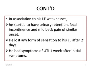
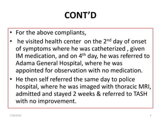
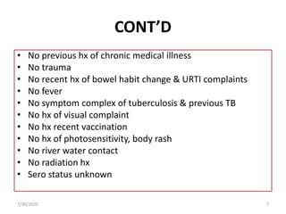
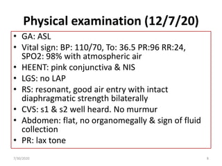
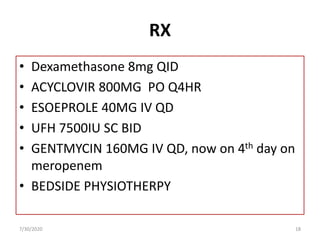
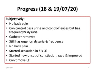
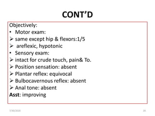
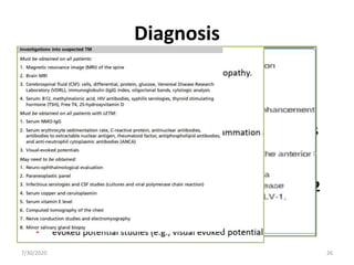
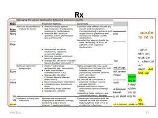
Case presentation on Transverse
Myelitis (TM)
By Dr. Mestet Yibeltal (Neurosurgery
resident)
Moderator: Dr. Teklil ( Consultant
Neurologist)
7/30/2020 1
OUTLINE
• Patient clinical profile
• Investigations
• Diagnosis
• Treatment
• Patient response
• Scientific background of TM
• Reference
7/30/2020 2
• Identification
Id:86035
Age: 20
Sex: M
Occupation: policeman
Residence: Arsi
Ethnicity: Oromo
• Chief complaint: bilateral lower extremity of 1
month duration
7/30/2020 3
HPI
• A 20 years old right handed policeman
 Healthy one month back
Started to have a sudden onset of weakness of
his LT LE while he was walking.
The weakness was more distally that then
progressed and incapacitated him to walk with in
one day.
The day after his LE weakness, he started to have
a right LE weakness and followed the same
progress like his LE.
7/30/2020 4
CONT’D
• In association to his LE weaknesses,
he started to have urinary retention, fecal
incontinence and mid back pain of similar
onset.
He lost any form of sensation to his LE after 2
days.
He had symptoms of UTI 1 week after initial
symptoms.
7/30/2020 5
CONT’D
• For the above compliants,
• he visited health center on the 2nd day of onset
of symptoms where he was catheterized , given
IM medication, and on 4th day, he was referred to
Adama General Hospital, where he was
appointed for observation with no medication.
• He then self referred the same day to police
hospital, where he was imaged with thoracic MRI,
admitted and stayed 2 weeks & referred to TASH
with no improvement.
7/30/2020 6
CONT’D
• No previous hx of chronic medical illness
• No trauma
• No recent hx of bowel habit change & URTI complaints
• No fever
• No symptom complex of tuberculosis & previous TB
• No hx of visual complaint
• No hx recent vaccination
• No hx of photosensitivity, body rash
• No river water contact
• No radiation hx
• Sero status unknown
7/30/2020 7
Physical examination (12/7/20)
• GA: ASL
• Vital sign: BP: 110/70, To: 36.5 PR:96 RR:24,
SPO2: 98% with atmospheric air
• HEENT: pink conjunctiva & NIS
• LGS: no LAP
• RS: resonant, good air entry with intact
diaphragmatic strength bilaterally
• CVS: s1 & s2 well heard. No murmur
• Abdomen: flat, no organomegally & sign of fluid
collection
• PR: lax tone
7/30/2020 8
CONT’D
• GUS: Catheterized, no CVAT, 1400ml/24hrs
UOP
• MSS: no tenderness , deformity
• Integumentary: no rash, pallor, no edema
7/30/2020 9
CONT’D
NS:
• GCS:15/15
• Pupil: mid size & reactive to light bilaterally
• Visual field & acuity: intact
• Eye moves in all direction
• Intact cornel reflex
• Face is symmetric
• Intact gag & swallowing reflex
• Central tongue with active movement
7/30/2020 10
Motor exam
Power Tone
LE RT + LT RT +LT
Dorsiflexors 0/5 Atonic
Plantar flexors 0/5
Knee flexors 0/5
Knee extensors 0/5
Hip adductors 0/5
Hip abductors 0/5
Hip flexors 0/5
Hip extensors 0/5
UE 5/5 2/5
7/30/2020 11
CONT’D
Sensory exam
• Sensory level: at T6/7
• Sweat level: at T6/7
• Position sensation: absent
Reflex:
• Areflexic LE bilaterally & normal reflex at UE
• Plantar reflex: equivocal bilaterally
• Abdominal &bulbocavernous reflex: absent
Meningeal signs: -ve
7/30/2020 12
Localization
• Is it neurologic?
7/30/2020 13
Investigations summary
IX Date
6/11/12 8/11/12
CBC WBC 9.9k 9.3k
neut
lym
Hct 48.2% 46.3%
PLT 295k 295k
ESR 25mm/hr
RFT normal
LFT
electrolyte
PIHCT Non reactive
ANA, RF -ve
7/30/2020 14
MRI
7/30/2020 15
DDX of TM
7/30/2020 16
DX
• PARAPLEGIA 20 LONGITUDINAL EXTENSIVE
TRANSVERSE MYELITIS + SPINAL SHOCK+UTI
7/30/2020 17
RX
• Dexamethasone 8mg QID
• ACYCLOVIR 800MG PO Q4HR
• ESOEPROLE 40MG IV QD
• UFH 7500IU SC BID
• GENTMYCIN 160MG IV QD, now on 4th day on
meropenem
• BEDSIDE PHYSIOTHERPY
7/30/2020 18
Progress (18 & 19/07/20)
Subjectively:
• No back pain
• Can control pass urine and control feaces but has
frequency& dysuria
• Catheter removed
• Still has urgency, dysuria & frequency
• No back pain
• Started sensation in his LE
• Started new onset of constiption, rxed & improved
• Can't move LE
7/30/2020 19
CONT’D
Objectively:
• Motor exam:
 same except hip & flexors:1/5
 areflexic, hypotonic
• Sensory exam:
 intact for crude touch, pain& To.
 Position sensation: absent
 Plantar reflex: equivocal
 Bulbocavernous reflex: absent
 Anal tone: absent
Asst: improving
7/30/2020 20
Transverse myelitis
• is a segmental spinal cord injury caused by
acute inflammation of d/t etiology.
Epidemiology
• 1-4 new cases per million people per year.
• peaks ages 10-19 years and 30-39 years .
• no sex or familial predisposition.
7/30/2020 21
Causes
• Post infectious
• Post vaccination
• multiple sclerosis
• Neuromyelitis optica
• Acute disseminated encephalomyelitis
• Myelopathies Associated with Other
• Connective tissue inflammatory disorders
(SLE,Sjogren’s syndrome, scleroderma, Behcet’s
disease, and sarcoidosis)
• idiopathic
7/30/2020 22
Pathophysiology
Three hypotheses:
1. Cell mediated autoimmune response
2. Autoimmune vasculitis
3. Direct viral invasion of spinal cord.
7/30/2020 23
Clinical presentation
• Variable but typically evolves over hrs or days.
• Combination of
1. Sensory
2. Motor
3. bladder symptoms
• Usually bilateral
• Complete vs Incomplete
• Band like sensation (pressure, pain,
numbness) over the trunk. Why pain?
7/30/2020 24
Course of ATM
• proceeds through three stages:
1. initial motor loss precedes sphincter
dysfunction in most patients, sensory loss
usually over 2 to 3 days
2. plateau phase: mean duration is 1 week
3. recovery phase.
7/30/2020 25
Diagnosis
• Laboratory Aids
• MRI and CSF analysis: two most important
tests in ATM.
• Enhancing spinal cord lesion or pleocytosis
or increased IgG index is required
for the diagnosis.
• If both tests are negative, repeat tests in 2
to 7 days is recommended.
7/30/2020 26
Rx
• First line: high dose iv steroids
(methylprednisolone)
• Second line: If poor response to high-dose steroids
after 5 to 7days use .
Plasma Exchange (PLEX)
Intravenous immunoglobulin
Plasma exchanges
 Rituximab
 Cyclophosphamide.
• Care of the paraplegic patient
7/30/2020 27
Natural History and Prognosis
• The progression of symptoms in ATM
 Often slows within 2 to 3 weeks of onset
 With a corresponding improvement in CSF
and MRI abnormalities
• 1/3 pts have a complete recovery
• 1/3 pts have some residual deficit
• 1/3 pts have no improvement from nadir
• Majority monophasic disease without
recurrence.
7/30/2020 28
Prognosticators
• Unfavorable outcomes
1. Rapid progression to maximal neurologic deficit (<24
hrs)
2. Severe motor weakness
3. Spinal shock
4. Back pain as the initial complaint, and
5. Sensory disturbances at the cervical level
• Better outcome
1. Older age
2. Increased deep tendon reflexes
3. Presence of the babinski sign
7/30/2020 29
Sample references
• Localization in clinical neurology, 5 ed.
• Harrison’s neurology in clinical medicine 2nd ed.
• Pandey S, Garg RK, Malhotra HS, Jain A, Malhotra
KP, Kumar N, Verma R, Sharma PK. Etiologic
spectrum and prognosis in noncompressive acute
transverse myelopathies: An experience of 80
patients at a tertiary care facility. Neurol India
2018;66:65-70
• Tan W, Lim CT. Dengue-related Longitudinally
Extensive Transverse Myelitis. Neurol India
2019;67:1116-7
7/30/2020 30
7/30/2020 31

Case presentation on transverse myelitis

  • 1.
    Case presentation onTransverse Myelitis (TM) By Dr. Mestet Yibeltal (Neurosurgery resident) Moderator: Dr. Teklil ( Consultant Neurologist) 7/30/2020 1
  • 2.
    OUTLINE • Patient clinicalprofile • Investigations • Diagnosis • Treatment • Patient response • Scientific background of TM • Reference 7/30/2020 2
  • 3.
    • Identification Id:86035 Age: 20 Sex:M Occupation: policeman Residence: Arsi Ethnicity: Oromo • Chief complaint: bilateral lower extremity of 1 month duration 7/30/2020 3
  • 4.
    HPI • A 20years old right handed policeman  Healthy one month back Started to have a sudden onset of weakness of his LT LE while he was walking. The weakness was more distally that then progressed and incapacitated him to walk with in one day. The day after his LE weakness, he started to have a right LE weakness and followed the same progress like his LE. 7/30/2020 4
  • 5.
    CONT’D • In associationto his LE weaknesses, he started to have urinary retention, fecal incontinence and mid back pain of similar onset. He lost any form of sensation to his LE after 2 days. He had symptoms of UTI 1 week after initial symptoms. 7/30/2020 5
  • 6.
    CONT’D • For theabove compliants, • he visited health center on the 2nd day of onset of symptoms where he was catheterized , given IM medication, and on 4th day, he was referred to Adama General Hospital, where he was appointed for observation with no medication. • He then self referred the same day to police hospital, where he was imaged with thoracic MRI, admitted and stayed 2 weeks & referred to TASH with no improvement. 7/30/2020 6
  • 7.
    CONT’D • No previoushx of chronic medical illness • No trauma • No recent hx of bowel habit change & URTI complaints • No fever • No symptom complex of tuberculosis & previous TB • No hx of visual complaint • No hx recent vaccination • No hx of photosensitivity, body rash • No river water contact • No radiation hx • Sero status unknown 7/30/2020 7
  • 8.
    Physical examination (12/7/20) •GA: ASL • Vital sign: BP: 110/70, To: 36.5 PR:96 RR:24, SPO2: 98% with atmospheric air • HEENT: pink conjunctiva & NIS • LGS: no LAP • RS: resonant, good air entry with intact diaphragmatic strength bilaterally • CVS: s1 & s2 well heard. No murmur • Abdomen: flat, no organomegally & sign of fluid collection • PR: lax tone 7/30/2020 8
  • 9.
    CONT’D • GUS: Catheterized,no CVAT, 1400ml/24hrs UOP • MSS: no tenderness , deformity • Integumentary: no rash, pallor, no edema 7/30/2020 9
  • 10.
    CONT’D NS: • GCS:15/15 • Pupil:mid size & reactive to light bilaterally • Visual field & acuity: intact • Eye moves in all direction • Intact cornel reflex • Face is symmetric • Intact gag & swallowing reflex • Central tongue with active movement 7/30/2020 10
  • 11.
    Motor exam Power Tone LERT + LT RT +LT Dorsiflexors 0/5 Atonic Plantar flexors 0/5 Knee flexors 0/5 Knee extensors 0/5 Hip adductors 0/5 Hip abductors 0/5 Hip flexors 0/5 Hip extensors 0/5 UE 5/5 2/5 7/30/2020 11
  • 12.
    CONT’D Sensory exam • Sensorylevel: at T6/7 • Sweat level: at T6/7 • Position sensation: absent Reflex: • Areflexic LE bilaterally & normal reflex at UE • Plantar reflex: equivocal bilaterally • Abdominal &bulbocavernous reflex: absent Meningeal signs: -ve 7/30/2020 12
  • 13.
    Localization • Is itneurologic? 7/30/2020 13
  • 14.
    Investigations summary IX Date 6/11/128/11/12 CBC WBC 9.9k 9.3k neut lym Hct 48.2% 46.3% PLT 295k 295k ESR 25mm/hr RFT normal LFT electrolyte PIHCT Non reactive ANA, RF -ve 7/30/2020 14
  • 15.
  • 16.
  • 17.
    DX • PARAPLEGIA 20LONGITUDINAL EXTENSIVE TRANSVERSE MYELITIS + SPINAL SHOCK+UTI 7/30/2020 17
  • 18.
    RX • Dexamethasone 8mgQID • ACYCLOVIR 800MG PO Q4HR • ESOEPROLE 40MG IV QD • UFH 7500IU SC BID • GENTMYCIN 160MG IV QD, now on 4th day on meropenem • BEDSIDE PHYSIOTHERPY 7/30/2020 18
  • 19.
    Progress (18 &19/07/20) Subjectively: • No back pain • Can control pass urine and control feaces but has frequency& dysuria • Catheter removed • Still has urgency, dysuria & frequency • No back pain • Started sensation in his LE • Started new onset of constiption, rxed & improved • Can't move LE 7/30/2020 19
  • 20.
    CONT’D Objectively: • Motor exam: same except hip & flexors:1/5  areflexic, hypotonic • Sensory exam:  intact for crude touch, pain& To.  Position sensation: absent  Plantar reflex: equivocal  Bulbocavernous reflex: absent  Anal tone: absent Asst: improving 7/30/2020 20
  • 21.
    Transverse myelitis • isa segmental spinal cord injury caused by acute inflammation of d/t etiology. Epidemiology • 1-4 new cases per million people per year. • peaks ages 10-19 years and 30-39 years . • no sex or familial predisposition. 7/30/2020 21
  • 22.
    Causes • Post infectious •Post vaccination • multiple sclerosis • Neuromyelitis optica • Acute disseminated encephalomyelitis • Myelopathies Associated with Other • Connective tissue inflammatory disorders (SLE,Sjogren’s syndrome, scleroderma, Behcet’s disease, and sarcoidosis) • idiopathic 7/30/2020 22
  • 23.
    Pathophysiology Three hypotheses: 1. Cellmediated autoimmune response 2. Autoimmune vasculitis 3. Direct viral invasion of spinal cord. 7/30/2020 23
  • 24.
    Clinical presentation • Variablebut typically evolves over hrs or days. • Combination of 1. Sensory 2. Motor 3. bladder symptoms • Usually bilateral • Complete vs Incomplete • Band like sensation (pressure, pain, numbness) over the trunk. Why pain? 7/30/2020 24
  • 25.
    Course of ATM •proceeds through three stages: 1. initial motor loss precedes sphincter dysfunction in most patients, sensory loss usually over 2 to 3 days 2. plateau phase: mean duration is 1 week 3. recovery phase. 7/30/2020 25
  • 26.
    Diagnosis • Laboratory Aids •MRI and CSF analysis: two most important tests in ATM. • Enhancing spinal cord lesion or pleocytosis or increased IgG index is required for the diagnosis. • If both tests are negative, repeat tests in 2 to 7 days is recommended. 7/30/2020 26
  • 27.
    Rx • First line:high dose iv steroids (methylprednisolone) • Second line: If poor response to high-dose steroids after 5 to 7days use . Plasma Exchange (PLEX) Intravenous immunoglobulin Plasma exchanges  Rituximab  Cyclophosphamide. • Care of the paraplegic patient 7/30/2020 27
  • 28.
    Natural History andPrognosis • The progression of symptoms in ATM  Often slows within 2 to 3 weeks of onset  With a corresponding improvement in CSF and MRI abnormalities • 1/3 pts have a complete recovery • 1/3 pts have some residual deficit • 1/3 pts have no improvement from nadir • Majority monophasic disease without recurrence. 7/30/2020 28
  • 29.
    Prognosticators • Unfavorable outcomes 1.Rapid progression to maximal neurologic deficit (<24 hrs) 2. Severe motor weakness 3. Spinal shock 4. Back pain as the initial complaint, and 5. Sensory disturbances at the cervical level • Better outcome 1. Older age 2. Increased deep tendon reflexes 3. Presence of the babinski sign 7/30/2020 29
  • 30.
    Sample references • Localizationin clinical neurology, 5 ed. • Harrison’s neurology in clinical medicine 2nd ed. • Pandey S, Garg RK, Malhotra HS, Jain A, Malhotra KP, Kumar N, Verma R, Sharma PK. Etiologic spectrum and prognosis in noncompressive acute transverse myelopathies: An experience of 80 patients at a tertiary care facility. Neurol India 2018;66:65-70 • Tan W, Lim CT. Dengue-related Longitudinally Extensive Transverse Myelitis. Neurol India 2019;67:1116-7 7/30/2020 30
  • 31.

Editor's Notes

  • #17 Magnetic resonance imaging (MRI) evaluation of the entire spinal cord axis is mandatory in all myelopathic patients. Nb:The classical "owl eye sign" on axial images involving central-anterior cord substance is suggestive of anterior spinal artery syndrome [Figure 1]. Anterior spinal artery syndrome is a rare presentation of acute spinal cord infarction. The etiology is varied ranging from atherosclerosis, vasculitis, surgery, aortic dissection, and acute trauma. It has to be differentiated from spinal multiple sclerosis (MS), the main points of differentiation being the involvement of antero-central cord substance corresponding to the vascular territory of anterior spinal artery and vertical extension over two spinal segments. MS plaques involve peripheral cord substance and seldom extend over two spinal cord segments. [1],[2] Neuromyelitis ophtlmic T1 hypointense follow-up scans may demonstrate cord atrophy and low T1 signal 5 T2 hyperintense (often >3 vertebral body lengths)  central grey matter involvement bright spotty lesions (see above) T1 C+ (Gd) enhancement is common and variable in appearance ring-enhancement seen in a third of patients 17 both on sagittal and axial imaging often ring extends over multiple vertebral levels  patchy "cloud-like" enhancement of the aforementioned T2 bright lesions may be present thin ependymal enhancement similar to ependymitis lens-shaped enhancement on sagittal images 11
  • #18 The presence of spinal shock seems to be prognostic only as it applies to the temporal profile for the mechanism of injury. Spinal cord injury with concomitant spinal shock usually has a worse associated prognosis than does the same degree of spinal cord injury without spinal shock because the injury is inflicted during a shorter period.4, 36
  • #19 Antiviral medications may help those individuals who have a viral infection of the spinal cord.
  • #25  The thoracic region was the most common level of cord damage. Band like sensation (pressure, pain, numbness) over the trunk 2. Bladder symptoms (incontinence, difficulty urinating, retention) Pain in TM may be as a result of 1. neuropathic pain from nerve root inflammation 2. nociceptive pain from dural inflammation 3. muscle spasm from motor dysfunction 4. bladder distension from dysautonomia 5. psychological distress from loss of motor control 6. dysesthesia from demyelination of spinothalamic tract.
  • #27 IgG index = : (CSF IgG + serum IgG) (CSF albumin + serum albumin)