case presentation..
•
0 likes•391 views

A 14-year-old female patient presented with irregularly placed and crowded upper and lower front teeth with poor smile display. Her examination revealed Class I malocclusion with crowding in the maxillary and mandibular anterior segments, retroclined lower incisors, and maxillary midline shift. Treatment objectives were to relieve crowding, procline the retroclined teeth, extrude maxillary incisors to improve smile display, and correct the midline shift through non-extraction therapy over 11 months.

Recommended
More Related Content
What's hot
What's hot (20)
Viewers also liked
Viewers also liked (15)
Similar to case presentation..
Similar to case presentation.. (20)
More from Indian dental academy
More from Indian dental academy (20)
Recently uploaded
Recently uploaded (20)
case presentation..
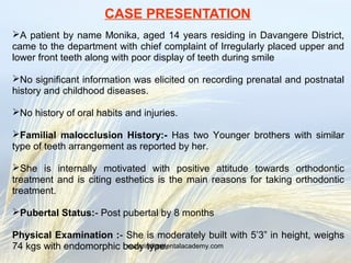
- 1. CASE PRESENTATION A patient by name Monika, aged 14 years residing in Davangere District, came to the department with chief complaint of Irregularly placed upper and lower front teeth along with poor display of teeth during smile No significant information was elicited on recording prenatal and postnatal history and childhood diseases. No history of oral habits and injuries. Familial malocclusion History:- Has two Younger brothers with similar type of teeth arrangement as reported by her. She is internally motivated with positive attitude towards orthodontic treatment and is citing esthetics is the main reasons for taking orthodontic treatment. Pubertal Status:- Post pubertal by 8 months Physical Examination :- She is moderately built with 5’3” in height, weighs 74 kgs with endomorphic body type.www.indiandentalacademy.com
- 2. Extraoral Examination :- Mesocephalic, Brachyfacial, Interlabial gap of 0mm, Decreased Length of nose, Decreased lower anterior face height Cuspid type of smile, Maxillary midline shifted to right by 1mm, Poor display of teeth during smile characterized by 4mm of crown length exposure with no exposure of gingiva Concave profile, anterior divergence, Nasolabial angle of 1030 , Deep mentolabial sulcus , Hyperactive Mentalis , obliterated throat neck angle due to sub mental fat deposition, Decreased length of nose Dished - in appearance of lower anterior face due to prominent chin and excessive deposition of fat in cheeks www.indiandentalacademy.com
- 3. Lower anterior face height is decreased by 4mm www.indiandentalacademy.com
- 4. Functional Examination :- She is found to have Nasal respiration Food Lodgment in maxillary and mandibular anterior segments No abnormality is detected in deglutition. TM Joint appears normal She has 3mm of freeway space and 1mm of incisor crown length exposure during speech and 4mm of crown length exposure during smile. Intraoral Examination Oral Hygiene status is fair , Brushes once daily, Maxillary midline shifted to right by 1mm, Crowding seen in anterior segment with overlapping right maxillary central incisor. Class –I molar relation & canine relation, Retroclined mandibular incisors with crowding 7654321 1234567 7654321 1234567 # Maxillary left central incisor (Ellis class -II ) Teeth Present www.indiandentalacademy.com
- 5. Class I molar and canine relation, # Maxillary central incisor, retrocline mandibular incisors. Maxillary Dental Midline shift to right by 1mm, Crowding in anterior segment with overlapping central incisors, tapered arch form , Normal depth of palatal vault. crowding in anterior segment, retroclined incisors , square arch form. www.indiandentalacademy.com
- 6. RADIOGRAPHIC RECORD Examination of O.P.G. Hand wrist radiograph Closed apex of canines approximate dental age 14 to 15 years PP3u completed, start of MP3u , 8th stage of pubertal growth spurt, approx. age 13.9 www.indiandentalacademy.com
- 7. CEPHALOMETRIC ANALYSIS Soft Tissue Analysis Facial Angle : 990 Nose tip to H-Line : 7 mm Upper Sulcus depth : 4mm Skeletal convexity at point – A : -2mm Upper lip strain : 2mm Upper lip curvature : 4mm Soft Tissue chick Thickness : 15mm Relation to TVL Upper lip anterior : + 3mm Lower lip Anterior : 0mm A Point ‘ : 0mm B Point’ : - -5.5mm Pogonion’ : +1.5mm S Line Upper lip : 0.5mm retrusive Lower LIP : -3mm retrusive Skeletal Relationship of maxilla SNA : 80.50, , Ext.of Max.Base : 46mm N Perp. To Pt. A : +1mm, Eff.Max Lng:87 NA – TH : 900 , N-A (11 HP) –3mm PNS-ANS : 48mm Mandibular Incisors 1 – NB : 1mm 1 – NB : 140 1 – APO Line : 0mm IMPA : 89 0 TVL Md1 : -15mm Max.1 to Mand. 1 - 1360 Relationship of Mandible N Perp. Pog : +5 mm N Pog – TH : 920, SNB : 80.5, Eff.Mand.Lng: 114mm SND : 770 , N-B (11 HP) –3mm,Go-Pg : 72 N-Pg(11HP) 0mm, Ext. of Mand.: 73mm Articular angle : 1370, Saddle Angle 1250 B-Pg : 8.2mm Relationship of Maxilla, and Mandible Go Me – FH : 150 Go Gn – Sn : 250 Gonial angle : 1180 Basal Plane angle : 150 Anterior to Posterior face height ratio : 76% Pn to Occlusal Plane : 800 Dental - Maxillary Incisors 1 – NA : 5mm 1 – NA : 290 1 – Pt A : 5mm 1 – SN : 1050 1 – PP : 660 TVL Mx1 : -11mm Upper 1 – NF : 22mm www.indiandentalacademy.com
- 8. MODEL ANALYSIS Bolton Tooth Ratio Anterior ratio (81%) indicates mandibular excess of 2mm Overall Tooth ratio (93.1%) indicates Mandibular excess of 1.4mm Careys / Arch Perimeter Analysis Crowding in Maxillary arch of 5mm and mandibular arch of 7.2mm Ashley Howe’s Analysis P.M.B.A.W % = 40% , (Border line case ) DIAGNOSIS 14 year old post pubertal female patient by name Monika is diagnosed as a case of skeletal class-I, hypodivergent jaw pattern characterized by anticlockwise rotating mandible,decreased lower anterior face height, prominent soft tissue chin button, crowding in mandibular and maxillary anterior segment with intruded maxillary incisors, Maxillary midline shift to the right by 1mm. DIFFERENTIAL DIAGNOSIS Class – III skeletal base www.indiandentalacademy.com
- 9. Treatment objectives To relieve crowding To procline retroclined mandibular and also maxillary incisors To extrude the intruded maxillary incisors and incisor display To correct maxillary dental midline shifts. To reduce the prominence of chin Muscle exercises to relieve hyperactive mentallis activity Treatment plan : Non-extraction line of treatment. Banding of Ist and IInd molars Continuous arch Mechanics to be employed to proclined and extrude the maxillary incisors, Brackets on anterior teeth to be placed gingivally In mandibular arch proximal stripping to be done in premolars followed by tight lace back and segmental approach followed by continious arch mechanic to procline retroclined lower anteriors. Opening arch wires – 0.014” HANT arch wires, without lace back and bend backs in maxillary arch Reshaping pogonion with myotomy to reduce the prominence of chin button. www.indiandentalacademy.com
- 10. Max1 -PP 61o Mand1 - MP 99o Max1-Mand1 - 124o Nasolabial angle - 96o Treatment duration : > Leveling aligning --> 4+2 months. > Extruding Anteriors --> 2 months > Midline correction --> 1 month. > Finishing and detailing --> 2 months. Total duration -> 11 months. Retention : Removable wraparound retainer in maxillary & mandibular arches along with fixed retainer in mandibular arch 6 months full time wear, 4 months night only, 2 months alternate night, 2 months once a week. Maxillary Mandibular R L R L -2.5 -2.5 -3.5 -3.5 +2.0 +2.0 +3.5 +3.5 +1.5 +1.5 0 0 +1.0 -1.0 0 0 0 0 www.indiandentalacademy.com
- 11. ANTICIPATED TREATMENT CHANGE 0 1 1 0 0 1 0 1 0 1 www.indiandentalacademy.com