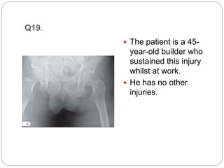
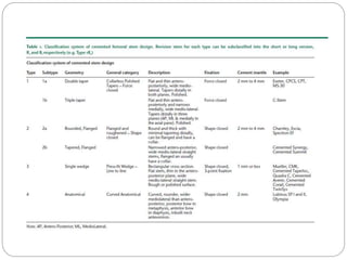
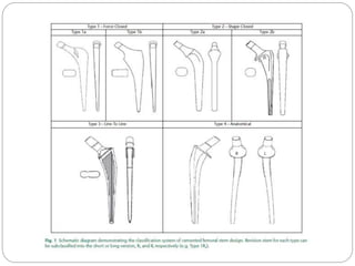
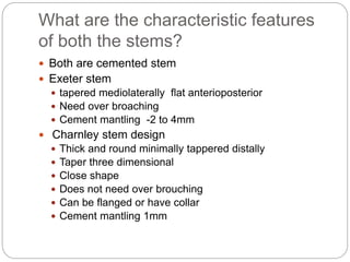
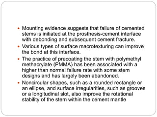
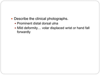
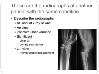
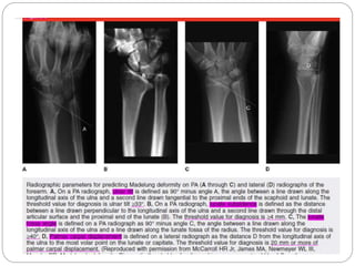
Gashaye T.
7th Case discussion
Q19.
 The patient is a 45-
year-old builder who
sustained this injury
whilst at work.
 He has no other
injuries.
Questions
 Describe what you can see
in these radiographs.
 AP X ray of pelvis
 Left femur neck fracture
 Garden Type IV
 Seems had commutation
What would be your management
plan for treatment of this fracture?
 Internal fixation with
cannulated screw
 Percutaneous or
Open
 Cannulated screw
fixation
 3partially threaded screw
in an inverted triangle
confinguration
 4screw in diamond
configuration
Others
 DHS/cephalomedilary
nail
 Basicervical #
 Additional screw above
sliding hip screw to
prevent rotation
Could you describe the blood supply
to the femoral head?
 The blood supply has three
sources:
 The medial circumflex
femoral artery (MCFA) is the
most important supply; it is a
branch of the profunda
femoris artery.
 The lateral circumflex femoral
artery (LCFA) supplies the
inferior portion; it is a branch
of the profunda femoris.
 The artery of the ligamentum
teres is a minor blood supply
 When consenting this patient for surgery what
particular risks would you warn him about?
 If we are going to do it percutaneous being open
reduction
 Subsequent development of non union and AVN
 REVISION SURGERY THR MAY NEED
 Can you quote the incidence of these complications
and any literature to back this up?
Title: Operative treatment of femoral neck fractures in patients
between the ages of fifteen and fifty years.
 Study Design: Retrospective Review
 83 femoral neck fractures in 82 patients between the ages of 15 and 50 years who were treated by ORIF at the Mayo Clinic
between 1975 and 2000.
 73 fractures were followed to union, until conversion to total hip arthroplasty (THA), or for a minimum of 2 years.
 Average follow-up: 6.6 years (3 months to 23 years).
 Fracture pattern
 51 displaced
 22 nondisplaced
 Evaluated effect of various factors on outcome
 Fracture displacement
 Reduction quality
 Capsular decompression
 Results
 53 fractures (73%) healed with no evidence of osteonecrosis.
 17 fractures (23%) developed osteonecrosis
 Osteonecrosis developed in 14/51 (27%) displaced fractures
 Osteonecrosis developed in 3/22 (14%) nondisplaced fractures
 There was a strong trend for displaced fractures to demonstrate a higher rate of osteonecrosis development, but the difference
was not significant (P = .17)
• 6 fractures (8%) developed nonunion
 4 nonunions later healed following a second procedure
 Nonunion developed in 5/51 (10%) displaced fractures
 Nonunion developed in 1/22 (4.5%) nondisplaced fractures
 Influence of reduction accuracy on outcome
 11/46 (24%) cases with good-to-excellent reduction developed osteonecrosis
 3/5 (60%) cases with fair-to-poor reduction developed osteonecrosis (2 of these also developed a nonunion)
 2/46 (4%) cases with good-to-excellent reduction developed nonunion
 3/5 (60%) cases with fair-to-poor reduction developed osteonecrosis
 Only 1/5 cases with fair to poor reduction healed without complication
 Influence of capsulotomy on outcome
 No statistical effect of capsulotomy on development of osteonecrosis (P = .50)
 14 displaced fractures underwent open reduction, with direct visualization and therefore had a
capsulotomy performed
 4 displaced fractures treated successfully by closed reduction were treated with a capsulotomy
 3 nondisplaced fractures were treated with a capsulotomy, and 1 with aspiration
 4/22 (18%) cases that underwent capsulotomy/decompression developed osteonecrosis
 13/51 (25%) cases without capsulotomy developed osteonecrosis
• At the time of final follow-up, 13 fractures (18%) had undergone conversion to THA
 11 due to osteonecrosis
 1 due to nonunion
 1 due to osteonecrosis and nonunion
 Average time from injury to THA was 7.3 years (3 months to 15 years)
 Conclusions
 The 10-year hip survival rate following femoral neck fracture in young patients treated by
ORIF was 85%.
 Osteonecrosis was the main reason for conversion to THA.
 Not all patients with osteonecrosis required further surgery.
 Initial fracture displacement and quality of reduction influenced the outcome.
Q20. not clear question
 This is a photograph
of two types of
femoral stem used in
a total hip
replacement
 Can be cementless
and cemented or
 Modular and non
modular design
respectively.
Questions
What are they called?
 Exeter stem design
and charnley stem
design or
 taper slip / force
closed and shape
closed/composite
beam
What are the characteristic features
of both the stems?
 Both are cemented stem
 Exeter stem
 tapered mediolaterally flat anterioposterior
 Need over broaching
 Cement mantling -2 to 4mm
 Charnley stem design
 Thick and round minimally tappered distally
 Taper three dimensional
 Close shape
 Does not need over brouching
 Can be flanged or have collar
 Cement mantling 1mm
 Mounting evidence suggests that failure of cemented
stems is initiated at the prosthesis-cement interface
with debonding and subsequent cement fracture.
 Various types of surface macrotexturing can improve
the bond at this interface.
 The practice of precoating the stem with polymethyl
methacrylate (PMMA) has been associated with a
higher than normal failure rate with some stem
designs and has largely been abandoned.
 Noncircular shapes, such as a rounded rectangle or
an ellipse, and surface irregularities, such as grooves
or a longitudinal slot, also improve the rotational
stability of the stem within the cement mantle
 There is concern that even with surface modifications the
stem may not remain bonded to the cement.
 If debonding does occur, a stem with a roughened or
textured surface generates more debris with motion than a
stem with a smooth, polished surface.
 Higher rates of loosening and bone resorption were found
with the use of an Exeter stem with a matter surface than
with an identical stem with a polished surface
 Similar findings have been reported when comparing the
original polished Charnley stem with its subsequent
mattefinish modification. For this reason, interest has been
renewed in the use of polished stems for cemented
applications.
 Ling recommended a design that is collarless, polished,
and tapered in two planes to allow a small amount of
subsidence and to maintain compressive stresses within
the cement mantle.
Which material are they composed of
and why?
 A femoral stem most
commonly composed of
 titanium
 ceramic
 cobalt chromium
(CoCr)or stainless-steel
alloy with insertion in
interference fit or with
PMMA as means of
fixation connected to a
modular femoral head
of Co Cr alloy or
ceramic
 Cobalt chromium
advantage
 Stronger than titanium
 cobalt disadvantage
 More bone resorbtion
proximally
 Toxic to body
 Titanium advantage
 Elastic modulus near to
bone
 Better bone ingrowth
 Better tolerated at
cellular level
 Titanium disadvantage
 Increased risk of
fracture
 Notch sensitivity
Why?
 material composition need to be
 resist cyclic loading in a demanding environment
 well tolerated by the body /inert
 needs to be corrosion resistant
 have adequate material strength
 inexpensive to manufacture
 available in large quantities
What is the engineering principle behind the success of
each of these stems? Please describe with an
illustration.
 The good long-term outcomes for the flat-back Charnley
and the polished Exeter stems can be attributed to the
taper slip principle
 Taper-slip principle
 depends on shortening or subsidence in order to obtain and
maintain a tight fit
 the taper should be polished and that the cement should allow
some subsidence.
 The system is then held together by the resultants from
axial forces and the greater the load, the tighter is the fit of
the taper
 When there is subsidence, radial compressive forces are
created in the adjacent cement, and transferred to bone as
hoop stress
Cont.
 The polished double
tapered Exeter stem
creates radial
compressive loading
as the predominant
force, unlike a non-
polished surface non
tapered stem, which
creates greater shear
force as stem
migrates distally
What are their long-term clinical
results?
 The long term survival of hip implants depends on
how well the implant becomes fixed from the
outset of the procedure.
 Two methods are now routinely used to attain
initial implant fixation:
 cementing the implant in the bone using
polymethylmethacryV late(PMMA)
 press fitting the implant into the prepared bone site
with subsequent bone ingrowth into the implant
porosity
Long term Out come
 Overall, long-term results of Type 1 /exeter stems
are excellent.
 proven to be reliable in both young and old
patients, and for multiple pathologies
 high incidence of early per prosthetic fracture than
other may act as a wedge, splitting the femur
following a fall.
 Aseptic loosening 0.6%
 Overall long-term results of Type 2 /Charnley
stems are excellent and variable with generation
and design
 exhibiting higher revision rates compared to others
at the 10 to 15-year follow up
 6% rate of aseptic loosening
Q21.
 10-year-old girl
presented to the
outpatients clinic with a
progressively worsening
deformity of her wrist
joint.
 There was no history of
trauma or of recent
infection.
 These are her clinical
photographs
 Describe the clinical photographs.
 Prominent distal dorsal ulna
 Mild deformity… volar displaced wrist or hand fall
forwardly
 What is the diagnosis?
 Madlung deformity
 Female
 Adolescent
 Spontaneous deformity
and what are the classical clinical findings in
these patients?
 absence or underdevelopment of ulnar and volar
portion of growth plate of the radius
 articular surface directed ulnar ward and volar ward
 uninvolved radial and dorsal portions of the physis
continue to grow
 faster growing, newly formed bone bends toward area
of slower growth, causing the articular surface of distal
radius to slant in palmar and ulnar direction
Cont.
 ulna is unaffected and remains in its usual dorsal
position
 Clinical feature
 Wrist deformity- prominent dorsal ulna
 pain from radioulnar subluxation or radiolunate
impingement (insidious onset )
 Worse at activity
 usually becomes less severe at maturity
 wrist motion
 extension and supination, is limited
These are the radiographs of another
patient with the same condition
 Describe the radiographs
 AP and lat x ray of wrist
 Ap view
 Posetive ulnar variance
 Significant
 ulnar tilt
 Lunate subsidence
 Lat view
 Palmar carpal displacement
McCarron et al
 4 radiographic parameters for early MD
 Ulnar tilt >330
 Lunate subsidence > 4mm
 Lunate fossa angle > 400
 Palmar carpal displacement > 20mm
 Controversy
 Symptoms are mild & the condition is often self
limiting
 Symptoms are significant and prolonged in to
adulthood
 Deformity and cosmetic issue
 Conservative
• If mild or intermittent pain in skeletally mature patient
• She is young progressive deformity expected
• Need follow-up in the mean time surgery
surgery
 Goals for surgical
 primarily of pain relief and correction of the
cosmetic deformity.
 A secondary goal is to increase range of motion
 decision is based on the following four factors:
 Patient's age and the growth remaining in the distal
radius
 Severity of the deformity
 Severity of the symptoms
 Clinical and radiographic findings
 Three broad categories
 Procedures that correct primary deformity of
radius
 Change the growth or anatomy of the physis
 Change the bony anatomy of metaphysis
 Salvage procedures
 Procedures that attempt to decrease pain and
increase ROM by making compensatory change
to ulna
 Procedures that address both
 Darrach procedure – ulnar resection arthroplasty
 Sauve Kapandji procedure
 DRUJ arthrodesis + distal ulna osteotomy creating
pseudoarthrosis
Combine radial and ulnar osteotomies
 Dorsolateral closing wedge osteotomy of radius
 Ulnar osteotomy +/- DRUJ arthrodesis
 Ligamentous release
and dome osteotomy
 Apex distal or
proximal Dome
 Dome in both
directions
 Radial to ulnar and
volar to dorsal
 Rotation reorients
lunate facet in three
dimension
 From anterior ulnar
stance to dorsal ulnar
position
 Distal – radial and dorsal translation
 Proximal – volar and ulnar
 Pins or plate fixation
 Ronguer to limit cortical prominence of proximal
fragment
Others
 Physiolysis with release of Vickers ligament
 wrist pain or decreased range of motion
 efficacy of prophylactic release of Vickers ligament
in mild deformity in skeletally immature patients
unknown
 DRUJ arthroplasty and arthrodesis
 highly controversial
 painful DRUJ instability and limited
supination/pronation
 significant deformity may require staged procedures
Reference
 Campbell's operative orthopedics
 orthobullet
 Rapid_reference_review_in_orthopedic_trauma_pivot
al_papers_revealed
 Current concepts and outcomes in cemented femoral
stem design and cementation techniques: the
argument for a new classification system
 Exeter total hip system
 FEMORAL STEM FIXATION An engineering
interpretation of the long-term outcome of Charnley
and Exeter stems
 Madelung Deformity Andrew C. Ghatan, MD Douglas
P. Hanel, MD
 Thank you!

Case discussion 7

  • 1.
  • 2.
    Q19.  The patientis a 45- year-old builder who sustained this injury whilst at work.  He has no other injuries.
  • 3.
    Questions  Describe whatyou can see in these radiographs.  AP X ray of pelvis  Left femur neck fracture  Garden Type IV  Seems had commutation
  • 4.
    What would beyour management plan for treatment of this fracture?  Internal fixation with cannulated screw  Percutaneous or Open  Cannulated screw fixation  3partially threaded screw in an inverted triangle confinguration  4screw in diamond configuration
  • 5.
    Others  DHS/cephalomedilary nail  Basicervical#  Additional screw above sliding hip screw to prevent rotation
  • 6.
    Could you describethe blood supply to the femoral head?  The blood supply has three sources:  The medial circumflex femoral artery (MCFA) is the most important supply; it is a branch of the profunda femoris artery.  The lateral circumflex femoral artery (LCFA) supplies the inferior portion; it is a branch of the profunda femoris.  The artery of the ligamentum teres is a minor blood supply
  • 7.
     When consentingthis patient for surgery what particular risks would you warn him about?  If we are going to do it percutaneous being open reduction  Subsequent development of non union and AVN  REVISION SURGERY THR MAY NEED
  • 8.
     Can youquote the incidence of these complications and any literature to back this up?
  • 9.
    Title: Operative treatmentof femoral neck fractures in patients between the ages of fifteen and fifty years.  Study Design: Retrospective Review  83 femoral neck fractures in 82 patients between the ages of 15 and 50 years who were treated by ORIF at the Mayo Clinic between 1975 and 2000.  73 fractures were followed to union, until conversion to total hip arthroplasty (THA), or for a minimum of 2 years.  Average follow-up: 6.6 years (3 months to 23 years).  Fracture pattern  51 displaced  22 nondisplaced  Evaluated effect of various factors on outcome  Fracture displacement  Reduction quality  Capsular decompression  Results  53 fractures (73%) healed with no evidence of osteonecrosis.  17 fractures (23%) developed osteonecrosis  Osteonecrosis developed in 14/51 (27%) displaced fractures  Osteonecrosis developed in 3/22 (14%) nondisplaced fractures  There was a strong trend for displaced fractures to demonstrate a higher rate of osteonecrosis development, but the difference was not significant (P = .17) • 6 fractures (8%) developed nonunion  4 nonunions later healed following a second procedure  Nonunion developed in 5/51 (10%) displaced fractures  Nonunion developed in 1/22 (4.5%) nondisplaced fractures  Influence of reduction accuracy on outcome  11/46 (24%) cases with good-to-excellent reduction developed osteonecrosis  3/5 (60%) cases with fair-to-poor reduction developed osteonecrosis (2 of these also developed a nonunion)  2/46 (4%) cases with good-to-excellent reduction developed nonunion  3/5 (60%) cases with fair-to-poor reduction developed osteonecrosis  Only 1/5 cases with fair to poor reduction healed without complication
  • 10.
     Influence ofcapsulotomy on outcome  No statistical effect of capsulotomy on development of osteonecrosis (P = .50)  14 displaced fractures underwent open reduction, with direct visualization and therefore had a capsulotomy performed  4 displaced fractures treated successfully by closed reduction were treated with a capsulotomy  3 nondisplaced fractures were treated with a capsulotomy, and 1 with aspiration  4/22 (18%) cases that underwent capsulotomy/decompression developed osteonecrosis  13/51 (25%) cases without capsulotomy developed osteonecrosis • At the time of final follow-up, 13 fractures (18%) had undergone conversion to THA  11 due to osteonecrosis  1 due to nonunion  1 due to osteonecrosis and nonunion  Average time from injury to THA was 7.3 years (3 months to 15 years)  Conclusions  The 10-year hip survival rate following femoral neck fracture in young patients treated by ORIF was 85%.  Osteonecrosis was the main reason for conversion to THA.  Not all patients with osteonecrosis required further surgery.  Initial fracture displacement and quality of reduction influenced the outcome.
  • 11.
    Q20. not clearquestion  This is a photograph of two types of femoral stem used in a total hip replacement  Can be cementless and cemented or  Modular and non modular design respectively.
  • 12.
    Questions What are theycalled?  Exeter stem design and charnley stem design or  taper slip / force closed and shape closed/composite beam
  • 15.
    What are thecharacteristic features of both the stems?  Both are cemented stem  Exeter stem  tapered mediolaterally flat anterioposterior  Need over broaching  Cement mantling -2 to 4mm  Charnley stem design  Thick and round minimally tappered distally  Taper three dimensional  Close shape  Does not need over brouching  Can be flanged or have collar  Cement mantling 1mm
  • 16.
     Mounting evidencesuggests that failure of cemented stems is initiated at the prosthesis-cement interface with debonding and subsequent cement fracture.  Various types of surface macrotexturing can improve the bond at this interface.  The practice of precoating the stem with polymethyl methacrylate (PMMA) has been associated with a higher than normal failure rate with some stem designs and has largely been abandoned.  Noncircular shapes, such as a rounded rectangle or an ellipse, and surface irregularities, such as grooves or a longitudinal slot, also improve the rotational stability of the stem within the cement mantle
  • 17.
     There isconcern that even with surface modifications the stem may not remain bonded to the cement.  If debonding does occur, a stem with a roughened or textured surface generates more debris with motion than a stem with a smooth, polished surface.  Higher rates of loosening and bone resorption were found with the use of an Exeter stem with a matter surface than with an identical stem with a polished surface  Similar findings have been reported when comparing the original polished Charnley stem with its subsequent mattefinish modification. For this reason, interest has been renewed in the use of polished stems for cemented applications.  Ling recommended a design that is collarless, polished, and tapered in two planes to allow a small amount of subsidence and to maintain compressive stresses within the cement mantle.
  • 21.
    Which material arethey composed of and why?  A femoral stem most commonly composed of  titanium  ceramic  cobalt chromium (CoCr)or stainless-steel alloy with insertion in interference fit or with PMMA as means of fixation connected to a modular femoral head of Co Cr alloy or ceramic  Cobalt chromium advantage  Stronger than titanium  cobalt disadvantage  More bone resorbtion proximally  Toxic to body  Titanium advantage  Elastic modulus near to bone  Better bone ingrowth  Better tolerated at cellular level  Titanium disadvantage  Increased risk of fracture  Notch sensitivity
  • 22.
    Why?  material compositionneed to be  resist cyclic loading in a demanding environment  well tolerated by the body /inert  needs to be corrosion resistant  have adequate material strength  inexpensive to manufacture  available in large quantities
  • 23.
    What is theengineering principle behind the success of each of these stems? Please describe with an illustration.  The good long-term outcomes for the flat-back Charnley and the polished Exeter stems can be attributed to the taper slip principle  Taper-slip principle  depends on shortening or subsidence in order to obtain and maintain a tight fit  the taper should be polished and that the cement should allow some subsidence.  The system is then held together by the resultants from axial forces and the greater the load, the tighter is the fit of the taper  When there is subsidence, radial compressive forces are created in the adjacent cement, and transferred to bone as hoop stress
  • 24.
    Cont.  The polisheddouble tapered Exeter stem creates radial compressive loading as the predominant force, unlike a non- polished surface non tapered stem, which creates greater shear force as stem migrates distally
  • 25.
    What are theirlong-term clinical results?  The long term survival of hip implants depends on how well the implant becomes fixed from the outset of the procedure.  Two methods are now routinely used to attain initial implant fixation:  cementing the implant in the bone using polymethylmethacryV late(PMMA)  press fitting the implant into the prepared bone site with subsequent bone ingrowth into the implant porosity
  • 26.
    Long term Outcome  Overall, long-term results of Type 1 /exeter stems are excellent.  proven to be reliable in both young and old patients, and for multiple pathologies  high incidence of early per prosthetic fracture than other may act as a wedge, splitting the femur following a fall.  Aseptic loosening 0.6%  Overall long-term results of Type 2 /Charnley stems are excellent and variable with generation and design  exhibiting higher revision rates compared to others at the 10 to 15-year follow up  6% rate of aseptic loosening
  • 27.
    Q21.  10-year-old girl presentedto the outpatients clinic with a progressively worsening deformity of her wrist joint.  There was no history of trauma or of recent infection.  These are her clinical photographs
  • 28.
     Describe theclinical photographs.  Prominent distal dorsal ulna  Mild deformity… volar displaced wrist or hand fall forwardly
  • 29.
     What isthe diagnosis?  Madlung deformity  Female  Adolescent  Spontaneous deformity
  • 30.
    and what arethe classical clinical findings in these patients?  absence or underdevelopment of ulnar and volar portion of growth plate of the radius  articular surface directed ulnar ward and volar ward  uninvolved radial and dorsal portions of the physis continue to grow  faster growing, newly formed bone bends toward area of slower growth, causing the articular surface of distal radius to slant in palmar and ulnar direction
  • 31.
    Cont.  ulna isunaffected and remains in its usual dorsal position  Clinical feature  Wrist deformity- prominent dorsal ulna  pain from radioulnar subluxation or radiolunate impingement (insidious onset )  Worse at activity  usually becomes less severe at maturity  wrist motion  extension and supination, is limited
  • 32.
    These are theradiographs of another patient with the same condition  Describe the radiographs  AP and lat x ray of wrist  Ap view  Posetive ulnar variance  Significant  ulnar tilt  Lunate subsidence  Lat view  Palmar carpal displacement
  • 34.
    McCarron et al 4 radiographic parameters for early MD  Ulnar tilt >330  Lunate subsidence > 4mm  Lunate fossa angle > 400  Palmar carpal displacement > 20mm
  • 35.
     Controversy  Symptomsare mild & the condition is often self limiting  Symptoms are significant and prolonged in to adulthood  Deformity and cosmetic issue
  • 36.
     Conservative • Ifmild or intermittent pain in skeletally mature patient • She is young progressive deformity expected • Need follow-up in the mean time surgery
  • 37.
    surgery  Goals forsurgical  primarily of pain relief and correction of the cosmetic deformity.  A secondary goal is to increase range of motion  decision is based on the following four factors:  Patient's age and the growth remaining in the distal radius  Severity of the deformity  Severity of the symptoms  Clinical and radiographic findings
  • 38.
     Three broadcategories  Procedures that correct primary deformity of radius  Change the growth or anatomy of the physis  Change the bony anatomy of metaphysis  Salvage procedures  Procedures that attempt to decrease pain and increase ROM by making compensatory change to ulna  Procedures that address both
  • 39.
     Darrach procedure– ulnar resection arthroplasty
  • 40.
     Sauve Kapandjiprocedure  DRUJ arthrodesis + distal ulna osteotomy creating pseudoarthrosis
  • 41.
    Combine radial andulnar osteotomies  Dorsolateral closing wedge osteotomy of radius  Ulnar osteotomy +/- DRUJ arthrodesis
  • 42.
     Ligamentous release anddome osteotomy  Apex distal or proximal Dome  Dome in both directions  Radial to ulnar and volar to dorsal  Rotation reorients lunate facet in three dimension  From anterior ulnar stance to dorsal ulnar position
  • 43.
     Distal –radial and dorsal translation  Proximal – volar and ulnar  Pins or plate fixation  Ronguer to limit cortical prominence of proximal fragment
  • 44.
    Others  Physiolysis withrelease of Vickers ligament  wrist pain or decreased range of motion  efficacy of prophylactic release of Vickers ligament in mild deformity in skeletally immature patients unknown  DRUJ arthroplasty and arthrodesis  highly controversial  painful DRUJ instability and limited supination/pronation  significant deformity may require staged procedures
  • 45.
    Reference  Campbell's operativeorthopedics  orthobullet  Rapid_reference_review_in_orthopedic_trauma_pivot al_papers_revealed  Current concepts and outcomes in cemented femoral stem design and cementation techniques: the argument for a new classification system  Exeter total hip system  FEMORAL STEM FIXATION An engineering interpretation of the long-term outcome of Charnley and Exeter stems  Madelung Deformity Andrew C. Ghatan, MD Douglas P. Hanel, MD
  • 46.

Editor's Notes

  • #12 Collarless ,polished and coatted
  • #13 Cemented stems have classically been classified into two broad categories, taper slip or force closed, and composite beams or shaped closed designs. While these simplifications are acceptable general categories, they miss important design features, have different broaching techniques and make comparisons misleading. With the evolution of cemented implants, the introduction of newer implants which have hybrid properties, and the use of different broaching techniques, the classification of these implants into these simple categories becomes increasingly difficult. A more comprehensive classification system would aid in comparison of results and better understanding of the implants biomechanics. We propose the following classification system. Cemented stems can be classified according to their geometry, broaching technique, and biomechanics. We define four general types based on shape, broaching technique used and biomechanics, with all four categories having a revision version (Table 1 and Fig. 1). The revision stem can be subclassified into long and short versions of the primary stem. In this classification Type 1 and Type 2 stems use traditional broaching techniques which allow for a cement mantle of 2 mm or more. Type 3 implants use a line-to-line broaching technique often referred as the ‘French paradox’ with a cement mantle of 1 mm or less. Type 4 are anatomical stems and have mixed features when compared to the other types and have a consistent cement mantle of 2 mm along the length of the stem. While future prosthesis may not fit into one of these categories, this classification system represents the great majority of the cemented stems currently in use and with long-term follow up.
  • #24 ‘taper slip principle’ ie. the design and its surface finish allow subsidence to occur
  • #25 Roentegen stereophotogrammetric analysis (RSA) has demonstrated distal stem migration at the cementimplant interface with the polished Exeter stem. The nonpolished design migrated not only at the cement-implant interface, but also at the cement-bone interface. Migration at the cement-bone interface may interfere with Fixation RSA techniques have also demonstrated significant differences in rapid posterior head migration of the polished Exeter compared to that of a non-polished design. The subsidence of the polished, collarless, tapered stem within the cement mantle compresses the interfaces and renders them more able to resist shear forces generated by the posteriorly directed loads on the femoral head. Polished, collarless, tapered stems are more forgiving than conventional designs