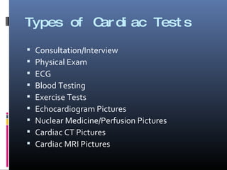
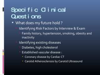
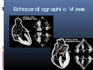
Robert E. Henson II, MD RM Cardiovascular Associates, PC Feb, 2009
Questions to be Answered What does the future hold? What is bothering me now? How is the course of my disease progressing?
Types of Cardiac Tests Consultation/Interview Physical Exam ECG Blood Testing Exercise Tests Echocardiogram Pictures Nuclear Medicine/Perfusion Pictures Cardiac CT Pictures Cardiac MRI Pictures
Characteristics of Testing True Positives  /  False Positives True Negatives  /  False Negatives
Specific Clinical Questions What does my future hold ? Identifying Risk Factors by Interview & Exam Family history, hypertension, smoking, obesity and inactivity Identifying existing diseases Diabetes, high cholesterol Established vascular disease : Coronary disease by Cardiac CT Carotid Atherosclerosis by Carotid Ultrasound
Specific Clinical Questions What is bothering me ? Stress Testing to screen for Obstructive Coronary Disease Chest pain/ shortness of breath Echo/Cardiac MR to evaluate for structural Heart Disease Shortness of breath, palpitations, murmurs ECG monitoring to look for arrythmias Palpitations, lightheadedness, fainting spells
Specific Imaging Tests
Echocardiographic Views
Nuclear Medicine Tests
 
Cardiac CT
Diseased vs Normal
Mollet, N. R. et al. J Am Coll Cardiol 2004;43:2265-2270 Volume-rendered multislice spiral computed tomography images (left) and corresponding conventional angiography images (right) of the left anterior descending (LAD) (A), circumflex (C), and right coronary artery (D) in a 69-year-old woman with stable angina and a positive bicycle test
3D Reconstruction of the Heart
Severe LAD Calcification
CABG
Raff, G. L. et al. J Am Coll Cardiol 2005;46:552-557 Visualization of soft and hard coronary atherosclerotic plaques by 64-slice multislice computed tomography (MSCT)
3D Reconstruction: s/p CABG
Cardiac MRI
Cardiac MR Comparable Views * SAX: 1 echo, 9 CMR views * “ Parasternal Long Axis” Short Axis 4 Chamber 2 Chamber
CE-CMR vs SPECT for Viability Wagner, et al.  Lancet 2003
Where to go for Information Heart Hub for Patients :  www.hearthub.org CardioSmart for Patients:  www.cardiosmart.org
Questions
Mollet, N. R. et al. J Am Coll Cardiol 2004;43:2265-2270 Colored image = volume rendered multislice spiral computed tomography (MSCT) image providing an overview of the anatomy of the main coronary arteries

Cardiac Testing for the Layman

  • 1.
    Robert E. HensonII, MD RM Cardiovascular Associates, PC Feb, 2009
  • 2.
    Questions to beAnswered What does the future hold? What is bothering me now? How is the course of my disease progressing?
  • 3.
    Types of CardiacTests Consultation/Interview Physical Exam ECG Blood Testing Exercise Tests Echocardiogram Pictures Nuclear Medicine/Perfusion Pictures Cardiac CT Pictures Cardiac MRI Pictures
  • 4.
    Characteristics of TestingTrue Positives / False Positives True Negatives / False Negatives
  • 5.
    Specific Clinical QuestionsWhat does my future hold ? Identifying Risk Factors by Interview & Exam Family history, hypertension, smoking, obesity and inactivity Identifying existing diseases Diabetes, high cholesterol Established vascular disease : Coronary disease by Cardiac CT Carotid Atherosclerosis by Carotid Ultrasound
  • 6.
    Specific Clinical QuestionsWhat is bothering me ? Stress Testing to screen for Obstructive Coronary Disease Chest pain/ shortness of breath Echo/Cardiac MR to evaluate for structural Heart Disease Shortness of breath, palpitations, murmurs ECG monitoring to look for arrythmias Palpitations, lightheadedness, fainting spells
  • 7.
  • 8.
  • 9.
  • 10.
  • 11.
  • 12.
  • 13.
  • 14.
    Mollet, N. R.et al. J Am Coll Cardiol 2004;43:2265-2270 Volume-rendered multislice spiral computed tomography images (left) and corresponding conventional angiography images (right) of the left anterior descending (LAD) (A), circumflex (C), and right coronary artery (D) in a 69-year-old woman with stable angina and a positive bicycle test
  • 15.
  • 16.
  • 17.
  • 18.
    Raff, G. L.et al. J Am Coll Cardiol 2005;46:552-557 Visualization of soft and hard coronary atherosclerotic plaques by 64-slice multislice computed tomography (MSCT)
  • 19.
  • 20.
  • 21.
    Cardiac MR ComparableViews * SAX: 1 echo, 9 CMR views * “ Parasternal Long Axis” Short Axis 4 Chamber 2 Chamber
  • 22.
    CE-CMR vs SPECTfor Viability Wagner, et al. Lancet 2003
  • 23.
    CE-CMR vs SPECTfor Viability Wagner, et al. Lancet 2003
  • 24.
    Where to gofor Information Heart Hub for Patients : www.hearthub.org CardioSmart for Patients: www.cardiosmart.org
  • 25.
  • 26.
    Mollet, N. R.et al. J Am Coll Cardiol 2004;43:2265-2270 Colored image = volume rendered multislice spiral computed tomography (MSCT) image providing an overview of the anatomy of the main coronary arteries