
Recommended
More Related Content
What's hot
What's hot (20)
Similar to CNS PERCUSSION & AUSCULTATION GUIDE
Similar to CNS PERCUSSION & AUSCULTATION GUIDE (20)
Recently uploaded
Recently uploaded (20)
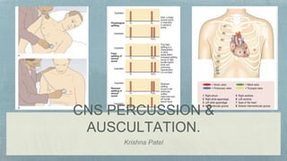
CNS PERCUSSION & AUSCULTATION GUIDE
- 2. PERCUSSION Determination of Heart Border Right heart border: Percuss from above downward in midclavicular line up to the liver dullness Start percussing one space above the liver dullness , from the right midclavicular line to the sternum keeping the pleximeter finger parallel to the sternal edge Repeat this in two more consecutive spaces above.
- 4. • Left heart border: • Palpate the apex • In the same ICS go to the mid axillary line and start percussing medially. • Direction of percussion should be parallel to the apparent left heart border.
- 6. For aortic area: Start percussing parallel to the right sternal edge and percuss laterally. For pulmonary area: Start percussing parallel to the left sternal edge and percuss laterally. Normally it is resonant.
- 9. Each heartbeat is approximately .8 seconds in which .5 second is a diastolic and .3 second is a systole.
- 10. The first heart sound (S1), ‘lub’, is caused by closure of the mitral and tricuspid valves at the onset of ventricular systole. It is best heard at the apex. In mitral stenosis the intensity of S1 is increased due to elevated left atrial pressure.
- 11. The second heart sound (S2), ‘dub’, is caused by closure of the pulmonary and aortic valves at the end of ventricular systole and is best heard at the left sternal edge. It is louder and higher-pitched than the S1 ‘lub’, and the aortic component is normally louder than the pulmonary component. Physiological splitting of S2 occurs because left ventricular contraction slightly precedes that of the right ventricle so that the aortic valve closes before the pulmonary valve.
- 12. This splitting increases at end-inspiration because increased venous filling of the right ventricle further delays pulmonary valve closure. The separation disappears on expiration . On auscultation, ‘lub d- dub’ (inspiration) ‘lub-dub’ (expiration) is heard. The aortic component of S2 is sometimes quiet or absent in calcific aortic stenosis and reduced in aortic regurgitation . The aortic component of S2 is loud in systemic hypertension, and the pulmonary component is increased in pulmonary hypertension.
- 13. Wide splitting of S2, but with normal respiratory variation, occurs in conditions that delay right ventricular emptying, such as right bundle branch block or pulmonary hypertension. Fixed splitting of S2, with no variation with respiration, is a feature of atrial septal defect . In this condition the right ventricular stroke volume is larger than the left, and the splitting is fixed because the defect equalises the pressure between the two atria throughout the respiratory cycle.
- 16. The third heart sound (S3) is a low-pitched early diastolic sound best heard with the bell at the apex. It coincides with rapid ventricular filling immediately after opening of the atrioventricular valves and is therefore heard after the second heart sound as ‘lub-dub-dum’. It is a normal physiological finding in children, young adults and febrile patients, and during pregnancy, but is usually pathological after the age of 40 years.
- 17. • The most common causes are left ventricular failure, when it is an early sign, and mitral regurgitation, due to volume loading of the ventricle. • In heart failure, S3 occurs with a tachycardia, referred to as a ‘gallop’ rhythm, and S1 and S2 are quiet (lub-da-dub).
- 18. The fourth heart sound (S4) is less common. It is soft and low- pitched, best heard with the bell at the apex. It occurs just before S1 (da-lub-dub). It is always pathological and is caused by forceful atrial contraction against a non-compliant or stiff ventricle. A S4 is most often heard with left ventricular hypertrophy (due to hypertension, aortic stenosis or hypertrophic cardiomyopathy). It cannot occur when there is atrial fibrillation.
- 19. Opening snap An opening snap is commonly heard in mitral (rarely, tricuspid) stenosis. It results from sudden opening of a stenosed valve and occurs early in diastole, just after the S2 . It is best heard with the diaphragm at the apex. Added sounds.
- 20. Ejection clicks are high-pitched sounds best heard with the diaphragm. They occur early in systole just after the S1, in patients with congenital pulmonary or aortic stenosis . The mechanism is similar to that of an opening snap. Ejection clicks do not occur in calcific aortic stenosis because the cusps are rigid.
- 21. Mid-systolic clicks are high-pitched and best heard at the apex with the diaphragm. They occur in mitral valve prolapse and may be associated with a late systolic murmur.
- 22. Pericardial rub (friction rub) is a coarse scratching sound, often with systolic and diastolic components. It is best heard using the diaphragm with the patient holding their breath in expiration. It may be audible over any part of the precordium but is often localised, varying in intensity over time and with the position of the patient. It is most often heard in acute pericarditis or a few days after an extensive myocardial infarction. A pleuropericardial rub is a similar sound that occurs in time with the cardiac cycle, but is also influenced by respiration and is pleural in origin. Occasionally, a ‘crunching’ noise can be heard, caused by gas in the pericardium (pneumopericardium).
- 23. Heart murmurs are produced by turbulent flow across an abnormal valve, septal defect or outflow obstruction. ‘Innocent’ murmurs are caused by increased velocity of flow through a normal valve and occur when stroke volume is increased, as in pregnant women, athletes with resting bradycardia or patients with fever. 1. Timing 2. Duration 3. Character & pitch 4. Intensity 5. Location 6. Radiation
- 24. Ejection systolic murmurs are caused by increased flow through a normal valve (flow or innocent murmur), or by turbulent flow through an abnormal valve, as in aortic or pulmonary stenosis.
- 26. Continuous murmurs are rare in adults. The most common cause is a patent ductus arteriosus. In the fetus this connects the upper descending aorta and pulmonary artery, and normally closes just after birth. The murmur is best heard at the upper left sternal border and radiates over the left scapula. Its continuous character is ‘machinery-like’; as aortic pressure always exceeds pulmonary pressure, there is continuous ductal flow, with the greatest pressure difference in systole, resulting in a louder systolic component
- 27. The murmurs of mitral and tricuspid regurgitation start with S1, sometimes muffling or obscuring it, and continue throughout systole (pansystolic). The murmur produced by mitral valve prolapse does not begin until the mitral valve leaflet has prolapsed during systole, producing a late systolic murmur . The ejection systolic murmur of aortic or pulmonary stenosis begins after S1 reaches maximal intensity in mid-systole, then fades, stopping before S2 .
- 28. The quality of a murmur is subjective but terms such as harsh, blowing, musical, rumbling and high- or low-pitched can be useful. High-pitched murmurs often correspond to high-pressure gradients, so the diastolic murmur of aortic regurgitation is higher-pitched than that of mitral stenosis.
- 29. Describe any murmur according to its grade of intensity. Diastolic murmurs are rarely louder than grade 3. The intensity of a murmur does not correlate with severity of valve dysfunction; for instance, the murmur of critical aortic stenosis can be quiet and occasionally inaudible. Changes in intensity with time are important, as they can denote progression of a valve lesion. Rapidly changing murmurs can occur with infective endocarditis because of valve destruction.
- 31. Record the site(s) where you hear the murmur best. This helps to differentiate diastolic murmurs (mitral stenosis at the apex, aortic regurgitation at the left sternal edge) but is less helpful with systolic murmurs, which are often audible across the precordium
- 32. Bell Diaphragm MDM of mitral stenosis and tricuspid stenosis Systolic murmur of mitral & tricuspid regurgitation and aortic stenosis. Diastolic Murmur Aortic regurgitation
- 33. Murmurs radiate in the direction of the blood flow to specific sites outside the precordium. Differentiate radiation from location. The pansystolic murmur of mitral regurgitation radiates towards the left axilla, the murmur of ventricular septal defect towards the right sternal edge, and that of aortic stenosis to the suprasternal notch and the carotid arteries.
- 36. Innocent Murmur are those murmurs is which are not due to recognisable lesions of the heart or blood vessels. They are most common in children and adolescents.
- 37. Patient in left lateral position Breath held in expiration Using bell of stethoscope Time the Murmur with carotid
- 38. Patient in supine position Breath held in inspiration Using diaphragm of stethoscope Murmur Increases on hepatic compression or passive leg raise
- 39. Patient in sitting up and leaning forward position Breath held in expiration Using diaphragm of stethoscope Time the murmur with carotid.
- 40. Thank you