SlideShare a Scribd company logo
CARBOHYDRATES
• Carbohydrates are the most abundant class of organic compounds found in
living organisms.
• A carbohydrate is a large biological molecule, or macromolecule, consisting only
of carbon (C), hydrogen (H), and oxygen (O), usually with a hydrogen : oxygen
atom ratio of 2:1.
• Carbohydrates are technically hydrates of carbon.
• Rhamnose is a carbohydrate which does not have Cx(H2O)y general formula. The
formula of Rhamnose is C6H12O5.
• Referred as Saccharides (Latin, Saccharam = Sugar)
• Common name “-ose”
UNIT -III
WHAT ARE CARBOHYDRATES ?
• The carbohydrates are
polyfunctional compounds
• They contain the following
functional groups:
• Alcoholic hydroxy groups, -
OH
• Aldehyde group -CHO
• Ketone group, >C=O
• A precise definition of the
term 'Carbohydrate' can be
given as :
“Polyhydroxyaldehydes or
C
H
HO
OH
H
OH
H
CH2OH
CH2OH
O
CHO
OH
H
H
HO
OH
H
OH
H
CH2OH
Glucose Fructose
C
H
O
O
H
H
C
H
2
O
H
C
H
O
H
H
O
C
H
2
O
H
D
-
G
l
y
c
e
r
a
l
d
e
h
y
d
e L
-
G
l
y
c
e
r
a
l
d
e
h
y
d
e
Carbohydrates originate as products of
photosynthesis, an endothermic reductive
condensation of carbon dioxide requiring light
energy and the pigment chlorophyll.
FORMATION OF CARBOHYDRATES
Carbohydrates
Simple
Monosaccharides Disaccharides Oligosaccharides
Complex
Polysaccharides
CLASSIFICATION OF CARBOHYDRATES
Monosaccharides:
 The simplest form of carbohydrates is the monosaccharide. 'Mono' means 'one' and 'saccharide' means
'sugar'. Monosaccharides are polyhydroxy aldehyde or ketone that cannot be hydrolyzed further to give
simpler sugar.
Disaccharides:
• They give two monosaccharide units on
hydrolysis, which may be the same or
different. For example, sucrose on hydrolysis
gives one molecule each of glucose and
fructose, whereas maltose gives two
molecules of glucose, While Lactose gives
glucose and galactose.
Trisaccharides:
• These carbohydrates yield three molecules
of monosaccharides units on hydrolysis.
• Raffinose ->Glucose + Fructose + Galactose
Raffinose
Polysaccharides:
• These carbohydrates give a large number of
monosaccharide units on hydrolysis.
• These monosaccharide units are joined
together by oxide bridges.
• These linkages are called glycosidic linkages.
• The common and widely distributed
polysaccharides correspond to the general
formula (C6H10O5)n .
• Polysaccharides are not sweet in taste, so
they are called non-sugars.
• Some common examples are starch,
cellulose, glycogen, etc
D and L Notations
• The notations D and L are used to describe the configurations
of carbohydrates. Glyceraldehyde has been chosen as arbitrary
standard for the D and L notation in sugar chemistry.
• Because this has an asymmetric carbon and can exist as a pair of
enantiomers.
• In a Fischer projection, the carbonyl group is always placed on
the top position for monosaccharide.
• From its structure, if the –OH group attached to the bottom-
most asymmetric center (the carbon that is second from the
bottom) is on the right, then, the compound is a D-sugar.
• If the –OH group is on the left, then, the compound is a L-
sugar. Almost all sugars found in nature are D-sugar.
CHO
OH
H
H
HO
OH
H
OH
H
CH2OH
D-Glucose
CHO
OH
H
H
HO
OH
H
H
HO
CH2OH
L-Glucose
C
H
O
O
H
H
C
H
2
O
H
C
H
O
H
H
O
C
H
2
O
H
D
-
G
l
y
c
e
r
a
l
d
e
h
y
d
eL
-
G
l
y
c
e
r
a
l
d
e
h
y
d
e
Epimers are stereoisomers that differ in configuration of only one asymmetric carbon of
enantiomers or diastereomers.
Example
D-glucose and D-mannose are C-2 epimers
D-glucose and D-Galactose are C-4 epimers.
Epimers
Anomers are cyclic monosaccharides or glycosides that are epimers, differing from each other in the
configuration of C-1 if they are aldoses or in the configuration at C-2 if they are ketoses.
Anomers
Mutarotation
Anomers are diastereomers and as expected the glucose isomers can
be isolated and characterized separately showing different physical
properties.
For example, the melting point of α-D-Glucose is 146 °C, while that of
β-D-Glucose reaches 150 °C.
An interesting phenomenon was observed when measuring
the specific rotation of glucose.
When the α anomer is dissolved in water, at first, it exhibits a specific
rotation of +112.2 °, but this changes with time to +52.6 °.
The same happens with the β anomer and where the specific rotation
changes from +18.7 ° to +52.6 °.
The fact that in both cases the final number is the same +52.6
° suggests that the molecules undergo a transformation and
eventually set at an equilibrium.
Mutarotation
Mutarotation
Hemiacetal
Hemiacetal is a molecule made up of a core carbon atom connected to four groups: –OR, –OH, –R, and –H.
Acetal
Acetal is a molecule made of a core carbon atom that is attached to two –OR groups, a –R group, and a –H.
Reducing and Non-reducing Sugars
Some cyclic acetals or ketals do not exist in equilibrium with their open chain carbonyl
group containing forms in neutral or basic aqueous solutions.
These cannot be oxidized by reagents such as Tollen's reagent (Ag(NH3)2OH) or Br2. So,
these are referred as non-reducing sugars.
Eg: Sucrose
Some hemiacetals or hemiketals exist in equilibrium with the open-chain sugars in
aqueous solution such as glucose.
These sugars can reduce an oxidizing agent such as Tollens reagent or aqueous bromine
and are thus classified as a reducing sugars. Infact, all aldehyde bearing compounds can
reduce tollens reagent and this includes aldoses such as glucose.
STRUCTURAL ELUCIDATION OF GLUCOSE
1.Mol. formula C6H12O6: Elemental analysis and mol.wt determinations formed the
molecular formula of glucose was C6H12O6.
2. Presence of 6-carbon chain:
The complete reduction of glucose with conc. HI and Red Phosphorus gives n-Hexane.
HI/Red P
This proves that glucose molecules is made of an unsaturated 6-carbon chain.
C H O
O H
H
H
H O
O H
H
O H
H
C H 2 O H
D - G l u c o s e
CH3-CH2-CH2-CH2-CH2-CH3
3. Presence of 5 OH groups
Glucose reacts with acetic anhydride to form a pent acetyl derivative.
• This shows the presence of 5 hydroxy groups.
• Since glucose is a stable compound, no 2 OH groups are attached to the same carbon.
• In other words, the five OH groups are on different carbons.
(
C
H
O
H
)
4
H
C
C
H
2
O
H
O
(
C
H
O
.
C
O
C
H
3
)
4
H
C
C
H
2
O
C
O
C
H
3
O
P
e
n
t
a
a
c
e
t
y
l
g
l
u
c
o
s
e
C
H
3
C
O
O
H
+
5
(
C
H
3
C
O
)
2
O
+
4. Presence of C=O group
Glucose reacts with hydroxylamine to form an oxime.
It suggests the presence of a carboxylic group
(
C
H
O
H
)
4
H
C
C
H
2
O
H
O
(
C
H
O
H
)
4
H
C
C
H
2
O
H
N
-
O
H
+
N
H
2
O
H
G
l
u
c
o
s
e
O
x
i
m
e
+
H
2
O
5 Presence of terminal CHO group :
On mild oxidation with bromine water, glucose is converted to gluconic acid which when reduced with excess HI yield
n-hexanoic acid.
C5H11O5.CHO→C5H11O5.COOH→CH3(CH2)4COOH
Glucose Gluconic acid n-Hexanoic acid
This shows that glucose contains a six carbon straight chain with CHO at one end, which has been oxidised to COOH.
6. Construction of open-chain formula
We know that
• Glucose has a straight 6-carbon chain with a terminal CHO
• 5 OH groups can be placed one each on the remaining 5 carbons
• Supplying hydrogen atoms to these carbons to satisfy their tetravalency.
The open chain structure of glucose can be written as
C H O
O H
H
H
H O
O H
H
O H
H
C H 2 O H
D - G l u c o s e
Kiliani-Fischer Synthesis
The Kiliani-Fischer Synthesis is a method for extending a carbohydrate chain by a single carbon.
This Synthesis involves addition of cyanide ion to an open-chain aldehyde (in the case of aldoses) which is then
partially reduced and then hydrolyzed to give a new aldehyde.
CHO
H
HO
OH
H
OH
H
CH2OH
H
C
N
CN
OH
H
H
HO
OH
H
OH
H
CH2OH
2 H 2 O
- N H 3
C O O H
O H
H
H
H O
O H
H
O H
H
C H 2 O H
- H 2 O
C O
O H
H
H
H O
O H
H
O
H
C H 2 O H
N
a
B
H
4
2
[
H
]
C H O
O H
H
H
H O
O H
H
O H
H
C H 2 O H
Aldopentose Cyanohydrine Aldonic acid Lactone Aldohexose
Wohl Degradation
Cyclic Structure of Glucose
Open-chain structure not wholly true:
• Fischer realized that the open-chain pentahydroxy aldehyde structure of glucose did not wholly
explain its chemical behaviour.
• Unlike simple aldehydes, glucose did not form the crystalline bisulphate compound and failed to
give the Schiff’s test.
• Furthermore, the pentaacetate and pentamethyl ether derivatives of glucose are not oxidized by
Tollens reagent or Fehling’s solution, indicating the absence of the CHO group.
The cyclic structure suggested explaining mutarotation:
• The French chemist, Tarnet established the existence of two crystalline forms of glucose, alpha-glucose and
beta-glucose, alpha-glucose has specific rotation +112⁰, while beta-glucose +18.7⁰.
• The optical rotation of each of these forms changed gradually with time till finally a constant value of +52.7⁰
was reached.
• To explain this phenomenon of mutarotation, it was visualized that the alpha and beta glucose were, in
reality, the cyclic hemiacetal forms of glucose which were interconvertible via the open-chain form.
• The constant value of +52.7⁰ represented the state of equilibrium between alpha-D-glucose and beta-D-
glucose.
Glycoside formation confirms cyclic structure:
• When we use methanol in the presence of dry HCl to treat
glucose, it gives two isomeric glycosides or acetals.
• These crystalline glycosides namely methyl-alpha-D-glucose
and methyl-beta-Glucoside and are actually isolated.
• These are optically active but do not give any reactions of free
CHO group.
• Evidently, the two glycosides are the methyl derivatives of
alpha and beta-D-glucose, formed as a result of the reaction
between the hemiacetal OH of these forms and methanol.
Determination of ring size: So far we have represented the
structure of cyclic hemiacetals or anomers of D-glucose as
having a ring of six members, five carbons and one oxygen.
This has been proved to be correct and the five-membered
ring has been ruled out.
Hirst in 1926, prepared tetra-O-methyl-D-glucose, by
treating methyl-D-glucoside with dimethyl sulphate and
subsequent acid hydrolysis of the pentamethyl derivative
formed.
The oxidation of tetra-O-methyl-D- glucose with nitric acid
yielded trimethoxy glutaric acid.
Obviously, the two carboxylic carbons (1,5) of the
trimethoxy glutaric acid are the ones originally involved in
ring formation.
Hence, there must have existed an oxide ring between C-1
and C-5.
Tracing back the reaction sequence, it stands proved that
D-glucose has a six-membered ring.
The presence of a 6-membered ring in D-glucose has also
been confirmed by X-ray analysis.
Fischer and Haworth structures of Fructose
• Fructose occurs in fruits, and it is called the fruit sugar.
• It is also present in honey and sweet fruits along with
glucose.
• In the combined state, it is also present in disaccharide and
polysaccharide (insulin).
• Its molecular formula is C6H12O6.
• It contains the keto group at C-2 and the six carbon atoms
are arranged in a straight chain.
• The Fischer projection of the fructose can be converted into
the cyclic structure.
Because sugars often contain alcohol and carbonyl functional groups, intramolecular hemiacetal
formation is common in carbohydrate chemistry.
The hemiacetal is formed by the intramolecular combination of C2 , keto group and −OH group
of C5 the atom. As a result, C2 atom becomes asymmetric and therefore D-fructose has two possible
isomers as a α -D-fructose and β -D-fructose.
• To write the Haworth structures for any monosaccharide first, draw a
pentagon with its oxygen atom at the top.
• The terminal -CH2OH group as shown in the figure is always placed
above the plane of the pentagon ring.
• Place all the groups which are present on the left-hand side in Fischer
projection above the plane of the ring and all those groups on the right
hand in Fischer projection below the plane of the ring.
• This formation goes through the hemiacetal. This structure is like the
furan ring. The hemiacetal structure is as shown here:
Thus, here we know that the five-membered rings of oxide are called the
furanose.
Due to the formation of an oxide ring, the new asymmetric carbon
atom is created at the carbonyl carbon, which is called an
anomeric carbon atom.
Two different configurations are possible at the anomeric carbon
atom, and they are called the anomers.
Here, the two structures shown here are similar except at the C-2
carbon atom.
In α -D-fructose the hydroxyl group below the plane and β -
D-fructose the hydroxyl group is above the plane.
Fischer and Haworth structures of Galactose
• Galactose is known as the brain sugar.
• It supports the brain development of infants.
• The monosaccharide sugar helps trigger long-term memory formation.
• Galactose also has been shown to inhibit tumor growth and stop its spread.
• Galactose exists in both open-chain and cyclic forms.
• We find it in dairy products, avocados, sugar beets
Disaccharides
Previously, you learned that
• Monosaccharides could form cyclic structures by the reaction of the carbonyl
group with an OH group.
• These cyclic molecules can in turn react with another alcohol.
• Disaccharides (C12H22O11) are sugars composed of two monosaccharide units that
are joined by a carbon–oxygen-carbon linkage known as a glycosidic linkage.
• This linkage is formed from the reaction of the anomeric carbon of one cyclic
monosaccharide with the OH group of a second monosaccharide.
Glycosidic linkage
•“The two monosaccharides are joined together by an
oxide linkage formed by the loss of a water molecule.
Such a linkage between two monosaccharide units
through an oxygen atom is called glycosidic linkage.”
•The two monosaccharides C1 of α-D-glucose
and C2 of β-D-fructose are held together by a glycosidic
linkage which is shown below:
Maltose
• Maltose is a sugar made from two glucose molecules
bound together.
• It's created in seeds and other parts of plants as they break
down their stored energy in order to sprout.
• Thus, foods like cereals, certain fruits and sweet potatoes
contain naturally high amounts of this sugar.
• Maltose is a disaccharide formed from two units of glucose
joined with an α(1→4) bond.
Lactose
Lactose is a sugar found in milk and milk products.
Lactose is known as milk sugar because it occurs in the
milk of humans, cows, and other mammals.
Lactose intolerance happens when your small intestine does
not make enough of a digestive enzyme called lactase.
Lactase breaks down the lactose in food so your body can
absorb it.
Pyrimidine
Pyrimidine is an aromatic, heterocyclic, organic compound similar to pyridine.
One of the three diazines (six-membered heterocyclics with two nitrogen atoms in
the ring), it has nitrogen atoms at positions 1 and 3 in the ring.
Purine
• Purine is a heterocyclic aromatic organic compound that consists of two rings
(pyrimidine and imidazole) fused together.
• Purine also gives its name to the wider class of molecules, purines, which
include substituted purines and their tautomers.
• They are the most widely occurring nitrogen-containing heterocycles in nature.
• Purines are biologically synthesized as nucleosides
+ =
Nucleic Acids
Diazine isomers: 1-Pyridazine, 2-Pyrimidine, 3-Pyrazine
Nitrogenous bases
• The nitrogenous base is either a purine or a pyrimidine.
• There are five major bases found in cells. The derivatives of purine are called adenine and
guanine, and the derivatives of pyrimidine are called thymine, cytosine and uracil.
• Purines include adenine and guanine and have two rings.
• Adenine has an ammonia group on its rings, whereas guanine has a ketone group.
• Pyrimidines include cytosine, thiamine, and uracil and have one ring.
• Thymine (found in DNA) and uracil (found in RNA) are similar in that they both have ketone
groups, but thymine has an extra methyl group on its ring.
• Bonds between guanine and cytosine (three hydrogen bonds) are
stronger than bonds between adenine and thymine (two hydrogen bonds).
Pentose Sugar
•The five-carbon sugar is either a ribose (in RNA) or a deoxyribose (in DNA) molecule.
•In nucleotides, both types of pentose sugars are in their beta-furanose (closed five-
membered ring) form.
Phosphoric acid
• It may occur also as phosphate and forms
the backbone of DNA molecule along with
sugar molecule.
• It links the nucleotides by joining the
deoxyribose (pentose sugar) of two adjacent
nucleotides with an ester-phosphate bond.
• These bonds connect carbon 3′ in one
nucleotide with carbon 5′ in next.
Structure of nucleosides and nucleotides
• Nucleosides can be thought of as nucleotides without a phosphate group.
• A nucleoside consists simply of a nucleobase (also termed a nitrogenous base) and a five-carbon
sugar (ribose or 2'-deoxyribose) whereas a nucleotide is composed of a nucleobase, a five-carbon
sugar, and one or more phosphate groups.
• In a nucleoside, the anomeric carbon is linked through a glycosidic bond to the N9 of a purine or the
N1 of a pyrimidine. Nucleotides are the molecular building-blocks of DNA and RNA.
• While a nucleotide is composed of a nucleobase, a five-carbon sugar, and one or more
phosphate groups, a nucleoside has only a nitrogenous base and a five-carbon sugar.
• In a nucleoside, the base is bound to either ribose or deoxyribose via a beta-glycosidic
linkage at 1’ position.
• Examples of nucleosides include cytidine, uridine, adenosine, guanosine, thymidine and
inosine.
Structure of
nucleosides and
nucleotides
• Nucleosides can be thought of as
nucleotides without a phosphate
group.
• A nucleoside consists simply of a
nucleobase (also termed a
nitrogenous base) and a five-carbon
sugar (ribose or 2'-deoxyribose)
whereas a nucleotide is composed of
a nucleobase, a five-carbon sugar,
and one or more phosphate groups.
• In a nucleoside, the anomeric
carbon is linked through a
glycosidic bond to the N9 of a
purine or the N1 of a pyrimidine.
Nucleotides are the molecular
building-blocks of DNA and RNA.
Structure of nucleosides and nucleotides
List of nucleosides and
corresponding nucleobases
Summary of Differences Between DNA and RNA
• DNA contains the sugar deoxyribose, while RNA contains the sugar ribose.
• The only difference between ribose and deoxyribose is that ribose has one more -OH group than deoxyribose, which
has -H attached to the second (2') carbon in the ring.
• DNA is a double-stranded molecule, while RNA is a single-stranded molecule.
• DNA is stable under alkaline conditions, while RNA is not stable.
• DNA and RNA perform different functions in humans. DNA is responsible for storing and transferring genetic
information, while RNA directly codes for amino acids and acts as a messenger between DNA and ribosomes to
make proteins.
• DNA and RNA base pairing is slightly different since DNA uses the bases adenine, thymine, cytosine, and guanine;
RNA uses adenine, uracil, cytosine, and guanine. Uracil differs from thymine in that it lacks a methyl group on its
ring.
• While both DNA and RNA are used to store genetic information, there are clear differences between them.
Main Differences Between DNA and RNA
Comparison DNA RNA
Name DeoxyriboNucleic Acid RiboNucleic Acid
Function Long-term storage of genetic information; transmission of genetic
information to make other cells and new organisms.
Used to transfer the genetic code from the nucleus to the ribosomes
to make proteins. RNA is used to transmit genetic information in
some organisms and may have been the molecule used to store
genetic blueprints in primitive organisms.
Structural Features B-form double helix. DNA is a double-stranded molecule consisting of a
long chain of nucleotides.
A-form helix. RNA usually is a single-strand helix consisting of
shorter chains of nucleotides.
Composition of Bases and Sugars deoxyribose sugar
phosphate backbone
adenine, guanine, cytosine, thymine bases
ribose sugar
phosphate backbone
adenine, guanine, cytosine, uracil bases
Propagation DNA is self-replicating. RNA is synthesized from DNA on an as-needed basis.
Base Pairing AT (adenine-thymine)
GC (guanine-cytosine)
AU (adenine-uracil)
GC (guanine-cytosine)
Reactivity The C-H bonds in DNA make it fairly stable, plus the body destroys
enzymes that would attack DNA. The small grooves in the helix also serve
as protection, providing minimal space for enzymes to attach.
The O-H bond in the ribose of RNA makes the molecule more
reactive, compared with DNA. RNA is not stable under alkaline
conditions, plus the large grooves in the molecule make it
susceptible to enzyme attack. RNA is constantly produced, used,
degraded, and recycled.
Ultraviolet Damage DNA is susceptible to UV damage. Compared with DNA, RNA is relatively resistant to UV damage.
Comparison of DNA and RNA
While both DNA and RNA are used to store genetic information, there are clear differences between them. This table summarizes the key points:
Methods of formation of inter nucleotide bonds - Phosphodiester
approach
This method involves the formation of an ester linkage between an
activated phosphate group of one nucleotide with the hydroxyl group of
another nucleoside, thus forming the natural phosphodiester bridge between
the 5′-OH of one nucleoside unit and the 3′-OH of the next.
Phosphodiester bonds make up the backbones of DNA and RNA. The phosphate
is attached to the 5' carbon. The 3' carbon of one sugar is bonded to the 5'
phosphate of the adjacent sugar. Specifically, the phosphodiester bond links the 3'
carbon atom of one sugar molecule and the 5' carbon atom of another, hence the
name, 3', 5' phosphodiester linkage. These saccharide groups are derived from
deoxyribose in DNA and ribose in RNA.
Protein–Nucleic Acid Interactions
• Protein and nucleic acid interactions are vital to cellular processes.
• Proteins associate with nucleic acids to mediate transcription and translation of DNA and RNA to
decode the information carried by genetic material.
• In addition, protein–nucleic acid interactions are required to maintain the integrity of DNA and
RNA throughout generations.
• To do so, proteins interact with nucleic acids in processes such as DNA replication, repair and
processing, as well as RNA processing and translocation.

More Related Content

Similar to Carbohydrates.pptx

د.مصطفى طه محمد (Carbohydrates) الكاربوهيدرات
 د.مصطفى طه محمد (Carbohydrates) الكاربوهيدرات    د.مصطفى طه محمد (Carbohydrates) الكاربوهيدرات
د.مصطفى طه محمد (Carbohydrates) الكاربوهيدرات
Mustafa Taha mohammed
 
Carbohydrates
CarbohydratesCarbohydrates
Carbohydrates
J K COLLEGE,PURULIA
 
Carbohydrates.ppt
Carbohydrates.pptCarbohydrates.ppt
Carbohydrates - the carbohydrates are optically active polyhydroxy aldehydes/...
Carbohydrates - the carbohydrates are optically active polyhydroxy aldehydes/...Carbohydrates - the carbohydrates are optically active polyhydroxy aldehydes/...
Carbohydrates - the carbohydrates are optically active polyhydroxy aldehydes/...
animationindia3
 
CARBOHYDRATES.ppt
CARBOHYDRATES.pptCARBOHYDRATES.ppt
CARBOHYDRATES.ppt
KamranLaeeq1
 
CARBOHYDRATES.ppt
CARBOHYDRATES.pptCARBOHYDRATES.ppt
CARBOHYDRATES.ppt
RashmyindaSendikumar
 
Lecture 14 carbohydrates complete to be taught
Lecture 14 carbohydrates complete to be taughtLecture 14 carbohydrates complete to be taught
Lecture 14 carbohydrates complete to be taughtVedpal Yadav
 
CARBOHYDRATE
CARBOHYDRATECARBOHYDRATE
Carbohydrate
CarbohydrateCarbohydrate
Biomolecules (chapter 12 chemistry)
Biomolecules  (chapter 12 chemistry)Biomolecules  (chapter 12 chemistry)
Biomolecules (chapter 12 chemistry)
VeenuGupta8
 
Carbohydrhshshbebehhxbbebensidcfates_.pdf
Carbohydrhshshbebehhxbbebensidcfates_.pdfCarbohydrhshshbebehhxbbebensidcfates_.pdf
Carbohydrhshshbebehhxbbebensidcfates_.pdf
AroojSheikh12
 
Chemistry of carbohydrates and their structure
Chemistry of carbohydrates and their structureChemistry of carbohydrates and their structure
Chemistry of carbohydrates and their structure
muti ullah
 
Carbohydrates
CarbohydratesCarbohydrates
Carbohydratesredahegn
 
lech205.pdf
lech205.pdflech205.pdf
lech205.pdf
byjusaits
 
Biochemistry of Carbohydrates
Biochemistry of CarbohydratesBiochemistry of Carbohydrates
Biochemistry of Carbohydrates
Bhagraj Godara
 
Carbohydrates And Monosaccharide Notes No# 1
Carbohydrates And Monosaccharide Notes No# 1Carbohydrates And Monosaccharide Notes No# 1
Carbohydrates And Monosaccharide Notes No# 1
SidraMahmood15
 
Lecture 14 carbohydrates complete to be taught
Lecture 14 carbohydrates complete to be taughtLecture 14 carbohydrates complete to be taught
Lecture 14 carbohydrates complete to be taughtVedpal Yadav
 

Similar to Carbohydrates.pptx (20)

د.مصطفى طه محمد (Carbohydrates) الكاربوهيدرات
 د.مصطفى طه محمد (Carbohydrates) الكاربوهيدرات    د.مصطفى طه محمد (Carbohydrates) الكاربوهيدرات
د.مصطفى طه محمد (Carbohydrates) الكاربوهيدرات
 
Carbohydrates
CarbohydratesCarbohydrates
Carbohydrates
 
Carbohydrates.ppt
Carbohydrates.pptCarbohydrates.ppt
Carbohydrates.ppt
 
Carbohydrates - the carbohydrates are optically active polyhydroxy aldehydes/...
Carbohydrates - the carbohydrates are optically active polyhydroxy aldehydes/...Carbohydrates - the carbohydrates are optically active polyhydroxy aldehydes/...
Carbohydrates - the carbohydrates are optically active polyhydroxy aldehydes/...
 
CARBOHYDRATES.ppt
CARBOHYDRATES.pptCARBOHYDRATES.ppt
CARBOHYDRATES.ppt
 
CARBOHYDRATES.ppt
CARBOHYDRATES.pptCARBOHYDRATES.ppt
CARBOHYDRATES.ppt
 
Lecture 14 carbohydrates complete to be taught
Lecture 14 carbohydrates complete to be taughtLecture 14 carbohydrates complete to be taught
Lecture 14 carbohydrates complete to be taught
 
Monosaccharides(2)
Monosaccharides(2)Monosaccharides(2)
Monosaccharides(2)
 
CARBOHYDRATE
CARBOHYDRATECARBOHYDRATE
CARBOHYDRATE
 
Carbohydrate
CarbohydrateCarbohydrate
Carbohydrate
 
Biomolecules (chapter 12 chemistry)
Biomolecules  (chapter 12 chemistry)Biomolecules  (chapter 12 chemistry)
Biomolecules (chapter 12 chemistry)
 
Carbohydrate
CarbohydrateCarbohydrate
Carbohydrate
 
Carbohydrate 2
Carbohydrate 2Carbohydrate 2
Carbohydrate 2
 
Carbohydrhshshbebehhxbbebensidcfates_.pdf
Carbohydrhshshbebehhxbbebensidcfates_.pdfCarbohydrhshshbebehhxbbebensidcfates_.pdf
Carbohydrhshshbebehhxbbebensidcfates_.pdf
 
Chemistry of carbohydrates and their structure
Chemistry of carbohydrates and their structureChemistry of carbohydrates and their structure
Chemistry of carbohydrates and their structure
 
Carbohydrates
CarbohydratesCarbohydrates
Carbohydrates
 
lech205.pdf
lech205.pdflech205.pdf
lech205.pdf
 
Biochemistry of Carbohydrates
Biochemistry of CarbohydratesBiochemistry of Carbohydrates
Biochemistry of Carbohydrates
 
Carbohydrates And Monosaccharide Notes No# 1
Carbohydrates And Monosaccharide Notes No# 1Carbohydrates And Monosaccharide Notes No# 1
Carbohydrates And Monosaccharide Notes No# 1
 
Lecture 14 carbohydrates complete to be taught
Lecture 14 carbohydrates complete to be taughtLecture 14 carbohydrates complete to be taught
Lecture 14 carbohydrates complete to be taught
 

Recently uploaded

Comparative structure of adrenal gland in vertebrates
Comparative structure of adrenal gland in vertebratesComparative structure of adrenal gland in vertebrates
Comparative structure of adrenal gland in vertebrates
sachin783648
 
platelets- lifespan -Clot retraction-disorders.pptx
platelets- lifespan -Clot retraction-disorders.pptxplatelets- lifespan -Clot retraction-disorders.pptx
platelets- lifespan -Clot retraction-disorders.pptx
muralinath2
 
Structural Classification Of Protein (SCOP)
Structural Classification Of Protein  (SCOP)Structural Classification Of Protein  (SCOP)
Structural Classification Of Protein (SCOP)
aishnasrivastava
 
THE IMPORTANCE OF MARTIAN ATMOSPHERE SAMPLE RETURN.
THE IMPORTANCE OF MARTIAN ATMOSPHERE SAMPLE RETURN.THE IMPORTANCE OF MARTIAN ATMOSPHERE SAMPLE RETURN.
THE IMPORTANCE OF MARTIAN ATMOSPHERE SAMPLE RETURN.
Sérgio Sacani
 
ESR_factors_affect-clinic significance-Pathysiology.pptx
ESR_factors_affect-clinic significance-Pathysiology.pptxESR_factors_affect-clinic significance-Pathysiology.pptx
ESR_factors_affect-clinic significance-Pathysiology.pptx
muralinath2
 
platelets_clotting_biogenesis.clot retractionpptx
platelets_clotting_biogenesis.clot retractionpptxplatelets_clotting_biogenesis.clot retractionpptx
platelets_clotting_biogenesis.clot retractionpptx
muralinath2
 
GBSN- Microbiology (Lab 3) Gram Staining
GBSN- Microbiology (Lab 3) Gram StainingGBSN- Microbiology (Lab 3) Gram Staining
GBSN- Microbiology (Lab 3) Gram Staining
Areesha Ahmad
 
Observation of Io’s Resurfacing via Plume Deposition Using Ground-based Adapt...
Observation of Io’s Resurfacing via Plume Deposition Using Ground-based Adapt...Observation of Io’s Resurfacing via Plume Deposition Using Ground-based Adapt...
Observation of Io’s Resurfacing via Plume Deposition Using Ground-based Adapt...
Sérgio Sacani
 
SCHIZOPHRENIA Disorder/ Brain Disorder.pdf
SCHIZOPHRENIA Disorder/ Brain Disorder.pdfSCHIZOPHRENIA Disorder/ Brain Disorder.pdf
SCHIZOPHRENIA Disorder/ Brain Disorder.pdf
SELF-EXPLANATORY
 
In silico drugs analogue design: novobiocin analogues.pptx
In silico drugs analogue design: novobiocin analogues.pptxIn silico drugs analogue design: novobiocin analogues.pptx
In silico drugs analogue design: novobiocin analogues.pptx
AlaminAfendy1
 
Lab report on liquid viscosity of glycerin
Lab report on liquid viscosity of glycerinLab report on liquid viscosity of glycerin
Lab report on liquid viscosity of glycerin
ossaicprecious19
 
Predicting property prices with machine learning algorithms.pdf
Predicting property prices with machine learning algorithms.pdfPredicting property prices with machine learning algorithms.pdf
Predicting property prices with machine learning algorithms.pdf
binhminhvu04
 
The ASGCT Annual Meeting was packed with exciting progress in the field advan...
The ASGCT Annual Meeting was packed with exciting progress in the field advan...The ASGCT Annual Meeting was packed with exciting progress in the field advan...
The ASGCT Annual Meeting was packed with exciting progress in the field advan...
Health Advances
 
EY - Supply Chain Services 2018_template.pptx
EY - Supply Chain Services 2018_template.pptxEY - Supply Chain Services 2018_template.pptx
EY - Supply Chain Services 2018_template.pptx
AlguinaldoKong
 
RNA INTERFERENCE: UNRAVELING GENETIC SILENCING
RNA INTERFERENCE: UNRAVELING GENETIC SILENCINGRNA INTERFERENCE: UNRAVELING GENETIC SILENCING
RNA INTERFERENCE: UNRAVELING GENETIC SILENCING
AADYARAJPANDEY1
 
Mammalian Pineal Body Structure and Also Functions
Mammalian Pineal Body Structure and Also FunctionsMammalian Pineal Body Structure and Also Functions
Mammalian Pineal Body Structure and Also Functions
YOGESH DOGRA
 
FAIR & AI Ready KGs for Explainable Predictions
FAIR & AI Ready KGs for Explainable PredictionsFAIR & AI Ready KGs for Explainable Predictions
FAIR & AI Ready KGs for Explainable Predictions
Michel Dumontier
 
insect morphology and physiology of insect
insect morphology and physiology of insectinsect morphology and physiology of insect
insect morphology and physiology of insect
anitaento25
 
Anemia_ different types_causes_ conditions
Anemia_ different types_causes_ conditionsAnemia_ different types_causes_ conditions
Anemia_ different types_causes_ conditions
muralinath2
 
Hemoglobin metabolism_pathophysiology.pptx
Hemoglobin metabolism_pathophysiology.pptxHemoglobin metabolism_pathophysiology.pptx
Hemoglobin metabolism_pathophysiology.pptx
muralinath2
 

Recently uploaded (20)

Comparative structure of adrenal gland in vertebrates
Comparative structure of adrenal gland in vertebratesComparative structure of adrenal gland in vertebrates
Comparative structure of adrenal gland in vertebrates
 
platelets- lifespan -Clot retraction-disorders.pptx
platelets- lifespan -Clot retraction-disorders.pptxplatelets- lifespan -Clot retraction-disorders.pptx
platelets- lifespan -Clot retraction-disorders.pptx
 
Structural Classification Of Protein (SCOP)
Structural Classification Of Protein  (SCOP)Structural Classification Of Protein  (SCOP)
Structural Classification Of Protein (SCOP)
 
THE IMPORTANCE OF MARTIAN ATMOSPHERE SAMPLE RETURN.
THE IMPORTANCE OF MARTIAN ATMOSPHERE SAMPLE RETURN.THE IMPORTANCE OF MARTIAN ATMOSPHERE SAMPLE RETURN.
THE IMPORTANCE OF MARTIAN ATMOSPHERE SAMPLE RETURN.
 
ESR_factors_affect-clinic significance-Pathysiology.pptx
ESR_factors_affect-clinic significance-Pathysiology.pptxESR_factors_affect-clinic significance-Pathysiology.pptx
ESR_factors_affect-clinic significance-Pathysiology.pptx
 
platelets_clotting_biogenesis.clot retractionpptx
platelets_clotting_biogenesis.clot retractionpptxplatelets_clotting_biogenesis.clot retractionpptx
platelets_clotting_biogenesis.clot retractionpptx
 
GBSN- Microbiology (Lab 3) Gram Staining
GBSN- Microbiology (Lab 3) Gram StainingGBSN- Microbiology (Lab 3) Gram Staining
GBSN- Microbiology (Lab 3) Gram Staining
 
Observation of Io’s Resurfacing via Plume Deposition Using Ground-based Adapt...
Observation of Io’s Resurfacing via Plume Deposition Using Ground-based Adapt...Observation of Io’s Resurfacing via Plume Deposition Using Ground-based Adapt...
Observation of Io’s Resurfacing via Plume Deposition Using Ground-based Adapt...
 
SCHIZOPHRENIA Disorder/ Brain Disorder.pdf
SCHIZOPHRENIA Disorder/ Brain Disorder.pdfSCHIZOPHRENIA Disorder/ Brain Disorder.pdf
SCHIZOPHRENIA Disorder/ Brain Disorder.pdf
 
In silico drugs analogue design: novobiocin analogues.pptx
In silico drugs analogue design: novobiocin analogues.pptxIn silico drugs analogue design: novobiocin analogues.pptx
In silico drugs analogue design: novobiocin analogues.pptx
 
Lab report on liquid viscosity of glycerin
Lab report on liquid viscosity of glycerinLab report on liquid viscosity of glycerin
Lab report on liquid viscosity of glycerin
 
Predicting property prices with machine learning algorithms.pdf
Predicting property prices with machine learning algorithms.pdfPredicting property prices with machine learning algorithms.pdf
Predicting property prices with machine learning algorithms.pdf
 
The ASGCT Annual Meeting was packed with exciting progress in the field advan...
The ASGCT Annual Meeting was packed with exciting progress in the field advan...The ASGCT Annual Meeting was packed with exciting progress in the field advan...
The ASGCT Annual Meeting was packed with exciting progress in the field advan...
 
EY - Supply Chain Services 2018_template.pptx
EY - Supply Chain Services 2018_template.pptxEY - Supply Chain Services 2018_template.pptx
EY - Supply Chain Services 2018_template.pptx
 
RNA INTERFERENCE: UNRAVELING GENETIC SILENCING
RNA INTERFERENCE: UNRAVELING GENETIC SILENCINGRNA INTERFERENCE: UNRAVELING GENETIC SILENCING
RNA INTERFERENCE: UNRAVELING GENETIC SILENCING
 
Mammalian Pineal Body Structure and Also Functions
Mammalian Pineal Body Structure and Also FunctionsMammalian Pineal Body Structure and Also Functions
Mammalian Pineal Body Structure and Also Functions
 
FAIR & AI Ready KGs for Explainable Predictions
FAIR & AI Ready KGs for Explainable PredictionsFAIR & AI Ready KGs for Explainable Predictions
FAIR & AI Ready KGs for Explainable Predictions
 
insect morphology and physiology of insect
insect morphology and physiology of insectinsect morphology and physiology of insect
insect morphology and physiology of insect
 
Anemia_ different types_causes_ conditions
Anemia_ different types_causes_ conditionsAnemia_ different types_causes_ conditions
Anemia_ different types_causes_ conditions
 
Hemoglobin metabolism_pathophysiology.pptx
Hemoglobin metabolism_pathophysiology.pptxHemoglobin metabolism_pathophysiology.pptx
Hemoglobin metabolism_pathophysiology.pptx
 

Carbohydrates.pptx

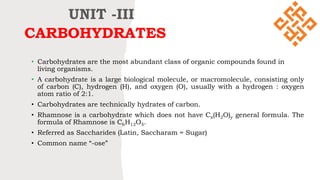
  • 1. CARBOHYDRATES • Carbohydrates are the most abundant class of organic compounds found in living organisms. • A carbohydrate is a large biological molecule, or macromolecule, consisting only of carbon (C), hydrogen (H), and oxygen (O), usually with a hydrogen : oxygen atom ratio of 2:1. • Carbohydrates are technically hydrates of carbon. • Rhamnose is a carbohydrate which does not have Cx(H2O)y general formula. The formula of Rhamnose is C6H12O5. • Referred as Saccharides (Latin, Saccharam = Sugar) • Common name “-ose” UNIT -III
  • 2. WHAT ARE CARBOHYDRATES ? • The carbohydrates are polyfunctional compounds • They contain the following functional groups: • Alcoholic hydroxy groups, - OH • Aldehyde group -CHO • Ketone group, >C=O • A precise definition of the term 'Carbohydrate' can be given as : “Polyhydroxyaldehydes or C H HO OH H OH H CH2OH CH2OH O CHO OH H H HO OH H OH H CH2OH Glucose Fructose C H O O H H C H 2 O H C H O H H O C H 2 O H D - G l y c e r a l d e h y d e L - G l y c e r a l d e h y d e
  • 3. Carbohydrates originate as products of photosynthesis, an endothermic reductive condensation of carbon dioxide requiring light energy and the pigment chlorophyll. FORMATION OF CARBOHYDRATES
  • 5. Monosaccharides:  The simplest form of carbohydrates is the monosaccharide. 'Mono' means 'one' and 'saccharide' means 'sugar'. Monosaccharides are polyhydroxy aldehyde or ketone that cannot be hydrolyzed further to give simpler sugar.
  • 6. Disaccharides: • They give two monosaccharide units on hydrolysis, which may be the same or different. For example, sucrose on hydrolysis gives one molecule each of glucose and fructose, whereas maltose gives two molecules of glucose, While Lactose gives glucose and galactose.
  • 7. Trisaccharides: • These carbohydrates yield three molecules of monosaccharides units on hydrolysis. • Raffinose ->Glucose + Fructose + Galactose Raffinose
  • 8. Polysaccharides: • These carbohydrates give a large number of monosaccharide units on hydrolysis. • These monosaccharide units are joined together by oxide bridges. • These linkages are called glycosidic linkages. • The common and widely distributed polysaccharides correspond to the general formula (C6H10O5)n . • Polysaccharides are not sweet in taste, so they are called non-sugars. • Some common examples are starch, cellulose, glycogen, etc
  • 9. D and L Notations • The notations D and L are used to describe the configurations of carbohydrates. Glyceraldehyde has been chosen as arbitrary standard for the D and L notation in sugar chemistry. • Because this has an asymmetric carbon and can exist as a pair of enantiomers. • In a Fischer projection, the carbonyl group is always placed on the top position for monosaccharide. • From its structure, if the –OH group attached to the bottom- most asymmetric center (the carbon that is second from the bottom) is on the right, then, the compound is a D-sugar. • If the –OH group is on the left, then, the compound is a L- sugar. Almost all sugars found in nature are D-sugar. CHO OH H H HO OH H OH H CH2OH D-Glucose CHO OH H H HO OH H H HO CH2OH L-Glucose C H O O H H C H 2 O H C H O H H O C H 2 O H D - G l y c e r a l d e h y d eL - G l y c e r a l d e h y d e
  • 10.
  • 11. Epimers are stereoisomers that differ in configuration of only one asymmetric carbon of enantiomers or diastereomers. Example D-glucose and D-mannose are C-2 epimers D-glucose and D-Galactose are C-4 epimers. Epimers
  • 12. Anomers are cyclic monosaccharides or glycosides that are epimers, differing from each other in the configuration of C-1 if they are aldoses or in the configuration at C-2 if they are ketoses. Anomers
  • 13.
  • 14. Mutarotation Anomers are diastereomers and as expected the glucose isomers can be isolated and characterized separately showing different physical properties. For example, the melting point of α-D-Glucose is 146 °C, while that of β-D-Glucose reaches 150 °C. An interesting phenomenon was observed when measuring the specific rotation of glucose. When the α anomer is dissolved in water, at first, it exhibits a specific rotation of +112.2 °, but this changes with time to +52.6 °. The same happens with the β anomer and where the specific rotation changes from +18.7 ° to +52.6 °. The fact that in both cases the final number is the same +52.6 ° suggests that the molecules undergo a transformation and eventually set at an equilibrium.
  • 17.
  • 18. Hemiacetal Hemiacetal is a molecule made up of a core carbon atom connected to four groups: –OR, –OH, –R, and –H. Acetal Acetal is a molecule made of a core carbon atom that is attached to two –OR groups, a –R group, and a –H.
  • 19.
  • 20. Reducing and Non-reducing Sugars Some cyclic acetals or ketals do not exist in equilibrium with their open chain carbonyl group containing forms in neutral or basic aqueous solutions. These cannot be oxidized by reagents such as Tollen's reagent (Ag(NH3)2OH) or Br2. So, these are referred as non-reducing sugars. Eg: Sucrose Some hemiacetals or hemiketals exist in equilibrium with the open-chain sugars in aqueous solution such as glucose. These sugars can reduce an oxidizing agent such as Tollens reagent or aqueous bromine and are thus classified as a reducing sugars. Infact, all aldehyde bearing compounds can reduce tollens reagent and this includes aldoses such as glucose.
  • 21. STRUCTURAL ELUCIDATION OF GLUCOSE 1.Mol. formula C6H12O6: Elemental analysis and mol.wt determinations formed the molecular formula of glucose was C6H12O6. 2. Presence of 6-carbon chain: The complete reduction of glucose with conc. HI and Red Phosphorus gives n-Hexane. HI/Red P This proves that glucose molecules is made of an unsaturated 6-carbon chain. C H O O H H H H O O H H O H H C H 2 O H D - G l u c o s e CH3-CH2-CH2-CH2-CH2-CH3
  • 22. 3. Presence of 5 OH groups Glucose reacts with acetic anhydride to form a pent acetyl derivative. • This shows the presence of 5 hydroxy groups. • Since glucose is a stable compound, no 2 OH groups are attached to the same carbon. • In other words, the five OH groups are on different carbons. ( C H O H ) 4 H C C H 2 O H O ( C H O . C O C H 3 ) 4 H C C H 2 O C O C H 3 O P e n t a a c e t y l g l u c o s e C H 3 C O O H + 5 ( C H 3 C O ) 2 O +
  • 23. 4. Presence of C=O group Glucose reacts with hydroxylamine to form an oxime. It suggests the presence of a carboxylic group ( C H O H ) 4 H C C H 2 O H O ( C H O H ) 4 H C C H 2 O H N - O H + N H 2 O H G l u c o s e O x i m e + H 2 O
  • 24. 5 Presence of terminal CHO group : On mild oxidation with bromine water, glucose is converted to gluconic acid which when reduced with excess HI yield n-hexanoic acid. C5H11O5.CHO→C5H11O5.COOH→CH3(CH2)4COOH Glucose Gluconic acid n-Hexanoic acid This shows that glucose contains a six carbon straight chain with CHO at one end, which has been oxidised to COOH.
  • 25. 6. Construction of open-chain formula We know that • Glucose has a straight 6-carbon chain with a terminal CHO • 5 OH groups can be placed one each on the remaining 5 carbons • Supplying hydrogen atoms to these carbons to satisfy their tetravalency. The open chain structure of glucose can be written as C H O O H H H H O O H H O H H C H 2 O H D - G l u c o s e
  • 26. Kiliani-Fischer Synthesis The Kiliani-Fischer Synthesis is a method for extending a carbohydrate chain by a single carbon. This Synthesis involves addition of cyanide ion to an open-chain aldehyde (in the case of aldoses) which is then partially reduced and then hydrolyzed to give a new aldehyde. CHO H HO OH H OH H CH2OH H C N CN OH H H HO OH H OH H CH2OH 2 H 2 O - N H 3 C O O H O H H H H O O H H O H H C H 2 O H - H 2 O C O O H H H H O O H H O H C H 2 O H N a B H 4 2 [ H ] C H O O H H H H O O H H O H H C H 2 O H Aldopentose Cyanohydrine Aldonic acid Lactone Aldohexose
  • 28.
  • 29. Cyclic Structure of Glucose Open-chain structure not wholly true: • Fischer realized that the open-chain pentahydroxy aldehyde structure of glucose did not wholly explain its chemical behaviour. • Unlike simple aldehydes, glucose did not form the crystalline bisulphate compound and failed to give the Schiff’s test. • Furthermore, the pentaacetate and pentamethyl ether derivatives of glucose are not oxidized by Tollens reagent or Fehling’s solution, indicating the absence of the CHO group.
  • 30. The cyclic structure suggested explaining mutarotation: • The French chemist, Tarnet established the existence of two crystalline forms of glucose, alpha-glucose and beta-glucose, alpha-glucose has specific rotation +112⁰, while beta-glucose +18.7⁰. • The optical rotation of each of these forms changed gradually with time till finally a constant value of +52.7⁰ was reached. • To explain this phenomenon of mutarotation, it was visualized that the alpha and beta glucose were, in reality, the cyclic hemiacetal forms of glucose which were interconvertible via the open-chain form. • The constant value of +52.7⁰ represented the state of equilibrium between alpha-D-glucose and beta-D- glucose.
  • 31. Glycoside formation confirms cyclic structure: • When we use methanol in the presence of dry HCl to treat glucose, it gives two isomeric glycosides or acetals. • These crystalline glycosides namely methyl-alpha-D-glucose and methyl-beta-Glucoside and are actually isolated. • These are optically active but do not give any reactions of free CHO group. • Evidently, the two glycosides are the methyl derivatives of alpha and beta-D-glucose, formed as a result of the reaction between the hemiacetal OH of these forms and methanol.
  • 32. Determination of ring size: So far we have represented the structure of cyclic hemiacetals or anomers of D-glucose as having a ring of six members, five carbons and one oxygen. This has been proved to be correct and the five-membered ring has been ruled out. Hirst in 1926, prepared tetra-O-methyl-D-glucose, by treating methyl-D-glucoside with dimethyl sulphate and subsequent acid hydrolysis of the pentamethyl derivative formed. The oxidation of tetra-O-methyl-D- glucose with nitric acid yielded trimethoxy glutaric acid.
  • 33. Obviously, the two carboxylic carbons (1,5) of the trimethoxy glutaric acid are the ones originally involved in ring formation. Hence, there must have existed an oxide ring between C-1 and C-5. Tracing back the reaction sequence, it stands proved that D-glucose has a six-membered ring. The presence of a 6-membered ring in D-glucose has also been confirmed by X-ray analysis.
  • 34. Fischer and Haworth structures of Fructose • Fructose occurs in fruits, and it is called the fruit sugar. • It is also present in honey and sweet fruits along with glucose. • In the combined state, it is also present in disaccharide and polysaccharide (insulin). • Its molecular formula is C6H12O6. • It contains the keto group at C-2 and the six carbon atoms are arranged in a straight chain. • The Fischer projection of the fructose can be converted into the cyclic structure.
  • 35. Because sugars often contain alcohol and carbonyl functional groups, intramolecular hemiacetal formation is common in carbohydrate chemistry. The hemiacetal is formed by the intramolecular combination of C2 , keto group and −OH group of C5 the atom. As a result, C2 atom becomes asymmetric and therefore D-fructose has two possible isomers as a α -D-fructose and β -D-fructose.
  • 36. • To write the Haworth structures for any monosaccharide first, draw a pentagon with its oxygen atom at the top. • The terminal -CH2OH group as shown in the figure is always placed above the plane of the pentagon ring. • Place all the groups which are present on the left-hand side in Fischer projection above the plane of the ring and all those groups on the right hand in Fischer projection below the plane of the ring. • This formation goes through the hemiacetal. This structure is like the furan ring. The hemiacetal structure is as shown here: Thus, here we know that the five-membered rings of oxide are called the furanose.
  • 37. Due to the formation of an oxide ring, the new asymmetric carbon atom is created at the carbonyl carbon, which is called an anomeric carbon atom. Two different configurations are possible at the anomeric carbon atom, and they are called the anomers. Here, the two structures shown here are similar except at the C-2 carbon atom. In α -D-fructose the hydroxyl group below the plane and β - D-fructose the hydroxyl group is above the plane.
  • 38. Fischer and Haworth structures of Galactose • Galactose is known as the brain sugar. • It supports the brain development of infants. • The monosaccharide sugar helps trigger long-term memory formation. • Galactose also has been shown to inhibit tumor growth and stop its spread. • Galactose exists in both open-chain and cyclic forms. • We find it in dairy products, avocados, sugar beets
  • 39. Disaccharides Previously, you learned that • Monosaccharides could form cyclic structures by the reaction of the carbonyl group with an OH group. • These cyclic molecules can in turn react with another alcohol. • Disaccharides (C12H22O11) are sugars composed of two monosaccharide units that are joined by a carbon–oxygen-carbon linkage known as a glycosidic linkage. • This linkage is formed from the reaction of the anomeric carbon of one cyclic monosaccharide with the OH group of a second monosaccharide.
  • 40.
  • 41. Glycosidic linkage •“The two monosaccharides are joined together by an oxide linkage formed by the loss of a water molecule. Such a linkage between two monosaccharide units through an oxygen atom is called glycosidic linkage.” •The two monosaccharides C1 of α-D-glucose and C2 of β-D-fructose are held together by a glycosidic linkage which is shown below:
  • 42. Maltose • Maltose is a sugar made from two glucose molecules bound together. • It's created in seeds and other parts of plants as they break down their stored energy in order to sprout. • Thus, foods like cereals, certain fruits and sweet potatoes contain naturally high amounts of this sugar. • Maltose is a disaccharide formed from two units of glucose joined with an α(1→4) bond.
  • 43.
  • 44.
  • 45. Lactose Lactose is a sugar found in milk and milk products. Lactose is known as milk sugar because it occurs in the milk of humans, cows, and other mammals. Lactose intolerance happens when your small intestine does not make enough of a digestive enzyme called lactase. Lactase breaks down the lactose in food so your body can absorb it.
  • 46.
  • 47. Pyrimidine Pyrimidine is an aromatic, heterocyclic, organic compound similar to pyridine. One of the three diazines (six-membered heterocyclics with two nitrogen atoms in the ring), it has nitrogen atoms at positions 1 and 3 in the ring. Purine • Purine is a heterocyclic aromatic organic compound that consists of two rings (pyrimidine and imidazole) fused together. • Purine also gives its name to the wider class of molecules, purines, which include substituted purines and their tautomers. • They are the most widely occurring nitrogen-containing heterocycles in nature. • Purines are biologically synthesized as nucleosides + = Nucleic Acids Diazine isomers: 1-Pyridazine, 2-Pyrimidine, 3-Pyrazine
  • 48.
  • 49. Nitrogenous bases • The nitrogenous base is either a purine or a pyrimidine. • There are five major bases found in cells. The derivatives of purine are called adenine and guanine, and the derivatives of pyrimidine are called thymine, cytosine and uracil. • Purines include adenine and guanine and have two rings. • Adenine has an ammonia group on its rings, whereas guanine has a ketone group. • Pyrimidines include cytosine, thiamine, and uracil and have one ring. • Thymine (found in DNA) and uracil (found in RNA) are similar in that they both have ketone groups, but thymine has an extra methyl group on its ring. • Bonds between guanine and cytosine (three hydrogen bonds) are stronger than bonds between adenine and thymine (two hydrogen bonds).
  • 50. Pentose Sugar •The five-carbon sugar is either a ribose (in RNA) or a deoxyribose (in DNA) molecule. •In nucleotides, both types of pentose sugars are in their beta-furanose (closed five- membered ring) form.
  • 51. Phosphoric acid • It may occur also as phosphate and forms the backbone of DNA molecule along with sugar molecule. • It links the nucleotides by joining the deoxyribose (pentose sugar) of two adjacent nucleotides with an ester-phosphate bond. • These bonds connect carbon 3′ in one nucleotide with carbon 5′ in next.
  • 52. Structure of nucleosides and nucleotides • Nucleosides can be thought of as nucleotides without a phosphate group. • A nucleoside consists simply of a nucleobase (also termed a nitrogenous base) and a five-carbon sugar (ribose or 2'-deoxyribose) whereas a nucleotide is composed of a nucleobase, a five-carbon sugar, and one or more phosphate groups. • In a nucleoside, the anomeric carbon is linked through a glycosidic bond to the N9 of a purine or the N1 of a pyrimidine. Nucleotides are the molecular building-blocks of DNA and RNA. • While a nucleotide is composed of a nucleobase, a five-carbon sugar, and one or more phosphate groups, a nucleoside has only a nitrogenous base and a five-carbon sugar. • In a nucleoside, the base is bound to either ribose or deoxyribose via a beta-glycosidic linkage at 1’ position. • Examples of nucleosides include cytidine, uridine, adenosine, guanosine, thymidine and inosine.
  • 53. Structure of nucleosides and nucleotides • Nucleosides can be thought of as nucleotides without a phosphate group. • A nucleoside consists simply of a nucleobase (also termed a nitrogenous base) and a five-carbon sugar (ribose or 2'-deoxyribose) whereas a nucleotide is composed of a nucleobase, a five-carbon sugar, and one or more phosphate groups. • In a nucleoside, the anomeric carbon is linked through a glycosidic bond to the N9 of a purine or the N1 of a pyrimidine. Nucleotides are the molecular building-blocks of DNA and RNA.
  • 54. Structure of nucleosides and nucleotides
  • 55. List of nucleosides and corresponding nucleobases
  • 56.
  • 57.
  • 58.
  • 59. Summary of Differences Between DNA and RNA • DNA contains the sugar deoxyribose, while RNA contains the sugar ribose. • The only difference between ribose and deoxyribose is that ribose has one more -OH group than deoxyribose, which has -H attached to the second (2') carbon in the ring. • DNA is a double-stranded molecule, while RNA is a single-stranded molecule. • DNA is stable under alkaline conditions, while RNA is not stable. • DNA and RNA perform different functions in humans. DNA is responsible for storing and transferring genetic information, while RNA directly codes for amino acids and acts as a messenger between DNA and ribosomes to make proteins. • DNA and RNA base pairing is slightly different since DNA uses the bases adenine, thymine, cytosine, and guanine; RNA uses adenine, uracil, cytosine, and guanine. Uracil differs from thymine in that it lacks a methyl group on its ring. • While both DNA and RNA are used to store genetic information, there are clear differences between them.
  • 60. Main Differences Between DNA and RNA Comparison DNA RNA Name DeoxyriboNucleic Acid RiboNucleic Acid Function Long-term storage of genetic information; transmission of genetic information to make other cells and new organisms. Used to transfer the genetic code from the nucleus to the ribosomes to make proteins. RNA is used to transmit genetic information in some organisms and may have been the molecule used to store genetic blueprints in primitive organisms. Structural Features B-form double helix. DNA is a double-stranded molecule consisting of a long chain of nucleotides. A-form helix. RNA usually is a single-strand helix consisting of shorter chains of nucleotides. Composition of Bases and Sugars deoxyribose sugar phosphate backbone adenine, guanine, cytosine, thymine bases ribose sugar phosphate backbone adenine, guanine, cytosine, uracil bases Propagation DNA is self-replicating. RNA is synthesized from DNA on an as-needed basis. Base Pairing AT (adenine-thymine) GC (guanine-cytosine) AU (adenine-uracil) GC (guanine-cytosine) Reactivity The C-H bonds in DNA make it fairly stable, plus the body destroys enzymes that would attack DNA. The small grooves in the helix also serve as protection, providing minimal space for enzymes to attach. The O-H bond in the ribose of RNA makes the molecule more reactive, compared with DNA. RNA is not stable under alkaline conditions, plus the large grooves in the molecule make it susceptible to enzyme attack. RNA is constantly produced, used, degraded, and recycled. Ultraviolet Damage DNA is susceptible to UV damage. Compared with DNA, RNA is relatively resistant to UV damage. Comparison of DNA and RNA While both DNA and RNA are used to store genetic information, there are clear differences between them. This table summarizes the key points:
  • 61. Methods of formation of inter nucleotide bonds - Phosphodiester approach This method involves the formation of an ester linkage between an activated phosphate group of one nucleotide with the hydroxyl group of another nucleoside, thus forming the natural phosphodiester bridge between the 5′-OH of one nucleoside unit and the 3′-OH of the next. Phosphodiester bonds make up the backbones of DNA and RNA. The phosphate is attached to the 5' carbon. The 3' carbon of one sugar is bonded to the 5' phosphate of the adjacent sugar. Specifically, the phosphodiester bond links the 3' carbon atom of one sugar molecule and the 5' carbon atom of another, hence the name, 3', 5' phosphodiester linkage. These saccharide groups are derived from deoxyribose in DNA and ribose in RNA.
  • 62.
  • 63. Protein–Nucleic Acid Interactions • Protein and nucleic acid interactions are vital to cellular processes. • Proteins associate with nucleic acids to mediate transcription and translation of DNA and RNA to decode the information carried by genetic material. • In addition, protein–nucleic acid interactions are required to maintain the integrity of DNA and RNA throughout generations. • To do so, proteins interact with nucleic acids in processes such as DNA replication, repair and processing, as well as RNA processing and translocation.