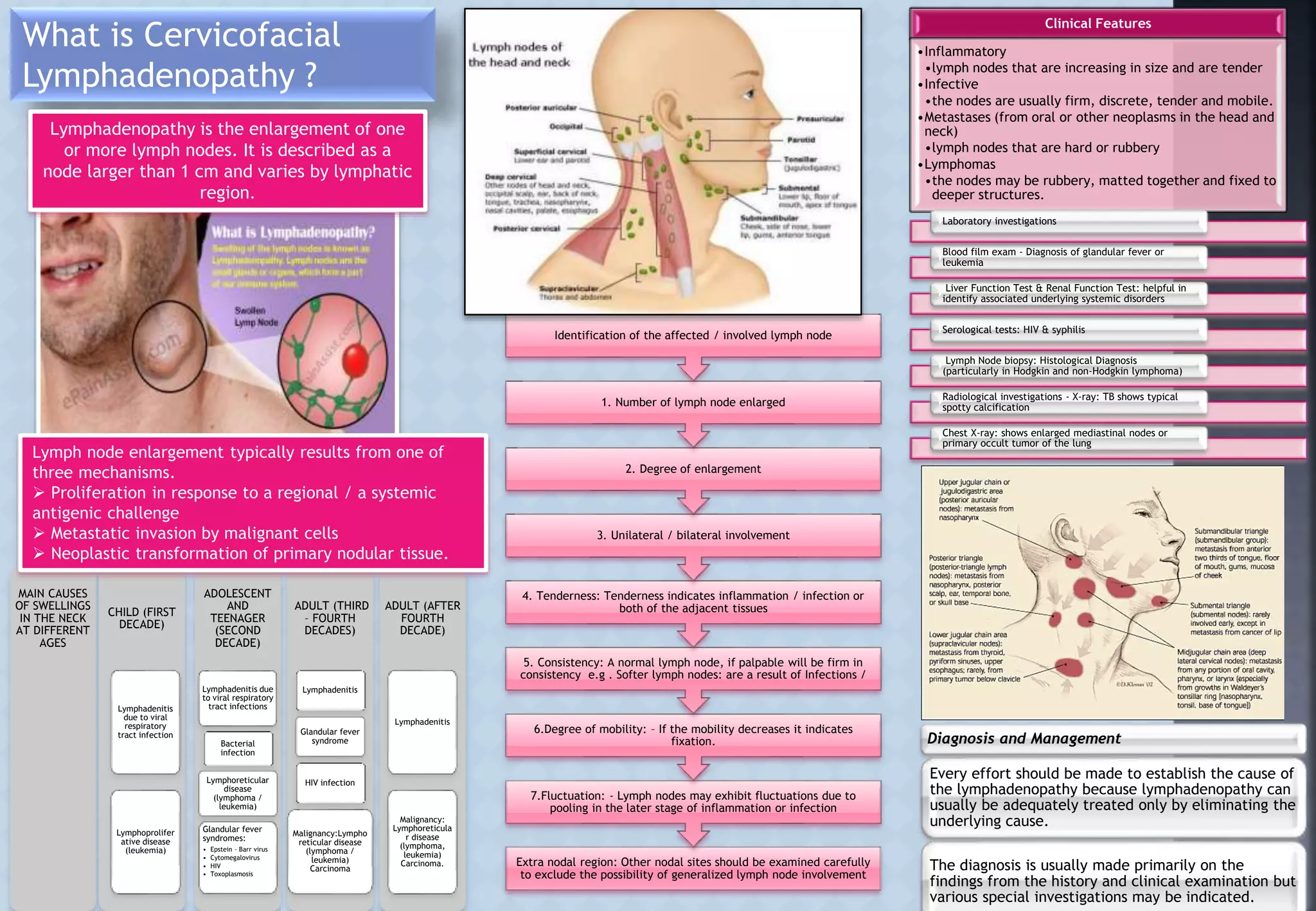
What is Cervicofacial
Lymphadenopathy ?
Extra nodal region: Other nodal sites should be examined carefully
to exclude the possibility of generalized lymph node involvement
7.Fluctuation: - Lymph nodes may exhibit fluctuations due to
pooling in the later stage of inflammation or infection
6.Degree of mobility: – If the mobility decreases it indicates
fixation.
5. Consistency: A normal lymph node, if palpable will be firm in
consistency e.g . Softer lymph nodes: are a result of Infections /
4. Tenderness: Tenderness indicates inflammation / infection or
both of the adjacent tissues
3. Unilateral / bilateral involvement
2. Degree of enlargement
1. Number of lymph node enlarged
Identification of the affected / involved lymph node
MAIN CAUSES
OF SWELLINGS
IN THE NECK
AT DIFFERENT
AGES
CHILD (FIRST
DECADE)
Lymphadenitis
due to viral
respiratory
tract infection
Lymphoprolifer
ative disease
(leukemia)
ADOLESCENT
AND
TEENAGER
(SECOND
DECADE)
Lymphadenitis due
to viral respiratory
tract infections
Bacterial
infection
Lymphoreticular
disease
(lymphoma /
leukemia)
Glandular fever
syndromes:
• Epstein - Barr virus
• Cytomegalovirus
• HIV
• Toxoplasmosis
ADULT (THIRD
– FOURTH
DECADES)
Lymphadenitis
Glandular fever
syndrome
HIV infection
Malignancy:Lympho
reticular disease
(lymphoma /
leukemia)
Carcinoma
ADULT (AFTER
FOURTH
DECADE)
Lymphadenitis
Malignancy:
Lymphoreticula
r disease
(lymphoma,
leukemia)
Carcinoma.
Lymph node enlargement typically results from one of
three mechanisms.
 Proliferation in response to a regional / a systemic
antigenic challenge
 Metastatic invasion by malignant cells
 Neoplastic transformation of primary nodular tissue.
Lymphadenopathy is the enlargement of one
or more lymph nodes. It is described as a
node larger than 1 cm and varies by lymphatic
region.
Clinical Features
•Inflammatory
•lymph nodes that are increasing in size and are tender
•Infective
•the nodes are usually firm, discrete, tender and mobile.
•Metastases (from oral or other neoplasms in the head and
neck)
•lymph nodes that are hard or rubbery
•Lymphomas
•the nodes may be rubbery, matted together and fixed to
deeper structures.
Laboratory investigations
Blood film exam - Diagnosis of glandular fever or
leukemia
Liver Function Test & Renal Function Test: helpful in
identify associated underlying systemic disorders
Serological tests: HIV & syphilis
Lymph Node biopsy: Histological Diagnosis
(particularly in Hodgkin and non-Hodgkin lymphoma)
Radiological investigations - X-ray: TB shows typical
spotty calcification
Chest X-ray: shows enlarged mediastinal nodes or
primary occult tumor of the lung
Diagnosis and Management
Every effort should be made to establish the cause of
the lymphadenopathy because lymphadenopathy can
usually be adequately treated only by eliminating the
underlying cause.
The diagnosis is usually made primarily on the
findings from the history and clinical examination but
various special investigations may be indicated.

Cervicofacial Lymphadenopathy

  • 1.
    What is Cervicofacial Lymphadenopathy? Extra nodal region: Other nodal sites should be examined carefully to exclude the possibility of generalized lymph node involvement 7.Fluctuation: - Lymph nodes may exhibit fluctuations due to pooling in the later stage of inflammation or infection 6.Degree of mobility: – If the mobility decreases it indicates fixation. 5. Consistency: A normal lymph node, if palpable will be firm in consistency e.g . Softer lymph nodes: are a result of Infections / 4. Tenderness: Tenderness indicates inflammation / infection or both of the adjacent tissues 3. Unilateral / bilateral involvement 2. Degree of enlargement 1. Number of lymph node enlarged Identification of the affected / involved lymph node MAIN CAUSES OF SWELLINGS IN THE NECK AT DIFFERENT AGES CHILD (FIRST DECADE) Lymphadenitis due to viral respiratory tract infection Lymphoprolifer ative disease (leukemia) ADOLESCENT AND TEENAGER (SECOND DECADE) Lymphadenitis due to viral respiratory tract infections Bacterial infection Lymphoreticular disease (lymphoma / leukemia) Glandular fever syndromes: • Epstein - Barr virus • Cytomegalovirus • HIV • Toxoplasmosis ADULT (THIRD – FOURTH DECADES) Lymphadenitis Glandular fever syndrome HIV infection Malignancy:Lympho reticular disease (lymphoma / leukemia) Carcinoma ADULT (AFTER FOURTH DECADE) Lymphadenitis Malignancy: Lymphoreticula r disease (lymphoma, leukemia) Carcinoma. Lymph node enlargement typically results from one of three mechanisms.  Proliferation in response to a regional / a systemic antigenic challenge  Metastatic invasion by malignant cells  Neoplastic transformation of primary nodular tissue. Lymphadenopathy is the enlargement of one or more lymph nodes. It is described as a node larger than 1 cm and varies by lymphatic region. Clinical Features •Inflammatory •lymph nodes that are increasing in size and are tender •Infective •the nodes are usually firm, discrete, tender and mobile. •Metastases (from oral or other neoplasms in the head and neck) •lymph nodes that are hard or rubbery •Lymphomas •the nodes may be rubbery, matted together and fixed to deeper structures. Laboratory investigations Blood film exam - Diagnosis of glandular fever or leukemia Liver Function Test & Renal Function Test: helpful in identify associated underlying systemic disorders Serological tests: HIV & syphilis Lymph Node biopsy: Histological Diagnosis (particularly in Hodgkin and non-Hodgkin lymphoma) Radiological investigations - X-ray: TB shows typical spotty calcification Chest X-ray: shows enlarged mediastinal nodes or primary occult tumor of the lung Diagnosis and Management Every effort should be made to establish the cause of the lymphadenopathy because lymphadenopathy can usually be adequately treated only by eliminating the underlying cause. The diagnosis is usually made primarily on the findings from the history and clinical examination but various special investigations may be indicated.