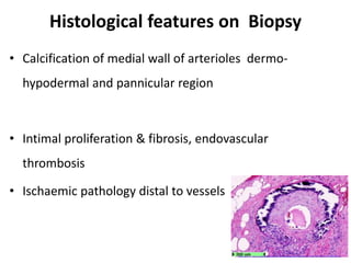
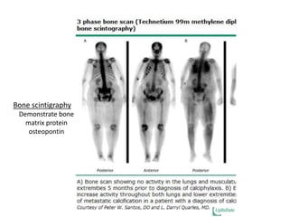
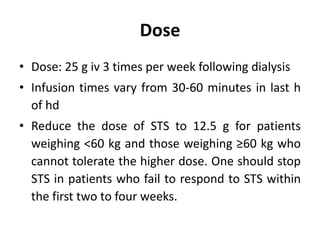
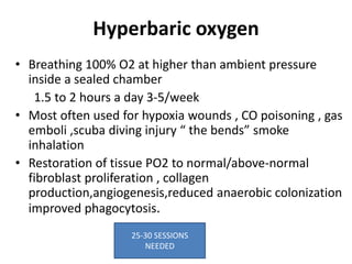
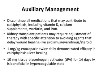
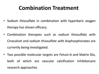
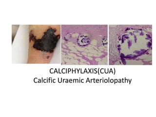
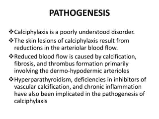
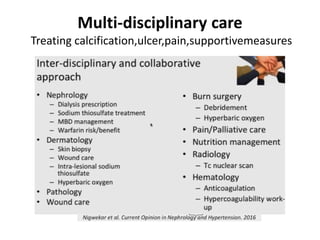
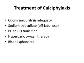
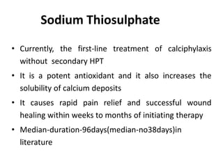
CALCIPHYLAXIS(CUA)
Calcific Uraemic Arteriolopathy
ADD SOME
RELATED
PICTURE
OUTLINES
• INTRODUCTION
• PATHOGENESIS
• EPIDEMIOLOGY
• RISK FACTORS
• NON ESRD POPULATION
• CLINICALFEATURES
• DIAGNOSIS AND DD
• LAB MANIF/BIOPSY/ RADIOLOGY
• TREATMENT
• OUTCOME
CALCIFIC URAEMIC ARTERIOLOPATHY
Introduction
• CUA or calciphylaxis is a rare disorder (Orphan disease)of
microvascular occlusionof subcutaneous adipose tissue &
dermis leading to painful skin lesions- Calciphylaxis Cutis
/Visceral Calciphylaxis.
• Associated histologically with media calcification of small
& medium-size vessels in dermis & subcutaneous adipose
tissue
• Has an estimated incidence of <1% among dialysis patients
who are predominantly affected cohort
EPIDEMIOLOGY-RELEVANCE
• Calciphylaxis most commonly occurs in patients of ESRD
and are on dialysis but may also occur in kidney tx
recipients & in non-ESRD patients
• Is a lethal disease with an estimated six-month survival
of approx 50% with no approved RX
NON UREMIC CALCIPHLAXIS
60% had been previously treated with glucocorticoids, and 25% with warfarin
•Nigwekar SU, Wolf M, Sterns RH, Hix JK. Calciphylaxis from nonuremic causes: a systematic review. Clin J Am Soc
Nephrol 2008; 3:1139.
Severe sec HPT /vascular calcif
PATHOGENESIS
Calciphylaxis is a poorly understood disorder.
The skin lesions of calciphylaxis result from
reductions in the arteriolar blood flow.
Reduced blood flow is caused by calcification,
fibrosis, and thrombus formation primarily
involving the dermo-hypodermic arterioles
Hyperparathyroidism, deficiencies in inhibitors of
vascular calcification, and chronic inflammation
have also been implicated in the pathogenesis of
calciphylaxis
PATOGENESIS
• Microvascular calcification occurs first likely via an
active process involving up regulation of factors
involved in osteogenesis and bone remodeling,
including bone morphogenetic protein 2 (BMP-2),
runt-related transcription factor 2 (RUNX-2) and
osteopontin
• Adipocytes may also play an important role in the
development of vascular calcification of calciphylaxis .
• Ongoing vascular endothelial injury causes cutaneous
arteriolar narrowing and thrombosis leading to tissue
infarction
PATHOGENETIC FACTORS
• Factors presumed to be involved in the widespread
calcification are
- Chronic kidney disease-mineral bone disorder (CKD-
MBD) and its treatment (including hyper
parathyroidism, abnormalities in calcium and
phosphate, and vitamin D administration)
- Deficiencies of the inhibitors of vascular calcifications
and chronic inflammation
Clinical recognition
• Typical sites- high fat tissue area
• Typical appearance – early red to violet/later
black
• Typical symptom- extremely painful
• Typical risk factors- esrd/warfarin/ autoimm
• Atypical sites need DD
What do we see and where?
Time of presentation – Early -late.
• Mottled skin (livedo reticularis)- Violaceous/red
• Purpuric plaque-like subcutaneous nodules, indurations
• Bullae over plaque ,Necrotic ulcers with eschars
(skin/subcutaneous fat/ muscle)
• Typical lesions seen more in central body area than on
finger and toes (areas with high adipose tissue content)
Followed the path of the vasculature
Intense pain is a constant finding
Digital gangrene in a dialysis patient due to calciphylaxis and systemic
polyarteritis nodosa: A diagnostic dilemma
Jain N; Sethi J; Ramachandran R; Kumar V; Rathi,M, et al.
Ind J of Nephrology 2019
Left foot gangrenous changes and
redness swelling of all toes with
ulcer on dorsal aspect
Differential diagnosis
 Many dermatologic conditions resemble the superficial lesions of
Calciphylaxis
Diagnosis Distinguishing characteristic(s)
Antiphospholipid
syndrome
Serum antiphospholipid antibodies
Radiation arteritis Radiation arteritis
Vasculitis Significant inflammatory infiltrate
Dystrophic
calcification
Localized tissue damage secondary to calcium and
phosphorous deposition; normal serum calcium and
phosphorous levels
Cholesterol emboli Acute onset; biopsy shows cholesterol crystals embedded
within intramural thrombi; lesions distributed in extremities
with acral involvement
Differential diagnosis
Diagnosis Distinguishing characteristic(s)
Panniculitis Biopsy reveals inflammatory infiltrate in the adipose tissue
without calcification of vessels
Purpura fulminans Sepsis and disseminated intravascular coagulation often
present; increased partial thromboplastin time, prothrombin
time, fibrinogen, D-dimers
Warfarin necrosis History of warfarin use
Heparin necrosis History of heparin use; occurs uniquely at sites of
intramuscular heparin injection
Atherosclerosis
obliterans
Affects intimal layer of the abdominal aorta and small- to
medium-sized arteries in the lower extremities, often
resulting in elimination of the popliteal, posterior tibial,
and/or dorsalis pedis pulses
Histological features on Biopsy
• Calcification of medial wall of arterioles dermo-
hypodermal and pannicular region
• Intimal proliferation & fibrosis, endovascular
thrombosis
• Ischaemic pathology distal to vessels
Role of Imaging in diagnosis
• Plain X Ray/3D CT/ Mammography-limited role
Demonstrates an arborisation of vascular calcification within
the dermis and the subcutaneous tissue &numerous
echogenic foci suggestive of large calcifications-best detected
by mammoographic technique or CT reconstruction rather
than plain xray
• BONE SCAN OR SCINTIGRAPHY
Three-phase technetium 99m methylene diphosphate bone
scan to identify cutaneous calcifications
Role of Imaging in diagnosis
• X-ray : Detects vascular calcification within the dermis
and subcutis
- “ Net-like”
- Mammography film better detects small vessels
• Bone scintigraphy :
-Scans entire body
-Detects micro calcificatio of soft tissue
-Evaluates extent of disease and response to treatment
-89% sensitivity,97% specificity
Bone scintigraphy
Demonstrate bone
matrix protein
osteopontin
Multi-disciplinary care
Treating calcification,ulcer,pain,supportivemeasures
Treatment of Calciphylaxis
• Optimising dialysis adequacy
• Sodium thiosulfate (off-label use)
• PD to HD transition
• Hyperbaric oxygen therapy
• Bisphosphonates
Sodium Thiosulphate
• Currently, the first-line treatment of calciphylaxis
without secondary HPT
• It is a potent antioxidant and it also increases the
solubility of calcium deposits
• It causes rapid pain relief and successful wound
healing within weeks to months of initiating therapy
• Median-duration-96days(median-no38days)in
literature
Dose
• Dose: 25 g iv 3 times per week following dialysis
• Infusion times vary from 30-60 minutes in last h
of hd
• Reduce the dose of STS to 12.5 g for patients
weighing <60 kg and those weighing ≥60 kg who
cannot tolerate the higher dose. One should stop
STS in patients who fail to respond to STS within
the first two to four weeks.
Hyperbaric oxygen
• Breathing 100% O2 at higher than ambient pressure
inside a sealed chamber
1.5 to 2 hours a day 3-5/week
• Most often used for hypoxia wounds , CO poisoning , gas
emboli ,scuba diving injury “ the bends” smoke
inhalation
• Restoration of tissue PO2 to normal/above-normal
fibroblast proliferation , collagen
production,angiogenesis,reduced anaerobic colonization
improved phagocytosis.
25-30 SESSIONS
NEEDED
Auxiliary Management
• Discontinue all medications that may contribute to
calciphylaxis, including vitamin D, calcium
supplements, warfarin, and iron.
• Kidney transplant patients may require adjustment of
therapy with specific attention to avoiding agents that
delay wound healing-like sirolimus/everolimus/steroid
• 1 mg/kg enoxaparin twice daily demonstrated efficacy in
calciphylaxis ulcer healing.
• 10 mg tissue plasminogen activator (tPA) for 14 days iv.
is beneficial in hypercoagulable state
Combination Treatment
• Sodium thiosulfate in combination with hyperbaric oxygen
therapy has shown efficacy.
• Combination therapies such as sodium thiosulfate with
Cinacalcet and sodium thiosulfate with bisphosphonates are
currently being investigated.
• Two possible molecular targets are Fetuin-A and Matrix Gla,
both of which are vascular calcification inhibitorsare
research approaches
Summary of treatment approaches
Complications
• Complications of calciphylaxis range from moderate
interference with activity to death.
• Lesions of calciphylaxis frequently result in nonhealing ulcers
and cutaneous gangrene.
• Acral lesions may fail to heal with conservative therapy and
lead to amputation. Sepsis may result from the non healing
wounds.
• Patients with internal involvement may develop
gastrointestinal haemorrhage, infarction, or organ failure.
• Patients who do not die of sepsis or organ failure frequently
undergo amputation of an involved limb.
Take home message
• Clinical picture is sufficient to diagnose CUA in
most cases
• There are high number of missed,
undiagnosed cases
• Optimize the dialysis prescription
• Sodium thiosulfate is a first-line treatment
option
• Bisphosphonates is another treatment option
Take home message
• Parathyroidectomy /cinacalcet are options in CUA
• CUA treatment should be multimodal
• Stopping vitamin K antagonist should be
considered
• Vitamin K supplementation may be considered
• Prognosis of CUA is poor(1&5y survival rates
estimated to be 45% & 35%)
• Lack of evidence does not justify neglect:how can
we address unmet needs in calciphylaxis?
THANK YOU

Calciphylaxis

  • 1.
  • 2.
    OUTLINES • INTRODUCTION • PATHOGENESIS •EPIDEMIOLOGY • RISK FACTORS • NON ESRD POPULATION • CLINICALFEATURES • DIAGNOSIS AND DD • LAB MANIF/BIOPSY/ RADIOLOGY • TREATMENT • OUTCOME
  • 3.
    CALCIFIC URAEMIC ARTERIOLOPATHY Introduction •CUA or calciphylaxis is a rare disorder (Orphan disease)of microvascular occlusionof subcutaneous adipose tissue & dermis leading to painful skin lesions- Calciphylaxis Cutis /Visceral Calciphylaxis. • Associated histologically with media calcification of small & medium-size vessels in dermis & subcutaneous adipose tissue • Has an estimated incidence of <1% among dialysis patients who are predominantly affected cohort
  • 5.
    EPIDEMIOLOGY-RELEVANCE • Calciphylaxis mostcommonly occurs in patients of ESRD and are on dialysis but may also occur in kidney tx recipients & in non-ESRD patients • Is a lethal disease with an estimated six-month survival of approx 50% with no approved RX
  • 8.
    NON UREMIC CALCIPHLAXIS 60%had been previously treated with glucocorticoids, and 25% with warfarin •Nigwekar SU, Wolf M, Sterns RH, Hix JK. Calciphylaxis from nonuremic causes: a systematic review. Clin J Am Soc Nephrol 2008; 3:1139.
  • 11.
    Severe sec HPT/vascular calcif
  • 12.
    PATHOGENESIS Calciphylaxis is apoorly understood disorder. The skin lesions of calciphylaxis result from reductions in the arteriolar blood flow. Reduced blood flow is caused by calcification, fibrosis, and thrombus formation primarily involving the dermo-hypodermic arterioles Hyperparathyroidism, deficiencies in inhibitors of vascular calcification, and chronic inflammation have also been implicated in the pathogenesis of calciphylaxis
  • 13.
    PATOGENESIS • Microvascular calcificationoccurs first likely via an active process involving up regulation of factors involved in osteogenesis and bone remodeling, including bone morphogenetic protein 2 (BMP-2), runt-related transcription factor 2 (RUNX-2) and osteopontin • Adipocytes may also play an important role in the development of vascular calcification of calciphylaxis . • Ongoing vascular endothelial injury causes cutaneous arteriolar narrowing and thrombosis leading to tissue infarction
  • 14.
    PATHOGENETIC FACTORS • Factorspresumed to be involved in the widespread calcification are - Chronic kidney disease-mineral bone disorder (CKD- MBD) and its treatment (including hyper parathyroidism, abnormalities in calcium and phosphate, and vitamin D administration) - Deficiencies of the inhibitors of vascular calcifications and chronic inflammation
  • 17.
    Clinical recognition • Typicalsites- high fat tissue area • Typical appearance – early red to violet/later black • Typical symptom- extremely painful • Typical risk factors- esrd/warfarin/ autoimm • Atypical sites need DD
  • 18.
    What do wesee and where? Time of presentation – Early -late. • Mottled skin (livedo reticularis)- Violaceous/red • Purpuric plaque-like subcutaneous nodules, indurations • Bullae over plaque ,Necrotic ulcers with eschars (skin/subcutaneous fat/ muscle) • Typical lesions seen more in central body area than on finger and toes (areas with high adipose tissue content)
  • 19.
    Followed the pathof the vasculature Intense pain is a constant finding
  • 21.
    Digital gangrene ina dialysis patient due to calciphylaxis and systemic polyarteritis nodosa: A diagnostic dilemma Jain N; Sethi J; Ramachandran R; Kumar V; Rathi,M, et al. Ind J of Nephrology 2019 Left foot gangrenous changes and redness swelling of all toes with ulcer on dorsal aspect
  • 23.
    Differential diagnosis  Manydermatologic conditions resemble the superficial lesions of Calciphylaxis Diagnosis Distinguishing characteristic(s) Antiphospholipid syndrome Serum antiphospholipid antibodies Radiation arteritis Radiation arteritis Vasculitis Significant inflammatory infiltrate Dystrophic calcification Localized tissue damage secondary to calcium and phosphorous deposition; normal serum calcium and phosphorous levels Cholesterol emboli Acute onset; biopsy shows cholesterol crystals embedded within intramural thrombi; lesions distributed in extremities with acral involvement
  • 24.
    Differential diagnosis Diagnosis Distinguishingcharacteristic(s) Panniculitis Biopsy reveals inflammatory infiltrate in the adipose tissue without calcification of vessels Purpura fulminans Sepsis and disseminated intravascular coagulation often present; increased partial thromboplastin time, prothrombin time, fibrinogen, D-dimers Warfarin necrosis History of warfarin use Heparin necrosis History of heparin use; occurs uniquely at sites of intramuscular heparin injection Atherosclerosis obliterans Affects intimal layer of the abdominal aorta and small- to medium-sized arteries in the lower extremities, often resulting in elimination of the popliteal, posterior tibial, and/or dorsalis pedis pulses
  • 25.
    Histological features onBiopsy • Calcification of medial wall of arterioles dermo- hypodermal and pannicular region • Intimal proliferation & fibrosis, endovascular thrombosis • Ischaemic pathology distal to vessels
  • 26.
    Role of Imagingin diagnosis • Plain X Ray/3D CT/ Mammography-limited role Demonstrates an arborisation of vascular calcification within the dermis and the subcutaneous tissue &numerous echogenic foci suggestive of large calcifications-best detected by mammoographic technique or CT reconstruction rather than plain xray • BONE SCAN OR SCINTIGRAPHY Three-phase technetium 99m methylene diphosphate bone scan to identify cutaneous calcifications
  • 27.
    Role of Imagingin diagnosis • X-ray : Detects vascular calcification within the dermis and subcutis - “ Net-like” - Mammography film better detects small vessels • Bone scintigraphy : -Scans entire body -Detects micro calcificatio of soft tissue -Evaluates extent of disease and response to treatment -89% sensitivity,97% specificity
  • 29.
  • 30.
  • 31.
    Treatment of Calciphylaxis •Optimising dialysis adequacy • Sodium thiosulfate (off-label use) • PD to HD transition • Hyperbaric oxygen therapy • Bisphosphonates
  • 32.
    Sodium Thiosulphate • Currently,the first-line treatment of calciphylaxis without secondary HPT • It is a potent antioxidant and it also increases the solubility of calcium deposits • It causes rapid pain relief and successful wound healing within weeks to months of initiating therapy • Median-duration-96days(median-no38days)in literature
  • 33.
    Dose • Dose: 25g iv 3 times per week following dialysis • Infusion times vary from 30-60 minutes in last h of hd • Reduce the dose of STS to 12.5 g for patients weighing <60 kg and those weighing ≥60 kg who cannot tolerate the higher dose. One should stop STS in patients who fail to respond to STS within the first two to four weeks.
  • 34.
    Hyperbaric oxygen • Breathing100% O2 at higher than ambient pressure inside a sealed chamber 1.5 to 2 hours a day 3-5/week • Most often used for hypoxia wounds , CO poisoning , gas emboli ,scuba diving injury “ the bends” smoke inhalation • Restoration of tissue PO2 to normal/above-normal fibroblast proliferation , collagen production,angiogenesis,reduced anaerobic colonization improved phagocytosis. 25-30 SESSIONS NEEDED
  • 35.
    Auxiliary Management • Discontinueall medications that may contribute to calciphylaxis, including vitamin D, calcium supplements, warfarin, and iron. • Kidney transplant patients may require adjustment of therapy with specific attention to avoiding agents that delay wound healing-like sirolimus/everolimus/steroid • 1 mg/kg enoxaparin twice daily demonstrated efficacy in calciphylaxis ulcer healing. • 10 mg tissue plasminogen activator (tPA) for 14 days iv. is beneficial in hypercoagulable state
  • 36.
    Combination Treatment • Sodiumthiosulfate in combination with hyperbaric oxygen therapy has shown efficacy. • Combination therapies such as sodium thiosulfate with Cinacalcet and sodium thiosulfate with bisphosphonates are currently being investigated. • Two possible molecular targets are Fetuin-A and Matrix Gla, both of which are vascular calcification inhibitorsare research approaches
  • 37.
  • 38.
    Complications • Complications ofcalciphylaxis range from moderate interference with activity to death. • Lesions of calciphylaxis frequently result in nonhealing ulcers and cutaneous gangrene. • Acral lesions may fail to heal with conservative therapy and lead to amputation. Sepsis may result from the non healing wounds. • Patients with internal involvement may develop gastrointestinal haemorrhage, infarction, or organ failure. • Patients who do not die of sepsis or organ failure frequently undergo amputation of an involved limb.
  • 39.
    Take home message •Clinical picture is sufficient to diagnose CUA in most cases • There are high number of missed, undiagnosed cases • Optimize the dialysis prescription • Sodium thiosulfate is a first-line treatment option • Bisphosphonates is another treatment option
  • 40.
    Take home message •Parathyroidectomy /cinacalcet are options in CUA • CUA treatment should be multimodal • Stopping vitamin K antagonist should be considered • Vitamin K supplementation may be considered • Prognosis of CUA is poor(1&5y survival rates estimated to be 45% & 35%) • Lack of evidence does not justify neglect:how can we address unmet needs in calciphylaxis?
  • 41.