bryopsida general characters in detail.ppt
•Download as PPT, PDF•
0 likes•52 views

describes the general characters of bryopsida in clear and lucid manner

Recommended
More Related Content
Similar to bryopsida general characters in detail.ppt
Similar to bryopsida general characters in detail.ppt (20)
More from babu kakumanu
Recently uploaded
Recently uploaded (20)
bryopsida general characters in detail.ppt
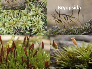
- 1. Bryopsida
- 2. General Characters: 1. It is the largest class in bryophyta and includes about 700 genera and 14,000 species. 2. The main plant body is gametophytic and can be differentiated into two stages- juvenile stage and leafy stage or gametophore. 3. Juvenile stage is represented by green, filamentous branched structures called protonema. It develops from the germination of the spore. 4. Gametophores are erect leafy branches which develop on the protonema. 5. Gametophores can be branched or un-branched and can be differentiated into three parts rhizoids, stem and leaves. 6. Branches arise below the leaves. 7. Leaves are with midrib, un-lobed and arranged spirally in three to eight rows on the axis. 8. Rhizoids are multicellular, filamentous, branched with oblique septa.
- 3. 9. The axis is differentiated into central conducting strand enclosed by cortex. 10. The sex organs (antheridia and archegonia) develop from the superficial cells of the gametophore. 11. The sporophyte is green in early stages and can be differentiated into foot, seta and capsule. 12. The seta is usually elongated. 13. The capsular wall remains interrupted by stomata at several places. 14. Columella is usually present and endothecial in origin. 16. Elaters are absent. 17. Dehiscence of capsule takes place by separation of lid or operculum. 18. Peristome helps in the dispersal of spores. 19. Spores on germination produce the protonema.
- 4. Classification • The class Bryopsida (Musci) has been divided into three sub-classes (1) Sphagnobrya (Sphagnidae); (2) Andreaeobrya (Andreaeidae), (3) Eubrya (Bryidae). I. Sub-class:Sphagnobrya: • The sub-class has a single order, the Sphagnales and a single family, the Sphagnaceae. (Single genus Sphagnum with 326 species). The characteristic features: 1. They are called 'bog mosses' or 'peat mosses'. 2. The protonema is broad and thallose and it produces one gametophore. 3. The leaves or gametophores lack mid-rib and usually composed of two types of cells- the narrow living green cells and large hyaline dead cells. 3. The branches arise in lateral clusters in the axis of the leaves. 4. The antheridia are borne in the axis of leaves on the antheridial branch. 5. The archegonia are terminal. 6. The sporogenous tissue of a sporophyte develops from the amphithecium. 7. The sporogonium remains elevated above the gametophyte.
- 5. II. Sub-class - Andreaeobrya: • This sub-class has a single order, the Andreaeales, and a singly family, the Andreaceceae. • The important genus is Andreaea. The characteristic features: 1. The gametophores are brittle, and can easily be broken. 2. There is practically no tissue differentiation in plant body. 3. The leaves are generally large, erect and convolute. 4. The archesporium and colonmella develop from the endothecium.
- 6. III. Sub-class Eubrya • 650 genera; 14,000 species • This sub-class has been further divided into fifteen orders. • The true mosses are included in this sub-class. The characteristic features: 1. The leaves of the gametophores are more than one cell in thickness and possess midrib on them. 2. The protonema is filamentous. 3. The sporophyte bears a well differentiated, elongated seta which pushes out the capsule from the gametophore. 4. The sporogenous tissue is derived from the endothecium. 5. The archesporium does not overarch the columella; 6. The columella continues upto the apex of the capsule; both columella and archesporium have been derived from the endothecium.
- 7. • Order- Sphagnales have single genus Sphagnum, which occupies a very distinct and isolated position among mosses. 1. The plants occur in large patches of a pale green or reddish colour. 2. Their growth has played a large part in the formation of peat. 3. The species are distributed in temperate and arctic climates, but in the tropics only occur at high levels. 4. The protonema forms a flat, lobed, thalloid structure attached to the soil by rhizoids. 5. The main shoot bears numerous branches which appear to stand in whorls. 6. The leaves have no midrib. 7. The antheridia are globular and have long stalks. 10. The archegonial groups occupy the apices of short branches. 11. The capsule, the wall of which bears rudimentary stomata, has a small operculum but no peristome.
- 8. Introduction to Sphagnum • Sphagnum is popularly known as bog moss, peat moss or turf moss because of its ecological importance in the development of peat or bog. • The plants are perennial and grow in swamps and moist habitat. • They grow along the bank of lakes. • Gradually encroach completely cover up the lake - bog. • Water of bogs becomes very acidic. • Upper portion of the Sphagnum gametophores grows indefinitely, while the basal part dies progressively. • The dead plant parts do not decompose easily in acidic soil. • Consequently a large mass of dead remains accumulated year after year followed by compression from plants on top, thus a compact, dark coloured substance rich in carbon is formed which is known as peat. • Since Sphagnum is the chief constituent of peat, it is often called peat moss.
- 10. Structure of Sphagnum: A. External Features: B. The gametophyte phase of Sphagnum is represented by two distinct stages namely, C. (a) juvenile protonema, and (b) mature leafy or gametophore stage. D. The mature plants grow in dense clumps and their shoots are of whitish or brownish green in colour. E. All species of Sphagnum accumulates water and often grow with bright colour (deep red, rose pink, etc.) due to the presence of water- soluble pigments, anthocyanin. F. They are perennial showing unlimited growth by means of an apical cell with three cutting faces. G. Very young gametophytes bear multicellular rhizoids with oblique septa. H. Mature gametophytes, however, do not bear rhizoids. I. It is differentiated into an upright branched axis and leaves.
- 11. Structure of Sphagnum: A. Main Axis and Branches: • The main axis is soft and weak at young stage, but becomes erect and stout at maturity. • However, the main axis is much longer in aquatic species, but is relatively short in terrestrial form. • The axis branches profusely on the lateral sides. • Single branch or in tufts of 3 to 8 branches arise from the axils of every fourth leaf of the main axis. • At the apex of the main stem, many small branches of limited growth are densely crowded forming a compact head called coma. • The coma is formed near the apex due to the condensed growth of apical internodes. • As the stem grows in length these short branches elongate and become normal branches.
- 13. • The submerged species (S. obesum, S. cuspidatum) have all the branches similar in form and structure, • Terrestrial species produce two types of branches viz., • (i) pendent branches, and (ii) upwardly divergent branches Pendent Branches: • These are long slender loosely arranged, turn downwards and then grow parallel to the main axis. • They are also termed flagella form or de-current branches. Divergent Branches: • These are short and stout branches which grow outwards and upwards. • They are also termed ex-current branches. • Sometimes, one divergent branch in each node develops strongly than others and ultimately gives rise to a new plant when it becomes detached from the mother plant.
- 15. Leaves: • The leaves occur both on the main axis as well as on the branches. • On the branches, the leaves are closely set and, therefore, overlapping and are placed apart on the main axis. • The leaves are arranged in spiral phyllotaxy. • Moreover, the leaves on the main axis differ from those on the branches in size, shape and details of cell structure. • In general, the leaves are small, sessile, entire, thin and scale-like with acute apex and without a midrib.
- 17. B. Internal Structure: • Stem: Internally, the stem shows three zones (a) Outer Cortex: • The cortex or the hyalodermis is the outermost region of the stem. • This is bounded externally by a single- layered epidermis. • It is composed of large hyaline cells. • The genus, Sphagnum has often been divided into two sub-genera based on the nature of hyaline cells. (b) Middle Hadrom: • It lies next to the cortex and consists of 4-6 layers of small thick-walled, prosenchymatous cells. • This part is called hadrom which gives mechanical support to the stem. (c) Central Cylinder or Medulla: • It is the innermost region of the stem, comprised of small, vertically elongated, thin-walled parenchymatous cells. • It functions as storage region.
- 19. Leaf: • In Sphagnum, the cross-section of leaf shows only one cell in thickness and composed of much elongated cells. • A young leaf is comprised of square or rectangular cells of uniform size, while a mature leaf is characterised by two types of cells, the ordinary type hyaline cells and the green chlorophyllous cells or the assimilatory cells. • The hyaline cells are large polygonal and become colourless or hyaline by losing their protoplasts. • Their walls are provided with pores and become spirally thickened. • The hyaline cells have a remarkable capacity of absorption and retention of water (hence called capillary cells), thus rhizoids are not necessary in the mature plants. • The chlorophyllous cells are small triangular or biconvex living cells with many discoid chloroplasts and have their photosynthetic ability. • The chlorophyllous and the hyaline cells are arranged in an alternate sequence to form a regular reticulate pattern and this leaf- feature alone can be used to identify the genus, Sphagnum.
- 23. Reproduction in Sphagnum • In Sphagnum, reproduction takes place both by vegetative and sexual methods. • The vegetative propagation is more common: Vegetative Reproduction: • Vegetatively, it reproduces by means of innovation. • Sometime, one of the divergent branches grows upwards and becomes as strong as the main stem. Such an apical branch is called innovation. • Due to the progressive death of the lower basal part of the main axis, the innovation gets detached from the mother plant and ultimately gives rise to a new plant. • This phenomenon is responsible for the extensive growth of Sphagnum in nature.
- 24. Sexual Reproduction: • Sphagnum may be monoecious or dioecious, • Antheridia and archegonia are always borne on the special separate antheridial and archegonial branches of the same plant. • These branches are much smaller than the vegetative branches. • In monoecious plants, the antheridial branches develop first. Antheridial Branch: • The antheridial branches first appear near the apex of the main stem. • These branches are usually shorter but stouter than the vegetative branches. • They are spindle-shaped and densely covered with yellow, red or dark green leaves generally smaller than the foliage leaves.
- 25. Sphagnum spp.(A) Antheridial Branch (B) Leaves and Antheridia (L.S. View) (C) Development of Antheridia (D) Mature Antheridium (E) Antherozoid
- 26. Antheridium Development and Structure of Antheridium: • The antheridia develop singly and acropetally below the leaves. • Each antheridium develops from a superficial antheridial initial of the stem. The Mature Antheridium: • It has a long stalk of two to four rows of cells and a globose body. • The body has a jacket of one layer of cells enclosing a mass of androcytes formed from the sperm mother cells. • Each androcyte cell metamorphoses into a spirally coiled biflagellate antherozoid or sperm. Dehiscence of the Antheridium: • The apical cells of the jacket of a mature antheridium swell through the absorption of water. • As a result of turgor pressure thus generated, the wall of the swollen antheridium breaks into a number of irregular lobes at the apex that eventually turns backwards. • The mass of androcytes comes out and the antherozoids are liberated immediately and swim freely in water.
- 28. Archegonial Branches: • Archegonia are borne at the apices of the archegonial branches which develop at the apex, or laterally. • The archegonial branches are very short and more or less ovoid in shape. • The leaves on these branches are larger than those present on the foliage leaves. • The upper leaves of these branches constitute the perichaetium enclosing the archegonia and thus protect archegonia from injury. Archegonium: The archegonia develop on the apex of the archegonial branches either singly or in groups. The apical cell of this branch forms the primary archegonium. Two to five secondary archegonia develop from derivatives of the apical cell. Usually, there are three archegonia in a group i.e., one primary archegonium at the apex and two secondary archegonia emerge from the base of primary archegonium.
- 29. Archegonial Branches: • Archegonia are borne at the apices of the archegonial branches which develop at the apex, or laterally. • The archegonial branches are very short and more or less ovoid in shape. • The leaves on these branches are larger than those present on the foliage leaves. • The upper leaves of these branches constitute the perichaetium enclosing the archegonia and thus protect archegonia from injury. Archegonium: The archegonia develop on the apex of the archegonial branches either singly or in groups. The apical cell of this branch forms the primary archegonium. Two to five secondary archegonia develop from derivatives of the apical cell. Mature Archegonium: The mature archegonium is a relatively large structure. It has a long stalk,
- 30. Mature Archegonium: • The mature archegonium is a relatively large structure. • It has a long stalk, a long twisted neck with 8 to 9 neck canal cells, a massive multilayered venter containing a ventral canal cell, and an egg. Fertilization of Archegonium: • The process of fertilization takes place only in the presence of water. • The antherozoids swim freely in water and reach the archegonia. • At maturity, the neck canal cells and the ventral canal cell disorganize and form a passage for the antherozoids. • The antherozoids reach near the archegonia attracted chemotactically and pass into the passage to reach the egg. • Ultimately, only one antherozoid fuses with the egg and forms a zygote.
- 31. Sphagnum Spp. (A) Mature Archegonia (B) Three Archegonia on the tip of a branch
- 32. The Sporophyte: • The diploid zygote is the first cell of the sporophytic generation. • Among the few archegonia only one is developed to form embryo in an archegonial branch. Structure of the Mature Sporophyte: • The mature sporophyte consists of a bulbous foot, a neck-like inconspicuous seta and an almost spherical black to dark-brown capsule. • The whole sporophyte is covered by the calyptra. • The lowest part of the calyptra that covers the foot is called the vaginula. • The perichaetial leaves are present below the sporophyte. • The elongated archegonial branch at the base of the sporogonium is called pseudopodium. • It increases in length and pushes out the capsule above the perichaetial leaves to facilitate the spreading of spores.
- 33. • The capsule in longitudinal section shows an outer jacket and middle spore-sac with spores which overarches the dome-shaped inner columella. • The capsule wall (jacket) is several layers thick. • The outermost layer of the jacket is thick which bears several rudimentary non-functional stomata. • The circular biconvex disc-shaped lid, called operculum, is present at the top of the jacket. • The operculum is delimited from the rest of the jacket by a groove of thin-walled cells, called the annulus.
- 35. Sphagnum: Different Stages in Sporophyte Development
- 36. Dehiscence of the Capsule: • The capsule dehisces on a bright sunny day by an explosive mechanism. • The capsule wall and columella become dry and shrivel due to heat. • This results in the formation of a large air space below the spore-sac. • The spherical capsule gradually becomes cylindrical and, therefore, an overpressure of 4-6 atmospheres builds up inside the capsule. • Under this condition its operculum bursts open through the annulus with an audible sound. • The spores are catapulted up to 20 cm and release in the air. The process is known as air-gun mechanism of spore dispersal.
- 38. • Each spore (tetrade) has a distinct triradiate ridge. • The wall of the spore is differentiated into an outer smooth granular or papillate exine and an inner thin intine. • Spores may germinate within 2-3 days or may remain viable for 4-6 months. • The spore on falling on a moist substratum germinates to develop a small thalloid primary protonema. • A single bud is developed from the marginal cell of the primary protonema. • The bud eventually develops into a new leafy gametophyte.
- 40. Alternation of Generation in Sphagnum
- 41. • MORPHOLOGY, ANATOMY AND REPRODUCTION OF EUBRYALES • 1. The main plant body is gametophytic and can be differentiated into two stages-juvenile stage and leafy stage or gametophore. • 2. Gametophores are erect leafy branches which develop on the protonema. Branched or un-branched and can be differentiated into three parts- rhizoids, ‘stem’ and ‘leaves’. • 3. ‘Leaves’ are with midrib, un-lobed and arranged spirally in three to eight rows on the axis or • 4. Rhizoids are multicellular, filamentous, branched with oblique septa. • 5. The axis is differentiated into central conducting strand enclosed by cortex. • 6. Sex organs borne apically in the groups on main ‘stem’ or a branch. • 7. The sporophyte is green in early stages and can be differentiated into foot, seta and capsule. • 8. The seta is usually elongated and rigid. • 9. Columella is usually present and endothecial in origin. • 10. Archesporiurn (spore forming tissue) is differentiated only in spores. • 11. Elaters are absent. • 12. Dehiscence of capsule takes place by separation of lid or operculum. • 13. Peristome helps in the dispersal of spores. • 14. Spores on germination produce the protonema. • 10.6.1-Funaria Division - Bryophyte Subdivision - Musci Class - Bryopsida Order - Funariales Family - Funariaceae Genus - Funaria Plant body is gametophytic and consists of two different stages namely: (i) Juvenile stage represented by primary protonema and (ii) The leafy gametophore which represents the adult form. The adult gametophyte (gametophore) is differentiated into rhizoids, axis or ‘stem’ and ‘leaves’. Rhizoids arise from the base of the axis. They are slender, branched, and multicellular and have oblique septa. Axis is 1—3 cm. high, upright, slender and branched. Each branch is extra axillary i.e., arise below a leaf. Leaves are sessile, oblong-ovate with entire margin, pointed apex and are arranged spirally on the branches and ‘stem’. Each ‘leaf’ is traversed by a single mid rib. ‘Leaves’ are borne in 1/3 phyllotaxy which becomes 3/8 at maturity.
- 43. Funaria Plant and Leaf
- 44. • (ii) Internal Structure: • 1. Axis or ‘stem’ The transverse section (T. S.) of axis can be differentiated into three distinct regions: (i) Epidermis (ii) Cortex (iii) Central conducting strand or central cylinder. (i) Epidermis: It is the outer most single layered protective covering consisting of small tangentially elongated chlorophyll bearing cells. Cuticle and stomata are absent. (ii) Cortex: It is present between the epidermis and conducting tissue. It is made up to parenchymatous cells. Younger part of the cortex contains chloroplasts but in the older part they are lacking. At maturity few outer layers of cortex become thick walled and are reddish brown in colour but those of the inner layers become thin walled. (iii) Central Conducting Strand: It is made up of long, narrow thin walled dead cells which lack protoplasm. These cells are now commonly called as hydroids. Conducting strand besides providing a certain amount of mechanical support, functions in the upward conduction of water and solutes.
- 45. Transverse Section of Axis
- 46. • 2. Leaf: Transverse section (T. S.) of ‘leaf’ shows a well-defined midrib with two lateral wings. Except the midrib region, the ‘leaf’ is composed of single layer of parenchymatous polygonal cells. The cells contain many large and prominent chloroplasts. The central part of the mid rib has narrow conducting strand of thick walled cells which help in conduction.
- 48. • Reproduction in Funaria: Funaria reproduces by vegetative and sexual methods. Vegetative Reproduction in Funaria: Vegetative propagation in Funaria is performed by the following methods: (a) Fragmentation of Primary Protonema: The primary protonema is developed through the germination of the spore. Under certain circumstances, it breaks into several fragments. Each detached fragment bearing buds may grow into a new plant. (b) Secondary Protonema: The protonema developing from any part of the plant other than spores is called secondary protonema. Generally, they are formed on injured rhizoids, stems, leaves or reproductive structures. They bear buds that are capable of growing into a new plant (Fig. 6.48B). (c) Bulbil: The bulbils are multicellular, brown, bud-like structures that develop on the rhizoidal branches. The bulbils are useful for propagation during unfavourable environmental conditions by detaching them from the parent
- 50. • Sexual Reproduction in Funaria: Funaria is autociously monoecious, because the male (antheridium) and female (archegonium) reproductive structures develop on separate shoots of the same plant. Antheridia are borne on the main shoot of the plant. The female branch develops as a side shoot, which grows more vigorously and becomes longer than the male branches. Antheridium: The antheridia are borne in clusters at the apex of the main axis. A number of long multicellular hairs, called paraphyses are intermingled with the antheridia (Fig. 6.53). Both antheridia and paraphyses are surrounded by a number of bract-like leaves forming a rosette called the perichaetium. The paraphyses have swollen tips (capitate) and contain chloroplasts. Besides their photosynthetic function, paraphyses protect the young antheridia against desiccation. The paraphyses assist in the liberation of antherozoids.
- 52. • Development of the Antheridium: The antheridium develops from a superficial antheridial initial located at the tip of the male branch. It becomes papillose and projects above. It divides by a transverse wall to form an outer cell and a basal cell. The outer cell divides further by successive transverse divisions to form a linear filament of 2 to 4 cells. The terminal cell of the filament divides by two vertical intersecting walls to form a wedgeshaped apical cell with two cutting faces. It forms segments in two rows in alternate sequence. Each young segment of the upper 3 to 4 cells now divides by a vertically diagonal wall to form two unequal cells. The smaller peripheral cells are the first jacket initials. While, the larger sister cell, by a similar division, forms the outer second jacket initials and the inner primary androgonial cell. The primary androgonial cell divides and re-divides to form androcyte mother cells. Each androcyte mother cell divides to
- 55. • Archegonium: The archegonia are borne in clusters at the apex of the archegonial branch. Development of the Archegonium: A cell at the tip of the female shoot differentiates into the archegonial initial. It divides transversely to form a upper cell and a lower cell. The upper cell becomes the archegonial mother cell which divides by two intersecting oblique walls forming an apical cell with two cutting faces. The apical cell further divides by three
- 56. • Development of the Sporophyte: The zygote divides transversely to form an upper epibasal cell and a lower hypobasal cell. Both the hypobasal and epibasal cells divide repeatedly to form an young embryo with two growing points at the two opposite ends, each representing an apical cell with two cutting faces. The archegonial wall enlarges and forms calyptra which covers the capsule till maturity. A long slender sporophyte is then
- 57. • Structure of the Mature Sporophyte: The mature sporophyte of Funaria is differentiated into a foot, a long seta and a pear- shaped capsule at the tip. 1. Foot: It is a poorly developed conical structure, embedded in the apex of archegonial branch. 2. Seta: Seta is long, green in colour when young, but becomes reddish brown at maturity. T.S. of seta shows a single-layered epidermis, a central conducting strand of thin-walled cells
- 58. Funaria hygromatica (A) T.S. of Seta (B) Mature Capsule with Operculum (C) L.S. of Capsule
- 61. • (d) Columella: It is the central, axial part of the fertile zone, comprising of thin-walled, colourless, compact, parenchymatous cells, constricted at the base just above the apophysis. The distal part of the columella is cone-shaped which projects into the concavity of the operculum. The columella serves the purpose of conduction of water and nutrients to the growing sporophyte. The Apical Region: The apical region of the capsule is a
- 62. • The whole structure is called peristome which is epicranoid in nature, because the outer peristome teeth are superposed on the inner ring. The tapering distal ends of the outer peristome teeth are joined to a centrally placed disc of tissue. Dehiscence of the Capsule and the Dispersal of Spores: At maturity, the operculum begins to dry up due to the non-availability of water supply to the capsule. Consequently, the thin-walled cells of
- 63. Life Cycle of Funaria hygromatica
- 65. Funaria hygromatica: Graphic Presentation of Life Cycle
- 67. Polytrichum Occurrence: • Polytrichum have worldwide distribution. • They are very common in cool temperature and tropical regions. • Plants live in cool and shady places. General structure: • The main plant body is gametophyte. • The adult plant consists of two parts: rhizome and upright leafy shoot. 1. Rhizome: • It is horizontal portion and grows underground. • It bears three rows of small brown or colourless leaves. • It also bears rhizoids. • The cells are rich in protoplasm and oil globules. • The rhizoids may arise from base of the erect gametophores. • They serve as mechanical function by providing support in species in which gametophores grows to considerable heights.
- 69. 2. Upright leafy shoot: • The leafy shoots are much longer. • It is the most conspicuous part of the plant. • It arises from rhizome. • These branches consist of central axis. • These branches bear large leaves arranged spirally. 3. Leaves: • Leaves have broad bases. • Leaves in the upper portion are green. • But the lower ones are brown. • Each leaf has a broad colourless sheathing leaf base and narrow distal limb. • The midrib forms the major part of the leaf. • These leaves possess extra photosynthetic tissue in the form of closely set vertical plates of green cells. These are known as lamellae. • Green lamellae act as additional photosynthetic tissue
- 70. The leaves are of two types: a) Scale leaves:- • These are small brown or almost colourless leaves with a rudimentary blade. • These occur in a spiral arrangement around the central axis. b) Foliage leaves: - • These are only present in upper portion of the branches. • Each foliage leaf is 6-10mm long it is differentiated into the proximal sheath base and the distal diverging narrow limb or blade. Anatomy Leaf: • Polytrichum have complex internal structure. • The mid-rib region is thick. • But the margins are only one cell thick. • The lower surface is bounded by epidermis. • One or two layers of sclerenchymatous tissues are present above the epidermis.
- 72. • The central tissue of leaf is composed of thin-walled parenchymatous tissues. • Above this are again sclerenchymatous cells. • The upper surface is formed of a layer of large cells from which arise numerous lamellae. • This upper portion is the main photosynthetic region of the leaf. Stem: • The T.S. of stem shows three regions: • medulla, cortex and epidermis. • The medulla is again differentiated into two zones: central zone and peripheral zone. • The cortex consists of thick-walled cells. • The innermost layer of cortex around the conducting strands is known as a mantle. • Its cells contain starch grain. • Epidermis is present over the cortex.
- 76. Vegetative reproduction • Vegetative reproduction takes place by following methods: I. Protonema: • The spores germinate to form protonema. • Several buds grow on the protonema. • Each bud by of its apical cell develops into gametophyte. 2. Buds • These are also called vegetative buds. • They are formed on the rhizoids. 3. Fragmentation: • Death or breaking of shoots separates the erect branches. • These branches behave as independent plants.
- 77. Sexual reproduction • Polytrichum is dioecious. • Antheridia archegonia occur on different plants. Antheridial head: • The antheridia are borne in the axillary clusters at the tips of leafy stems. • They are surrounded by a rosette of leaves called perigonial leaves. • The perigonial leaves are spirally arranged. • The antheridia are produced in groups in the axils of these leaves. • Paraphyses also occur among the antheridia. • Mature antheridium is club shaped. • It is composed of a short stalk and a club-shaped body. • Jacket is present around the capsule. • Inside the jacket are present androcyte mother cells. • They give rise to biflagellate sperms.
- 79. Archegonial head • The flask-shaped archegonia are borne at the apices of leafy stems. • Archegonium is surrounded by perichaetial leaves. • These leaves overlap to form a closed bud-like structure. • The archegonia occur in cluster of 3 to 6. • Mature archegonium is flask-shaped. • It has a thick multicellular stalk. • The neck is long and twisted. • It contains neck canal cells. The neck consists of 6-vertical rows of cells. • Neck gradually merges into venter. • Venter contains upper small venter canal cell and lower large egg cell. • Paraphyses are absent.
- 81. Fertilization: • The sex organs dehisce in the presence of water. • The venter canal cell and the neck canal cells dissolve to form mucilage. • This mucilage exerts pressure and the neck opens out. • The mucilage comes out of the neck. • The sperms reached the archegonial heads by rain water. • They are attracted towards the archegonia. • One of the sperm swims down the open neck and reaches the base. • It fuses with the egg to form oospore. • Oospore is the first stage of sporophytic generation.
- 82. Structure of Mature Sporogonium (Sporophyte) • The mature sporogonium is differentiated into foot, seta and capsule. Foot: • The foot is buried deep in the tissue of gametophyte. • It is absorptive in function. • It consists of thin-walled narrow cells containing dense cytoplasm. Seta: • The seta is several inches long. • It carries the capsule high into the air. • It also conducts water and food. • It consists of epidermis, cortex and central conducting strands. Capsule: • The upper part is capsule with quadrangular outline. • It is differentiated into three regions: • apophysis, theca and operculum.
- 85. Apophysis • The apophysis is the lowermost part of the capsule (also called the neck). • It is a bulbous, sterile structure that connects the capsule with the seta below. • Structurally, the apophysis is composed of a single-layered, thick-walled epidermis, which is interrupted by stomata. • The sotmata consist of a single ring of guard cells. • Usually, two guard cells are present in each stoma. • Below the epidermis is the chlorenchyma, consisting of many thin-walled, loosely arranged parenchymatous cells. • There is no hypodermis in the apophysis region. • The central part of the apophysis is composed of numerous thin- walled cells, forming a conducting strand.
- 86. Theca • The theca is the middle, fertile region of the capsule. • It lies between the apophysis and the operculum. • A longitudinal section of the theca shows the following regions: Capsule Wall • The capsule wall, or jacket, is several layers thick and composed of parenchymatous cells. • The outermost layer of the capsule wall is the single-layered epidermis. • It consists of compactly arranged cells with thick outer walls. • The epidermis of the wall lacks stomata. • The epidermis is followed by typically two layers of spongy parenchyma. • The cells of the spongy parenchyma are thin-walled and chlorophyllous.
- 87. Spore Sacs • The spore sacs are situated below the outer air cavities on either side of the columella. • Each spore sac has an outer wall and an inner wall. • Both walls are made up of two thin-walled cell layers. • The spore sacs are derived from the single-layered archesporium (sporogenous tissue). • The archesporium first develops 4-6 layers of sporogenous cells. • The sporogenous cells divide to form many spore mother cells. • The spore mother cells divide by meiotic divisions and form haploid spores. • The spore sac in Polytrichum is surrounded by a double air space system, as the air cavities are found on both its outer and inner sides.
- 88. Air Cavities • The air cavities (air spaces) are cylindrical and are present on both sides of the spore sacs. • These air spaces are traversed radially by delicate strands (short filaments) of elongated green cells called trabeculae. Columella • The columella is the central part of the theca region. • It is made up of compactly arranged parenchymatous cells. • The columella is continuous with the central strand of the apophysis below and extends up to the epiphragm of operculum above. • Columella helps in the conduction of water and mineral nutrients.
- 89. Operculum: • This is the uppermost part of the capsule. • It is conical. The operculum is covered by calyptra. • The calyptra forms a hairy structure. So Polytrichum is also known as ‘ hair cap’ moss. • A constriction is present between operculum and theta. • A rim or diaphragm is present at the base of this constriction. • It expands into a fan-shaped epiphragm. • Peristome is present in the form of a thick rim. • It bears a number of rigid teeth. • The epiphragm fills the space inside the ring of peristome teeth and is attached to their tips. • Peristome teeth arise from the rim or diaphragm.
- 90. • Spores lie free in the centre of the capsule at maturity and come out through pores. • They are dispersed by wind. Structure and germination of spores • The spores are yellow. • Each spore is uninucleate and has two wall layers. • The outer layer is exosporium (exine). • The inner layer is endosporium. • The spore germinates under favourable conditions. • Exosporium ruptures and endosporium comes out. • It forms protonema. • Protonema develops many buds. • These buds produce new moss plants. • Alternation of generation Polytrichum shows heteromorphic alternation of generation.
- 91. • Andreaea Division – Bryophyte • Subdivision - Musci Class - Andreaeopsida Order - Andreaeales Family - Andreaeaceae Genus - Andreaea Andreaea is one of two moss genera belonging to the class of mosses known as Andreaeidae and the family Andreaceae. Andreaeidae, referred to as the granite mosses, are commonly found on granite rock faces in mountainous and arctic regions. Andreaea, a genus comprised of
- 92. (A) Andreaea rupestris Gametophyte with Sporophyte (B) T.S. Stem
- 93. • Sexual Reproduction: Andreaea is homothallic i.e. monoecious. Antheridia and archegonia are borne terminally in groups on the separate branches. A few species for instance A. nivalis and A. blyttii are heterothallic. Apical cell is involved in the formation of sex organs. Antheridia It occurs in the male branch in the terminal as cluster. The antheridium is surrounded by a number of male perigonial bracts. The mature antheridium has an
- 94. • wall. Further development is similar to other mosses and depends on the divisions of apical cell. Primary stalk cell undergoes repeated divisions to form the stalk. Archegonia Like the antheridia, the archegonia arise from surface cells at the apex of the female branches. A surface cell protrudes and becomes more or less papilliform. The first wall formed in it shows a little variation, but generally it is very oblique, so cutting off a two-sided apical cell,
- 95. • The Sporophyte The zygote divides by a transverse wall. The lower (hypobasal) cell divides irregularly and forms a haustorial tissue which later organizes into the foot. The upper (epibasal) cell is destined to form the capsule. It first functions as an apical cell with two cutting faces and soon, after periclinal divisions, develops an inner endothecium and an outer amphithecium. The amphithecium forms the jacket, 3 to 8 cells in thickness. The
- 96. Andreaea rupestris (A) Sporophyte (B) Section of Sporogonial Shoot (C) Sporangial Shoot bearing Sporophyte
- 97. • The capsule is easily recognized by its paper- lantern-like dehiscence. Lacking peristome teeth, the capsule splits along four longitudinal slits as a result cell weakness. The longitudinal valves are responsive to humidity, which allows the spores to be carried away when the air is dry and conditions are optimal, closing when the air becomes moist. This dehiscence type is a unique characteristic and occurs exclusively among the granite mosses.
- 98. Andreaea: (A-E) Germination of Spore (E) Ribbon like Protonema
- 99. • MORPHOLOGY, ANATOMY AND REPRODUCTION OF TAKAKIALES • It is also a monotypic group having single family Takakiaceae and single genus Takakia. The genus is characterized by: 1. Heterotrichous nature of plants, which are differentiated into prostrate growing rhizomatous axis and erect growing aerial axis or shoot. 2. Both the shoots are densely covered with mucilage papillae. 3. The leaves
- 100. • establishment in 1958 of the species Takakia lepidozioides, in a new genus Takakia, named to honor the man who rediscovered it and recognized its unique characteristics. The species originally described by Mitten was subsequently recognized by Grolle as belonging to this new genus, and accordingly renamed Takakia ceratophylla. All of the plants originally collected lacked any reproductive structures; they were sterile
- 101. Takakia spp. Gametophyte with Sporophyte
- 103. • From a distance, Takakia looks like a typical layer of moss or green algae on the rock where it grows. On closer inspection, tiny shoots of Takakia grow from a turf of slender, creeping rhizomes. The green shoots which grow up from the turf are seldom taller than 1 cm, and bear an irregular arrangement of short, finger-like leaves (1 mm long). These leaves are deeply divided into two or more filaments, a characteristic not found in any
- 104. • The central strand of the medullary cells is always surrounded by a single layered cortex of cells smaller in size. Stem Anatomy: Stem T.S. shows that it consists of two zones viz. outer cortical and inner medullary region. The cortical region is chlorophyllose. It is 1-2 stratose thick and consists of slightly strongly thick walled cortical cells with brownish wall. The medullary region shows differentiation into a small central core of small celled tissue
- 105. • All cells of the archegonium, including the egg cell, are produced by mitosis of haploid gametophyte cells. The disintegrating neck and ventral canal cells provide chemicals involved in sperm chemotaxis to fuse with the egg. After fusion of egg and sperm, zygote is formed which is diploid. After fertilization, the sporophyte grows out of the archegonium, and nutrients for the developing sporophyte are provided by the gametophyte.
- 107. • The lower part i.e. foot and seta, is protected by vaginula and the upper part of the capsule and some part of seta is protected by calyptra. Capsule is schizocarpous erect, elliptical, green in early stage of development. Thus sporophyte is autotrophic in nature. Capsule is thick in the middle region and symmetrically tapered at base and apex. Operculum, annulus and peristome are absent. The outermost layer of the capsule wall has characteristic
- 109. • Characteristic features of the order takakiales (i) The plant body is differentiated into small, creeping pale-yellow, leafless rhizome and erect, green, leafy 1- 1.5 cm tall shoot. These are known as the Gametophores. (ii) Both the rhizome and the leafy shoots are devoid of rhizoids. (iii) The thick, fleshly, erect, soft- textured gametophore axis bears green leaflike appendages in a 3 - ranked phyllotaxy. The Leaf - like appendages are small fleshy,
- 110. Takakia sp. A, Rhizomatous gametophore of T. ceratophylla; B. Portion of gametophore of T. lepidoziodies with phyllids; C. a single undivided phyllid; D, E, F, bifid, trifid and quadrified phyllids respectively; G, T.s. Simple Phyllid; H, T.S. Phyllid of Complex construction, I. Archegonal short.
- 111. • ceratopylla was reported from the pool in Sikkim in Eastern Himalayas ( India). • Thus both the species grow in cool, moist or wet shady places at higher altitutes. • They are hygrophytes. • 4B.1.B STRUCTURE : • Morphological Structure of Gametophyte - • The plant body is gametophyte which is differentiated into a cylindrical,
- 112. • more vertically descending leafless branches which grow downward into the • substratum. These positively geotropic, leafless axis is fleshy and soft textured • The leaf-like appendages borne on the erect gametophore axis are unique in • form. They are isophyllous in a spiral manner from the shoot tip which grows by • the activity of a 3-sided apical cell. The leaves are arranged in 3 - ranked phylotaxy.
- 113. • Slime Papillae - Proskauer (1942) reported the occurrence of slime papillae on • the axis of Takakia . These are of two types - (i) Non - Beaked and (ii) Beaked • slime Papillae. • (i) Non - Beaked slime Papillae - These occurs singly on the erect shoots in or • near the leaf axils. The non - beaked slime Papillae in T. ceratophylla cornsists
- 114. Takakia sp. A, Immature beaked open slime papillae; C, stalked “closed” slime papillae; D, cells from the leaf primordia showing chloroplasts & nuclei; E, T.S. aerial stem.