Microbiological Aspect of brucellosis
introduction
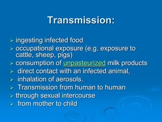
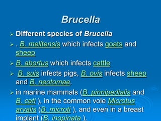
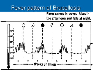
Brucella is a genus of Gram-negative
bacteria.
They are small (0.5 to 0.7 by 0.6 to
1.5 µm) non-motile, non-
encapsulated coccobacilli, which
function as facultative intracellular
parasites.
Transmission:
 ingesting infected food
 occupational exposure (e.g. exposure to
cattle, sheep, pigs)
 consumption of unpasteurized milk products
 direct contact with an infected animal,
 inhalation of aerosols.
 Transmission from human to human
 through sexual intercourse
 from mother to child
Brucella
 Different species of Brucella
 . B. melitensis which infects goats and
sheep
 B. abortus which infects cattle
 B. suis infects pigs, B. ovis infects sheep
and B. neotomae.
 in marine mammals (B. pinnipedialis and
B. ceti ), in the common vole Microtus
arvalis (B. microti ), and even in a breast
implant (B. inopinata ).
Fever pattern of Brucellosis
Brucillosis:Microbiological Aspects
 associated with many other febrile diseases, but
with emphasis on muscular pain and sweating.
 Duration of the disease: few weeks to many
months or even year
 In the first stage of the disease, septicaemia occurs
and leads to the classic triad of fever, sweating and
migratory arthralgia and myalgia.
 granulomatous hepatitis, arthritis, spondylitis,
anaemia, leukopenia, thrombocytopenia, meningitis,
uveitis, optic neuritis, endocarditis and various
neurological disorders collectively known as
neurobrucellosis
 endocarditis
Diagnosis
 Brucella is isolated from a blood culture on
Castaneda medium.
 Prolonged incubation (up to 6 weeks)
Castaneda bottle
Brucillosis:
 On Gram stain they appear as dense
clumps of Gram-negative coccobacilli and
are exceedingly difficult to see
 Treatment and prevention
 Antibiotics like tetracyclines, rifampicin and the
aminoglycosides streptomycin and gentamicin are
effective against Brucella bacteria. However, the use of
more than one antibiotic is needed for several weeks,
because the bacteria incubate within cells.
 The gold standard treatment for adults is daily
intramuscular injections of streptomycin 1 g for 14 days
and oral doxycycline 100 mg twice daily for 45 days
 Gentamicin 5 mg/kg by intramuscular injection once daily
for seven days is an acceptable substitute when
streptomycin is not available or difficult to obtain
 Another widely used regimen is doxycycline plus
rifampin twice daily for at least six weeks
 triple therapy of doxycycline, with rifampin and
cotrimoxazole, has been used successfully to treat
neurobrucellosis
 Prevention:
 by pasteurizing all milk that is to be
ingested by human beings, either in its
unaltered form or as a derivate, such as
cheese.
Diagnosis of brucellosis relies on:
 Demonstration of the agent: blood cultures in tryptose
broth,
 bone marrow cultures. The growth of brucellae is extremely
slow .the culture poses a risk to laboratory personnel due to
high infectivity of brucellae.
 Demonstration of antibodies against the agent either with
ELISA or the 2-mercaptoethanol assay for IgM antibodies
associated with chronic disease
 Histologic evidence of granulomatous hepatitis (hepatic
biopsy)
 Radiologic alterations in infected vertebrae: the Pedro Pons
sign (preferential erosion of antero-superior corner of lumbar
vertebrae) and marked osteophytosis are suspicious of
brucellic spondylitis.
 The disease's sequelae are highly variable and may include
granulomatous hepatitis, arthritis, spondylitis, anaemia,
leukopenia, thrombocytopenia, meningitis, uveitis, optic
neuritis, endocarditis and various neurological disorders
collectively known as neurobrucellosis.
 Identification and diagnosis
 From humans the organism is most commonly
isolated from blood or bone marrow, but may be
isolated from the liver, the spleen, cerebrospinal
fluid or focal abscess. From sheep, goats, and
cattle, the organism is most commonly cultured from
the reproductive tract or reproductive fluids,
including semen, uterine fluids and tissues, and
milk.
 Standard blood media may be used for blood or
bone marrow specimens (enriched culture, other
specimens may use Trypticase soy agar with 5%
sheep blood agar, MacConkey agar, or Martin Lewis
agar.
 Optimal Temperature: 35-37 degrees Celsius. Plate
cultures should be incubated in 5% carbon dioxide.
It takes 3-7 days to form colonies on plates.
 It is crucial to be able to differentiate
Brucella from Salmonella which could also
be isolated from blood cultures and are
Gram-negative. Testing for urease as it is
positive for the Brucella and negative for
the Salmonella.
Public health
 When brucellosis is suspected, clinicians should note "suspect or
rule out brucellosis" on the laboratory submission
 Review laboratory containment methods and microbiological
procedures to ensure compliance with recommendations in the
Biosafety in Microbiological and Biomedical Laboratories (BMBL).
 Use primary barriers: use safety centrifuge cups, personal protective
equipment, and class II or higher Biological Safety Cabinets (BSCs)
for procedures with a high likelihood of producing droplet splashes
or aerosols.
 Use secondary barriers: restrict access to the laboratory when work
is being performed and maintain the integrity of the laboratory’s air
handling system by keeping external doors and windows closed.
 Perform all procedures on unidentified isolates carefully to minimize
the creation of splashes or aerosols.
 Prohibit sniffing of opened culture plates to assist in the identification
of isolates.
 Manipulate isolates of small gram-negative or gram-variable rods
within a BSC.
 Serology for brucellosis
 Serology for brucellosis is a blood test to look for antibodies against
Brucella, the bacteria that causes the disease brucellosis.
 How the Test is Performed
 Blood is typically drawn from a vein, usually from the inside of the elbow or
the back of the hand. The site is cleaned with germ-killing medicine
(antiseptic). The health care provider wraps an elastic band around the
upper arm to apply pressure to the area and make the vein swell with blood.
 Next, the health care provider gently inserts a needle into the vein. The
blood collects into an airtight vial or tube attached to the needle. The elastic
band is removed from your arm. Once the blood has been collected, the
needle is removed, and the puncture site is covered to stop any bleeding.
 In infants or young children, a sharp tool called a lancet may be used to
puncture the skin and make it bleed. The blood collects into a small glass
tube called a pipette, or onto a slide or test strip. A bandage may be placed
over the area if there is any bleeding.
 The blood is then tested in a laboratory to look for antibodies. For Brucella,
the serum agglutination test (SAT) is the simplest and most widely used
testing method.
 DIAGNOSTIC WORKUP
 Routine studies include a CBC, sedimentation rate, chemistry panel,
urinalysis, chest x-rays, VDRL test, and tuberculin skin test. Serial blood
cultures should be done on all patients. Febrile agglutinins usually should be
done. An ASO titer or streptozyme test should be done to exclude rheumatic
fever. RNA, ANA, and DNA tests should be done to look for lupus and other
connective tissue disease. An HIV antibody titer may need to be ordered.
 The next step is to culture any discharge or various body fluids that might be
suspect. Thus, a urinalysis and urine culture should be done. A nose and throat
culture should be done. A sputum smear and culture may need to be done. The
next consideration is to do various serologic tests. A heterophile antibody titer
should be done in teenagers. Febrile agglutinin tests may need to be done.
Acute and convalescent phase sera for viral studies may need to be done.
 Next one should do skin testing. Thus, histoplasmin, coccidioidin, and
blastomycin skin testing should be done on patients with a cough. Trichinella
skin testing may need to be done, as well as brucellin skin testing. A Kveim test
might need to be done for suspected sarcoidosis.
 The next step is to do plain x-rays of suspected areas. For instance, x-rays of
the teeth may disclose an abscessed tooth. X-rays of the long bones may
disclose a metastatic carcinoma.
 The next step is contrast x-ray studies of various organ systems. An intravenous
pyelogram may show a hypernephroma. A cholecystogram may show
gallstones. An upper GI series and barium enema may show chronic
pancreatitis or diverticulitis. Angiography may disclose periarteritis nodosa,
aortitis or giant cell arteritis.
 The next step is to do a CT scan of the abdomen and pelvis. If this is negative,
consider a CT scan of the chest and mediastinum. Echocardiography may
disclose valvular vegetations or an atrial myxoma.
 Next, consider biopsying various organ systems. For instances, a lymph node
biopsy may disclose a lymphoma or sarcoidosis. A muscle biopsy may disclose
periarteritis nodosa, polymyositis, or trichinella.
 Next one should do bone scans and gallium scans for possible metastasis,
osteomyelitis, or localized abscesses.
 If all these procedures fail to turn up a lesion, then an exploratory laparotomy
may need to be done. A fibrin test may indicate Mediterranean fever, or urine for
etiocholanolone may also indicate a relapsing type of fever. A urine test for
porphobilinogen may diagnose porphyria.
 The wisest move is to conduct this investigation with the help of an infectious
disease specialist or a specialist in the body organ system most likely suspected
of harboring the infection

Brucella

  • 1.
  • 3.
    introduction Brucella is agenus of Gram-negative bacteria. They are small (0.5 to 0.7 by 0.6 to 1.5 µm) non-motile, non- encapsulated coccobacilli, which function as facultative intracellular parasites.
  • 4.
    Transmission:  ingesting infectedfood  occupational exposure (e.g. exposure to cattle, sheep, pigs)  consumption of unpasteurized milk products  direct contact with an infected animal,  inhalation of aerosols.  Transmission from human to human  through sexual intercourse  from mother to child
  • 5.
    Brucella  Different speciesof Brucella  . B. melitensis which infects goats and sheep  B. abortus which infects cattle  B. suis infects pigs, B. ovis infects sheep and B. neotomae.  in marine mammals (B. pinnipedialis and B. ceti ), in the common vole Microtus arvalis (B. microti ), and even in a breast implant (B. inopinata ).
  • 6.
    Fever pattern ofBrucellosis
  • 7.
    Brucillosis:Microbiological Aspects  associatedwith many other febrile diseases, but with emphasis on muscular pain and sweating.  Duration of the disease: few weeks to many months or even year  In the first stage of the disease, septicaemia occurs and leads to the classic triad of fever, sweating and migratory arthralgia and myalgia.  granulomatous hepatitis, arthritis, spondylitis, anaemia, leukopenia, thrombocytopenia, meningitis, uveitis, optic neuritis, endocarditis and various neurological disorders collectively known as neurobrucellosis  endocarditis
  • 8.
    Diagnosis  Brucella isisolated from a blood culture on Castaneda medium.  Prolonged incubation (up to 6 weeks)
  • 9.
  • 10.
    Brucillosis:  On Gramstain they appear as dense clumps of Gram-negative coccobacilli and are exceedingly difficult to see
  • 11.
     Treatment andprevention  Antibiotics like tetracyclines, rifampicin and the aminoglycosides streptomycin and gentamicin are effective against Brucella bacteria. However, the use of more than one antibiotic is needed for several weeks, because the bacteria incubate within cells.  The gold standard treatment for adults is daily intramuscular injections of streptomycin 1 g for 14 days and oral doxycycline 100 mg twice daily for 45 days  Gentamicin 5 mg/kg by intramuscular injection once daily for seven days is an acceptable substitute when streptomycin is not available or difficult to obtain  Another widely used regimen is doxycycline plus rifampin twice daily for at least six weeks  triple therapy of doxycycline, with rifampin and cotrimoxazole, has been used successfully to treat neurobrucellosis
  • 12.
     Prevention:  bypasteurizing all milk that is to be ingested by human beings, either in its unaltered form or as a derivate, such as cheese.
  • 13.
    Diagnosis of brucellosisrelies on:  Demonstration of the agent: blood cultures in tryptose broth,  bone marrow cultures. The growth of brucellae is extremely slow .the culture poses a risk to laboratory personnel due to high infectivity of brucellae.  Demonstration of antibodies against the agent either with ELISA or the 2-mercaptoethanol assay for IgM antibodies associated with chronic disease  Histologic evidence of granulomatous hepatitis (hepatic biopsy)  Radiologic alterations in infected vertebrae: the Pedro Pons sign (preferential erosion of antero-superior corner of lumbar vertebrae) and marked osteophytosis are suspicious of brucellic spondylitis.  The disease's sequelae are highly variable and may include granulomatous hepatitis, arthritis, spondylitis, anaemia, leukopenia, thrombocytopenia, meningitis, uveitis, optic neuritis, endocarditis and various neurological disorders collectively known as neurobrucellosis.
  • 14.
     Identification anddiagnosis  From humans the organism is most commonly isolated from blood or bone marrow, but may be isolated from the liver, the spleen, cerebrospinal fluid or focal abscess. From sheep, goats, and cattle, the organism is most commonly cultured from the reproductive tract or reproductive fluids, including semen, uterine fluids and tissues, and milk.  Standard blood media may be used for blood or bone marrow specimens (enriched culture, other specimens may use Trypticase soy agar with 5% sheep blood agar, MacConkey agar, or Martin Lewis agar.  Optimal Temperature: 35-37 degrees Celsius. Plate cultures should be incubated in 5% carbon dioxide. It takes 3-7 days to form colonies on plates.
  • 15.
     It iscrucial to be able to differentiate Brucella from Salmonella which could also be isolated from blood cultures and are Gram-negative. Testing for urease as it is positive for the Brucella and negative for the Salmonella.
  • 16.
    Public health  Whenbrucellosis is suspected, clinicians should note "suspect or rule out brucellosis" on the laboratory submission  Review laboratory containment methods and microbiological procedures to ensure compliance with recommendations in the Biosafety in Microbiological and Biomedical Laboratories (BMBL).  Use primary barriers: use safety centrifuge cups, personal protective equipment, and class II or higher Biological Safety Cabinets (BSCs) for procedures with a high likelihood of producing droplet splashes or aerosols.  Use secondary barriers: restrict access to the laboratory when work is being performed and maintain the integrity of the laboratory’s air handling system by keeping external doors and windows closed.  Perform all procedures on unidentified isolates carefully to minimize the creation of splashes or aerosols.  Prohibit sniffing of opened culture plates to assist in the identification of isolates.  Manipulate isolates of small gram-negative or gram-variable rods within a BSC.
  • 17.
     Serology forbrucellosis  Serology for brucellosis is a blood test to look for antibodies against Brucella, the bacteria that causes the disease brucellosis.  How the Test is Performed  Blood is typically drawn from a vein, usually from the inside of the elbow or the back of the hand. The site is cleaned with germ-killing medicine (antiseptic). The health care provider wraps an elastic band around the upper arm to apply pressure to the area and make the vein swell with blood.  Next, the health care provider gently inserts a needle into the vein. The blood collects into an airtight vial or tube attached to the needle. The elastic band is removed from your arm. Once the blood has been collected, the needle is removed, and the puncture site is covered to stop any bleeding.  In infants or young children, a sharp tool called a lancet may be used to puncture the skin and make it bleed. The blood collects into a small glass tube called a pipette, or onto a slide or test strip. A bandage may be placed over the area if there is any bleeding.  The blood is then tested in a laboratory to look for antibodies. For Brucella, the serum agglutination test (SAT) is the simplest and most widely used testing method.
  • 18.
     DIAGNOSTIC WORKUP Routine studies include a CBC, sedimentation rate, chemistry panel, urinalysis, chest x-rays, VDRL test, and tuberculin skin test. Serial blood cultures should be done on all patients. Febrile agglutinins usually should be done. An ASO titer or streptozyme test should be done to exclude rheumatic fever. RNA, ANA, and DNA tests should be done to look for lupus and other connective tissue disease. An HIV antibody titer may need to be ordered.  The next step is to culture any discharge or various body fluids that might be suspect. Thus, a urinalysis and urine culture should be done. A nose and throat culture should be done. A sputum smear and culture may need to be done. The next consideration is to do various serologic tests. A heterophile antibody titer should be done in teenagers. Febrile agglutinin tests may need to be done. Acute and convalescent phase sera for viral studies may need to be done.  Next one should do skin testing. Thus, histoplasmin, coccidioidin, and blastomycin skin testing should be done on patients with a cough. Trichinella skin testing may need to be done, as well as brucellin skin testing. A Kveim test might need to be done for suspected sarcoidosis.  The next step is to do plain x-rays of suspected areas. For instance, x-rays of the teeth may disclose an abscessed tooth. X-rays of the long bones may disclose a metastatic carcinoma.
  • 19.
     The nextstep is contrast x-ray studies of various organ systems. An intravenous pyelogram may show a hypernephroma. A cholecystogram may show gallstones. An upper GI series and barium enema may show chronic pancreatitis or diverticulitis. Angiography may disclose periarteritis nodosa, aortitis or giant cell arteritis.  The next step is to do a CT scan of the abdomen and pelvis. If this is negative, consider a CT scan of the chest and mediastinum. Echocardiography may disclose valvular vegetations or an atrial myxoma.  Next, consider biopsying various organ systems. For instances, a lymph node biopsy may disclose a lymphoma or sarcoidosis. A muscle biopsy may disclose periarteritis nodosa, polymyositis, or trichinella.  Next one should do bone scans and gallium scans for possible metastasis, osteomyelitis, or localized abscesses.  If all these procedures fail to turn up a lesion, then an exploratory laparotomy may need to be done. A fibrin test may indicate Mediterranean fever, or urine for etiocholanolone may also indicate a relapsing type of fever. A urine test for porphobilinogen may diagnose porphyria.  The wisest move is to conduct this investigation with the help of an infectious disease specialist or a specialist in the body organ system most likely suspected of harboring the infection