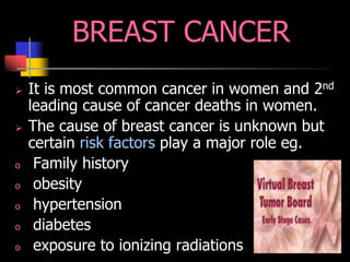
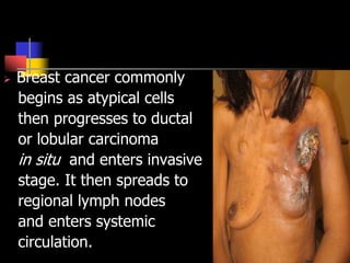
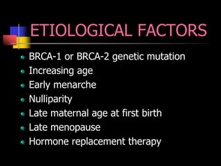
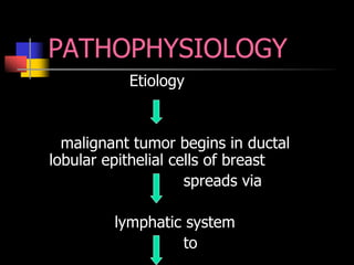
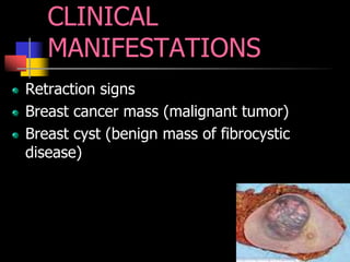
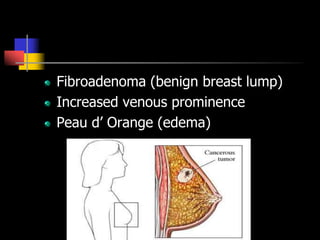
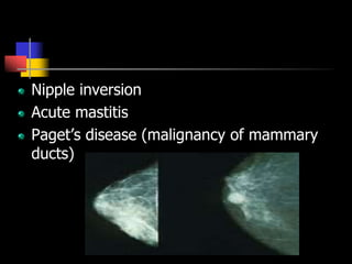
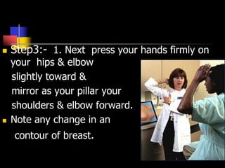
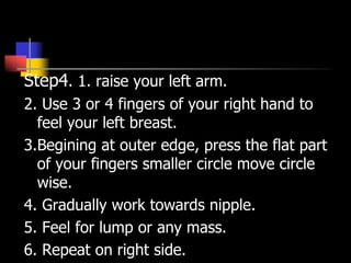
BREAST CANCER
DEFINITION
ANATOMY
RISK FACTORS
ETIOLOGICAL FACTORS
PATHOPHYSIOLOGY
CLINICAL MANIFESTATIONS
presented by-
Jasandeep
Kaur
INTRODUCTION
BREAST – Breast is a glandular and ductal
tissue along with fibrous tissue that binds
the lobes together and fatty tissue in
between the lobes. These paired mammary
glands are located between the 2nd and 6th
rib over pectoralis major muscle from
sternum to mid axillary line.
ANATOMY
 An area of breast tissue called “ tail of
spence” extends into axilla. Cooper’s
ligaments which are fascial bands support
the breast on chest wall. Each breast
consists of 12-20 cone shaped lobes that are
made up of lobules containing clusters of
acini, small structures ending in duct.
BREAST CANCER
 It is most common cancer in women and 2nd
leading cause of cancer deaths in women.
 The cause of breast cancer is unknown but
certain risk factors play a major role eg.
o Family history
o obesity
o hypertension
o diabetes
o exposure to ionizing radiations
o chemical carcinogens
o nulliparity
o lack of breast feeding
o use of estrgen replacement
therapy for menopausal
symptoms
o high fat diet.
o alcoholic
 Breast cancer commonly
begins as atypical cells
then progresses to ductal
or lobular carcinoma
in situ and enters invasive
stage. It then spreads to
regional lymph nodes
and enters systemic
circulation.
ETIOLOGICAL FACTORS
BRCA-1 or BRCA-2 genetic mutation
Increasing age
Early menarche
Nulliparity
Late maternal age at first birth
Late menopause
Hormone replacement therapy
PATHOPHYSIOLOGY
Etiology
malignant tumor begins in ductal
lobular epithelial cells of breast
spreads via
lymphatic system
to
axillary lymph nodes
metastasizes to
lungs, liver, bone, brain etc.
CLINICAL
MANIFESTATIONS
Retraction signs
Breast cancer mass (malignant tumor)
Breast cyst (benign mass of fibrocystic
disease)
Fibroadenoma (benign breast lump)
Increased venous prominence
Peau d’ Orange (edema)
Nipple inversion
Acute mastitis
Paget’s disease (malignancy of mammary
ducts)
STAGES
DIAGNOSTIC TESTS
Presented By –
Jaspreet Kaur
STAGES OF BREAST CANCER
Stage 1 : tumors < 2 cm.
Stage 2 : tumors are < 5 cm. , mobile and
involve axillary lymph nodes.
Stage 3 (a) : tumors >5 cm. with enlarged
axillary lymph nodes fixed to one another or
to the adjacent tissues.
Stage 3 (b) : advanced lesion with satellite
nodules, fixed to skin or chest
wall, ulceration, edema
supraclavicular
involvement.
Stage 5 : all tumors with
distant metastasis.
DIAGNOSTIC TESTS
Breast Self Examination (BSE)
Mammography
Galactography
Ultrasonography
Tissue analysis
Magnetic Resonance Imaging (MRI)
1. Fine Needle Aspiration
(FNA)
2. Stereotactic biopsy
3. Surgical biopsy
- Incisional biopsy
- Excisional biopsy
- Tru-cut core biopsy
- Wire needle loalization
TREATMENT AND
MANAGEMENT
Presented By –
Jatinder Kaur
TREATMENT AND
MANAGEMENT
MEDICAL MANAGEMENT
Treatment depends upon
tumor’s histopathology &
aggressiveness, hormonal
factors, extent of disease
and patient’s overall
health and treatment
preferences.
radiation is primary treatment for patients
who cannot tolerate anesthesia
or surgery and for those with
inflammatory carcinoma.
1. Primary Radiation Therapy with “boost”
to the tumor site (application of additional
needles in former tumor site) may be
used in early stages of breast cancer as
supplement to radiation therapy and
surgery.
2. Radiation Therapy to the breast and
lymph nodes followed by “boost” to the
tumor site is used after breast
preserving surgery (lumpectomy,
quadrantectomy & simple mastectomy.
3. Adjuvant chemotherapy is
recommended after primary treatment
for cell stages of breast to eliminate the
hidden micrometastatic spread of
disease.
4. Chemotherapeutic agents are
 Cyclophosphamide
 Methotrexate
5- fluorouracil
 Vincristine
 prednisone
5. Hormone therapy or
surgery is indicated for
patients with hormone
dependent breast cancer,
antiestrogen drug may
be prescribed.
SURGICAL MANAGEMENT
Lumpectomy
Tumorectomy
Quadrantectomy
Simple mastectomy
Modified Radical
Mastectomy (MRM)
Radical mastectomy
Nursing Management
 During a routine examination assess the
patient’s knowledge of risk factors & BSE
practices assess for contours, symmetry &
appearance of the breast tissue & nipples.
 BSE educate about breast
self examination & do
assessment of client .
 Step:-1 stand before a mirror.
 Check both breast for
anything unusual.
 Look for discharge from
nipples, puckering,
dimpling, scaling
of skin.
 Step2:- 1. watch closely in the mirror as
you claps your hand behind your head &
press your hands forward.
 2. note in any change in
contour of your breast.
 Step3:- 1. Next press your hands firmly on
your hips & elbow
slightly toward &
mirror as your pillar your
shoulders & elbow forward.
 Note any change in an
contour of breast.
Step4. 1. raise your left arm.
2. Use 3 or 4 fingers of your right hand to
feel your left breast.
3.Begining at outer edge, press the flat part
of your fingers smaller circle move circle
wise.
4. Gradually work towards nipple.
5. Feel for lump or any mass.
6. Repeat on right side.
 Step4:- 1 step 5 should repeated in lying down.
 Lie flat on your back with your left arm over your
head & pillow place below.
Nursing Diagnosis
 Deficient knowledge about breast cancer &
treatment options.
 Anxiety R/T cancer diagnosis.
 Fear R/T specific treatment body, image
change or possible death.
 Risk for impaired skin integrity R/T
surgery or radiation therapy.
 Disturbed body image R/T loss or alteration
of breast.
Pre-operative Nsg
Diagnosis:-
1.Deficient knowledge about breast
cancer & treatment option's.
Nsg interventions:-
 The knowledge about disease & its
treatment.
 Reduction of preoperative & pest opera
fear’s & emotional stress.
 Improving decision making ability .
 Pain management.
 2. Anxiety related to cancer diagnosis
 1. fear related to specific treatment, for
image change or possible death.
 Nsg Intervention’s
 1. explaining breast cancer & treatment
options.
 Prime ting decision making ability.
 Educate the client about that’s if early
treatment is done then it is controllable
disease.
 3. Fear related to specific
treatment, body image change or
possible death.
Nsg intervention’s
 1. allow patient to express her feeling &
emotion’s.
 Discuss strategies to improve body image.
 Explain possible body image concerns of
discharge.
 Encourage the self care activates.
 Explain about after surgery how do self care.
 Explain whole procedure to client.
 4. post-op Nsg diagnosis
1.Risk for impaired skin entirety related to
surgery or radiation therapy.
 Nsg Intervention’s
 .provide & explain & dressing & drain
care.
 Prevent skin complications following
radiating therapy.
2.Acute pain R/t surgical procedure.
 . relieving pain & discomfort by giving
I/V or I/M opioid analgesic agents
oral analgesics agent can be
effective in relieving pain.
 Patient should encourage to take
analgesic agent such opioid or non-
opioid analgesic medication such as
acetaminophen when before
exercise or at bed time.
 Take warm shower twice a daily.
5. Disturbed body image R/T to
loss or after action breast
 2. allowing her to pres her emotion's.
 3.Acknowledging her feelings.
 1. promoting positive body image.
 4. client will state feeling comfortable C
new body image that is going to occur.
Post-op exercises
 Wall hand climbing.
 Rod or broomstick lifting.
 Rope turning.
 Pulley tugging.
Client education
 Home care regarding client education .
(1) Needed care required of arm on surgical side.
(a) Avoid burn, insect bites, cuts, scrapes,& scratches if
there was a lost of lymph nods in the axial the grater
the no. of lymph nodes in the axial removed greater
the risk for infection.
(b) Avoid procedures an arm such is blood pressure
infection or blood drawing from affected are.
(c) Need to carry heavy objects in non-affected arm,
usually at weeks light parcels of <5/b are allowed to
be carried & driving is permitted .
Evaluation protocol
A. How will i know that my intervention’s were
effective?
1. client indicates pain is decrease or tolerable.
2. affected arm does not become edematous.
Client is able to perform assisted active range of
motion exercise effectively.
B what criteria will I use to change my interventions?
 1. affected arm become edematous.
 2. clients pain is increased or unrelieved.
 3. client is unable to perform assisted active range of
motion exercises.
(C) How will I know that my client teaching has been
effective?
1. Client wears medical alert bracelet.
2. Client carries package on unaffected side.
3. Client protects affected arm form trauma or from
exposure to infection.
Some objective type questions
 1. the nurse explain to a client that the
reason the client must net have injection .
Blood pressure or venipunctures in affected
arm following modified. Radical mastectomy
vis.
 2. the lymph glands have been removed from
axilla. Lymph drainage from arm is seriously
impended & may make the arm extremely
edematous with non absorption of any
medication if any of those procedure would
occur.
 B there is an incision in axilla that most
heal before any of those procedures
can be performed due to possible. In
faired healing of incision.
 C pressure from the tourniquet for
venipuncture & tied fluid load form
infection can impede healing of the
mastery scar.
 D blood pooling from surgery makes
view impossible to find on the affected
arm & injections impossible to absorb.
 (B) when positioning client immediately
post per, actively following a
lumpectomy the unrest would use
which position.
 A. supine with affected arm elevated on
pillow above the level of heart.
 B. side lying non-surgical side only C
affected arm elevated arm above level
of heart.
 C. low fouler with affected arm exulted
on 2 pillow ‘s above the level of heart .
 What ever position is comfortable for
client . The nurse know that a client has
grasped the concept of arm exercise
following a modified radical mastectomy
when client says she will.
 A. wear her sling all the time until her in
arm is healed.
 B . Perform wall walking exercises with
her fingertips above her head every 2
to 4 hours following pain medication .
 C. exercise only her wruits until she is
at time.
 D. limit her exercising to only her bath,
an only those parts that she can reach.
BREAST CANCER.ppt

BREAST CANCER.ppt

  • 1.
  • 2.
  • 3.
    INTRODUCTION BREAST – Breastis a glandular and ductal tissue along with fibrous tissue that binds the lobes together and fatty tissue in between the lobes. These paired mammary glands are located between the 2nd and 6th rib over pectoralis major muscle from sternum to mid axillary line.
  • 4.
    ANATOMY  An areaof breast tissue called “ tail of spence” extends into axilla. Cooper’s ligaments which are fascial bands support the breast on chest wall. Each breast consists of 12-20 cone shaped lobes that are made up of lobules containing clusters of acini, small structures ending in duct.
  • 6.
    BREAST CANCER  Itis most common cancer in women and 2nd leading cause of cancer deaths in women.  The cause of breast cancer is unknown but certain risk factors play a major role eg. o Family history o obesity o hypertension o diabetes o exposure to ionizing radiations
  • 7.
    o chemical carcinogens onulliparity o lack of breast feeding o use of estrgen replacement therapy for menopausal symptoms o high fat diet. o alcoholic
  • 8.
     Breast cancercommonly begins as atypical cells then progresses to ductal or lobular carcinoma in situ and enters invasive stage. It then spreads to regional lymph nodes and enters systemic circulation.
  • 9.
    ETIOLOGICAL FACTORS BRCA-1 orBRCA-2 genetic mutation Increasing age Early menarche Nulliparity Late maternal age at first birth Late menopause Hormone replacement therapy
  • 10.
    PATHOPHYSIOLOGY Etiology malignant tumor beginsin ductal lobular epithelial cells of breast spreads via lymphatic system to
  • 11.
    axillary lymph nodes metastasizesto lungs, liver, bone, brain etc.
  • 12.
    CLINICAL MANIFESTATIONS Retraction signs Breast cancermass (malignant tumor) Breast cyst (benign mass of fibrocystic disease)
  • 13.
    Fibroadenoma (benign breastlump) Increased venous prominence Peau d’ Orange (edema)
  • 14.
    Nipple inversion Acute mastitis Paget’sdisease (malignancy of mammary ducts)
  • 15.
  • 16.
    STAGES OF BREASTCANCER Stage 1 : tumors < 2 cm. Stage 2 : tumors are < 5 cm. , mobile and involve axillary lymph nodes. Stage 3 (a) : tumors >5 cm. with enlarged axillary lymph nodes fixed to one another or to the adjacent tissues. Stage 3 (b) : advanced lesion with satellite nodules, fixed to skin or chest
  • 17.
    wall, ulceration, edema supraclavicular involvement. Stage5 : all tumors with distant metastasis.
  • 18.
    DIAGNOSTIC TESTS Breast SelfExamination (BSE) Mammography Galactography Ultrasonography Tissue analysis
  • 19.
  • 20.
    1. Fine NeedleAspiration (FNA) 2. Stereotactic biopsy
  • 21.
    3. Surgical biopsy -Incisional biopsy - Excisional biopsy - Tru-cut core biopsy - Wire needle loalization
  • 23.
  • 24.
    TREATMENT AND MANAGEMENT MEDICAL MANAGEMENT Treatmentdepends upon tumor’s histopathology & aggressiveness, hormonal factors, extent of disease and patient’s overall health and treatment preferences.
  • 25.
    radiation is primarytreatment for patients who cannot tolerate anesthesia or surgery and for those with inflammatory carcinoma. 1. Primary Radiation Therapy with “boost” to the tumor site (application of additional needles in former tumor site) may be used in early stages of breast cancer as supplement to radiation therapy and surgery.
  • 26.
    2. Radiation Therapyto the breast and lymph nodes followed by “boost” to the tumor site is used after breast preserving surgery (lumpectomy, quadrantectomy & simple mastectomy. 3. Adjuvant chemotherapy is recommended after primary treatment for cell stages of breast to eliminate the hidden micrometastatic spread of disease.
  • 27.
    4. Chemotherapeutic agentsare  Cyclophosphamide  Methotrexate 5- fluorouracil  Vincristine  prednisone
  • 28.
    5. Hormone therapyor surgery is indicated for patients with hormone dependent breast cancer, antiestrogen drug may be prescribed.
  • 29.
  • 30.
  • 31.
    Nursing Management  Duringa routine examination assess the patient’s knowledge of risk factors & BSE practices assess for contours, symmetry & appearance of the breast tissue & nipples.  BSE educate about breast self examination & do assessment of client .
  • 32.
     Step:-1 standbefore a mirror.  Check both breast for anything unusual.  Look for discharge from nipples, puckering, dimpling, scaling of skin.
  • 33.
     Step2:- 1.watch closely in the mirror as you claps your hand behind your head & press your hands forward.  2. note in any change in contour of your breast.
  • 34.
     Step3:- 1.Next press your hands firmly on your hips & elbow slightly toward & mirror as your pillar your shoulders & elbow forward.  Note any change in an contour of breast.
  • 35.
    Step4. 1. raiseyour left arm. 2. Use 3 or 4 fingers of your right hand to feel your left breast. 3.Begining at outer edge, press the flat part of your fingers smaller circle move circle wise. 4. Gradually work towards nipple. 5. Feel for lump or any mass. 6. Repeat on right side.
  • 36.
     Step4:- 1step 5 should repeated in lying down.  Lie flat on your back with your left arm over your head & pillow place below.
  • 37.
    Nursing Diagnosis  Deficientknowledge about breast cancer & treatment options.  Anxiety R/T cancer diagnosis.  Fear R/T specific treatment body, image change or possible death.  Risk for impaired skin integrity R/T surgery or radiation therapy.  Disturbed body image R/T loss or alteration of breast.
  • 38.
    Pre-operative Nsg Diagnosis:- 1.Deficient knowledgeabout breast cancer & treatment option's. Nsg interventions:-  The knowledge about disease & its treatment.  Reduction of preoperative & pest opera fear’s & emotional stress.  Improving decision making ability .  Pain management.
  • 39.
     2. Anxietyrelated to cancer diagnosis  1. fear related to specific treatment, for image change or possible death.  Nsg Intervention’s  1. explaining breast cancer & treatment options.  Prime ting decision making ability.  Educate the client about that’s if early treatment is done then it is controllable disease.
  • 40.
     3. Fearrelated to specific treatment, body image change or possible death. Nsg intervention’s  1. allow patient to express her feeling & emotion’s.  Discuss strategies to improve body image.  Explain possible body image concerns of discharge.  Encourage the self care activates.  Explain about after surgery how do self care.  Explain whole procedure to client.
  • 41.
     4. post-opNsg diagnosis 1.Risk for impaired skin entirety related to surgery or radiation therapy.  Nsg Intervention’s  .provide & explain & dressing & drain care.  Prevent skin complications following radiating therapy. 2.Acute pain R/t surgical procedure.  . relieving pain & discomfort by giving
  • 42.
    I/V or I/Mopioid analgesic agents oral analgesics agent can be effective in relieving pain.  Patient should encourage to take analgesic agent such opioid or non- opioid analgesic medication such as acetaminophen when before exercise or at bed time.  Take warm shower twice a daily.
  • 43.
    5. Disturbed bodyimage R/T to loss or after action breast  2. allowing her to pres her emotion's.  3.Acknowledging her feelings.  1. promoting positive body image.  4. client will state feeling comfortable C new body image that is going to occur.
  • 44.
    Post-op exercises  Wallhand climbing.  Rod or broomstick lifting.  Rope turning.  Pulley tugging.
  • 45.
    Client education  Homecare regarding client education . (1) Needed care required of arm on surgical side. (a) Avoid burn, insect bites, cuts, scrapes,& scratches if there was a lost of lymph nods in the axial the grater the no. of lymph nodes in the axial removed greater the risk for infection. (b) Avoid procedures an arm such is blood pressure infection or blood drawing from affected are. (c) Need to carry heavy objects in non-affected arm, usually at weeks light parcels of <5/b are allowed to be carried & driving is permitted .
  • 46.
    Evaluation protocol A. Howwill i know that my intervention’s were effective? 1. client indicates pain is decrease or tolerable. 2. affected arm does not become edematous. Client is able to perform assisted active range of motion exercise effectively. B what criteria will I use to change my interventions?  1. affected arm become edematous.  2. clients pain is increased or unrelieved.
  • 47.
     3. clientis unable to perform assisted active range of motion exercises. (C) How will I know that my client teaching has been effective? 1. Client wears medical alert bracelet. 2. Client carries package on unaffected side. 3. Client protects affected arm form trauma or from exposure to infection.
  • 48.
    Some objective typequestions  1. the nurse explain to a client that the reason the client must net have injection . Blood pressure or venipunctures in affected arm following modified. Radical mastectomy vis.  2. the lymph glands have been removed from axilla. Lymph drainage from arm is seriously impended & may make the arm extremely edematous with non absorption of any medication if any of those procedure would occur.
  • 49.
     B thereis an incision in axilla that most heal before any of those procedures can be performed due to possible. In faired healing of incision.  C pressure from the tourniquet for venipuncture & tied fluid load form infection can impede healing of the mastery scar.  D blood pooling from surgery makes view impossible to find on the affected arm & injections impossible to absorb.
  • 50.
     (B) whenpositioning client immediately post per, actively following a lumpectomy the unrest would use which position.  A. supine with affected arm elevated on pillow above the level of heart.  B. side lying non-surgical side only C affected arm elevated arm above level of heart.  C. low fouler with affected arm exulted on 2 pillow ‘s above the level of heart .
  • 51.
     What everposition is comfortable for client . The nurse know that a client has grasped the concept of arm exercise following a modified radical mastectomy when client says she will.  A. wear her sling all the time until her in arm is healed.  B . Perform wall walking exercises with her fingertips above her head every 2 to 4 hours following pain medication .
  • 52.
     C. exerciseonly her wruits until she is at time.  D. limit her exercising to only her bath, an only those parts that she can reach.