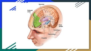
Brain Structures
Involved in Memory
Manvi Singh
Hippocampus
The hippocampus, located in the brain's temporal lobe, is where episodic memories are formed
and indexed for later access. Episodic memories are autobiographical memories from specific
events in our lives, like the coffee we had with a friend last weeK.
Henry Molaison Case 1953
Amygdala
The amygdala, an almond-shaped structure in the brain’s temporal lobe, attaches emotional
significance to memories. This is particularly important because strong emotional memories
(e.g. those associated with shame, joy, love or grief) are difficult to forget. The permanence of
these memories suggests that interactions between the amygdala, hippocampus and
neocortex are crucial in determining the ‘stability’ of a memory – that is, how effectively it is
retained over time.
The main job of the amygdala is to regulate emotions, such as fear and aggression. The
amygdala plays a part in how memories are stored because storage is influenced by stress
hormones. For example, one researcher experimented with rats and the fear response
(Josselyn, 2010).
Neocortex
The neocortex is the largest part of the cerebral cortex, the sheet of neural tissue that forms
the outside surface of the brain, distinctive in higher mammals for its wrinkly appearance. In
humans, the neocortex is involved in higher functions such as sensory perception, generation
of motor commands, spatial reasoning and language. Over time, information from certain
memories that are temporarily stored in the hippocampus can be transferred to the neocortex
as general knowledge – things like knowing that coffee provides a pick-me-up. Researchers
think this transfer from hippocampus to neocortex happens as we sleep.
Cerebellum
The cerebellum, a separate structure located at the rear base of the brain, is most important
in fine motor control, the type that allows us to use chopsticks or press that piano key a
fraction more softly. A well-studied example of cerebellar motor learning is the vestibulo-
ocular reflex, which lets us maintain our gaze on a location as we rotate our heads
Basal Ganglia
The basal ganglia are structures lying deep within the brain and are involved in a wide range of
processes such as emotion, reward processing, habit formation, movement and learning. They
are particularly involved in co-ordinating sequences of motor activity, as would be needed
when playing a musical instrument, dancing or playing basketball. The basal ganglia are the
regions most affected by Parkinson’s disease. This is evident in the impaired movements of
Parkinson’s patients.
Prefrontal Cortex
The prefrontal cortex (PFC) is the part of the neocortex that sits at the very front of the brain.
It is the most recent addition to the mammalian brain, and is involved in many complex
cognitive functions. Human neuroimaging studies using magnetic resonance imaging (MRI)
machines show that when people perform tasks requiring them to hold information in their
short-term memory, such as the location of a flash of light, the PFC becomes active. There also
seems to be a functional separation between left and right sides of the PFC: the left is more
involved in verbal working memory while the right is more active in spatial working memory,
such as remembering where the flash of light occurred.

Brain Structures involved in Memory.pptx

  • 1.
    Brain Structures Involved inMemory Manvi Singh
  • 3.
    Hippocampus The hippocampus, locatedin the brain's temporal lobe, is where episodic memories are formed and indexed for later access. Episodic memories are autobiographical memories from specific events in our lives, like the coffee we had with a friend last weeK. Henry Molaison Case 1953
  • 4.
    Amygdala The amygdala, analmond-shaped structure in the brain’s temporal lobe, attaches emotional significance to memories. This is particularly important because strong emotional memories (e.g. those associated with shame, joy, love or grief) are difficult to forget. The permanence of these memories suggests that interactions between the amygdala, hippocampus and neocortex are crucial in determining the ‘stability’ of a memory – that is, how effectively it is retained over time. The main job of the amygdala is to regulate emotions, such as fear and aggression. The amygdala plays a part in how memories are stored because storage is influenced by stress hormones. For example, one researcher experimented with rats and the fear response (Josselyn, 2010).
  • 5.
    Neocortex The neocortex isthe largest part of the cerebral cortex, the sheet of neural tissue that forms the outside surface of the brain, distinctive in higher mammals for its wrinkly appearance. In humans, the neocortex is involved in higher functions such as sensory perception, generation of motor commands, spatial reasoning and language. Over time, information from certain memories that are temporarily stored in the hippocampus can be transferred to the neocortex as general knowledge – things like knowing that coffee provides a pick-me-up. Researchers think this transfer from hippocampus to neocortex happens as we sleep.
  • 6.
    Cerebellum The cerebellum, aseparate structure located at the rear base of the brain, is most important in fine motor control, the type that allows us to use chopsticks or press that piano key a fraction more softly. A well-studied example of cerebellar motor learning is the vestibulo- ocular reflex, which lets us maintain our gaze on a location as we rotate our heads
  • 7.
    Basal Ganglia The basalganglia are structures lying deep within the brain and are involved in a wide range of processes such as emotion, reward processing, habit formation, movement and learning. They are particularly involved in co-ordinating sequences of motor activity, as would be needed when playing a musical instrument, dancing or playing basketball. The basal ganglia are the regions most affected by Parkinson’s disease. This is evident in the impaired movements of Parkinson’s patients.
  • 8.
    Prefrontal Cortex The prefrontalcortex (PFC) is the part of the neocortex that sits at the very front of the brain. It is the most recent addition to the mammalian brain, and is involved in many complex cognitive functions. Human neuroimaging studies using magnetic resonance imaging (MRI) machines show that when people perform tasks requiring them to hold information in their short-term memory, such as the location of a flash of light, the PFC becomes active. There also seems to be a functional separation between left and right sides of the PFC: the left is more involved in verbal working memory while the right is more active in spatial working memory, such as remembering where the flash of light occurred.