National Institute ofTechnology, Patna , 22 March 2021
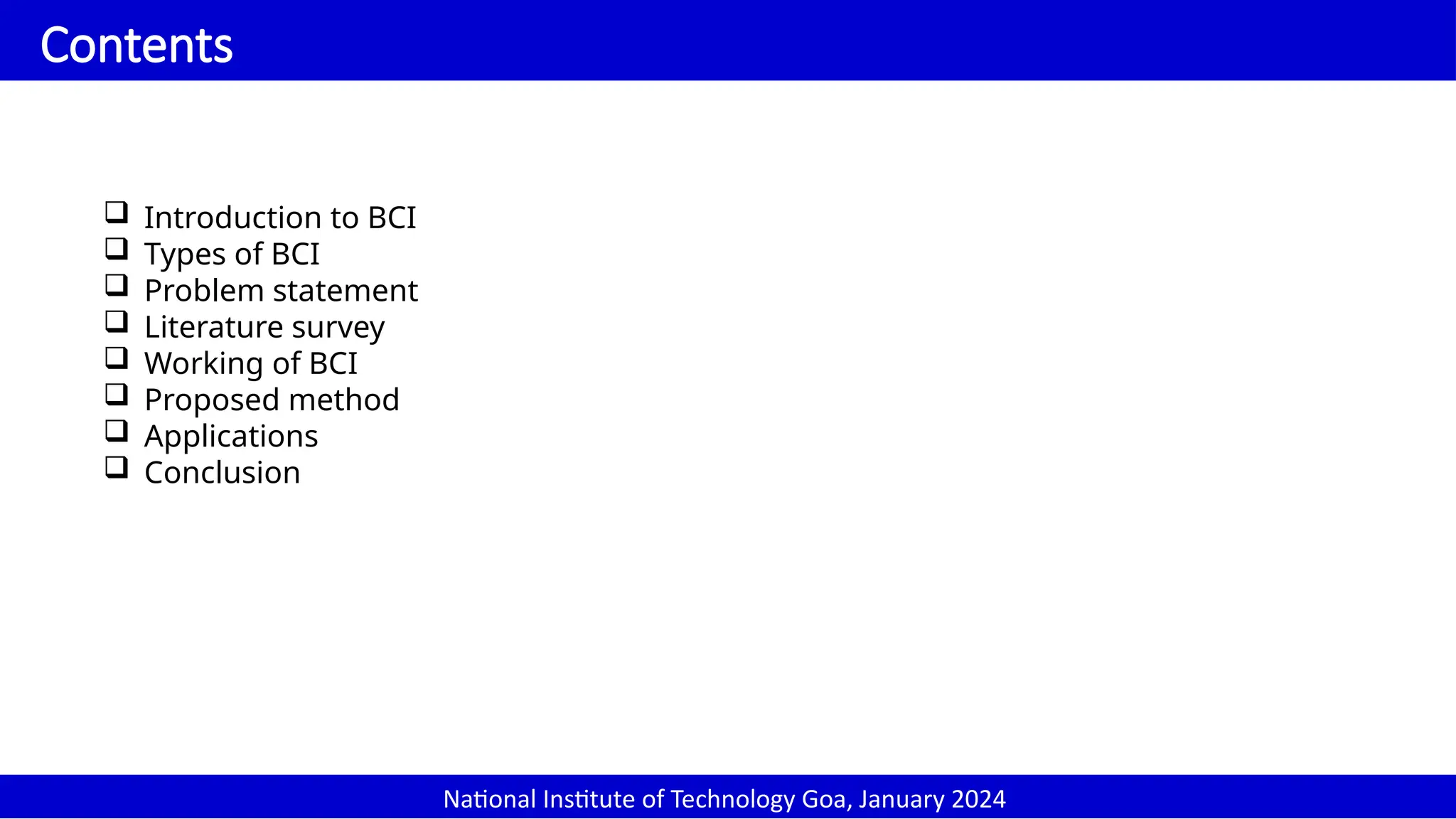
Contents
Introduction to BCI
Types of BCI
Problem statement
Literature survey
Working of BCI
Proposed method
Applications
Conclusion
National Institute of Technology Goa, January 2024
2.
National Institute ofTechnology, Patna , 22 March 2021
Introduction
National Institute of Technology Goa, January 2024
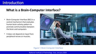
Figure 1: Brain Computer Interface
What is a Brain-Computer Interface?
• Brain-Computer Interface (BCI) is a
control mechanism that evaluates
human brain activity pattern to
promote communication between
the brain and computers
• It does not depend on input from
peripheral nerves or muscles.
3.
National Institute ofTechnology, Patna , 22 March 2021
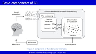
Basic components of BCI
National Institute of Technology Goa, January 2024
Figure 2: Components of Brain Computer Interfaces
4.
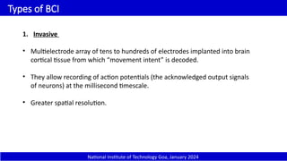
Types of BCI
1.Invasive
• Multielectrode array of tens to hundreds of electrodes implanted into brain
cortical tissue from which “movement intent” is decoded.
• They allow recording of action potentials (the acknowledged output signals
of neurons) at the millisecond timescale.
• Greater spatial resolution.
National Institute of Technology Goa, January 2024
5.
National Institute ofTechnology, Patna , 22 March 2021
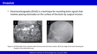
Invasive
National Institute of Technology Goa, January 2024
• Electrocorticography (ECoG) is a technique for recording brain signals that
involves placing electrodes on the surface of the brain by surgical incision.
Figure 3. (A) Electrode array is placed under the dura onto the brain surface (B) X-ray image of the skull showing the
location of the electrode array.
A B
6.
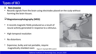
Types of BCI
2.Non-invasive
• Records signals from the brain using electrodes placed on the scalp without
harming the brain tissues.
Magnetoencephalography (MEG)
• It records magnetic fields produced as a result of
neural activity generated in response to a stimulus.
• High temporal resolution
• No distortions
• Expensive, bulky and not portable, require
magnetically shielded room Figure 4. Example MEG system
National Institute of Technology Goa, January 2024
7.
Non-invasive
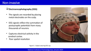
Electroencephalography (EEG)
•The signals are recorded by placing
metal electrodes on the scalp.
• EEG signals reflect the summation of
postsynaptic potentials from many
thousands of neurons
• Captures electrical activity in the
cerebral cortex.
• Poor spatial resolution
Figure 5. Subject wearing EEG cap
National Institute of Technology Goa, January 2024
8.
Non-invasive
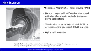
Functional MagneticResonance Imaging (fMRI)
• Detects changes in blood flow due to increased
activation of neurons in particular brain areas
during specific tasks
• The signal recorded by fMRI is called the blood
oxygenation level dependent (BOLD) response.
• High spatial resolution.
Figure 6. fMRI machine with a subject whose brain is being scanned while performing an experiment.
The subject is holding a button-press device for indicating choices or outputs
National Institute of Technology Goa, January 2024
9.
National Institute ofTechnology, Patna , 22 March 2021
Non-invasive
National Institute of Technology Goa, January 2024
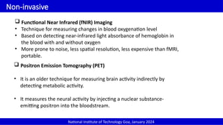
Functional Near Infrared (fNIR) Imaging
• Technique for measuring changes in blood oxygenation level
• Based on detecting near-infrared light absorbance of hemoglobin in
the blood with and without oxygen
• More prone to noise, less spatial resolution, less expensive than fMRI,
portable.
Positron Emission Tomography (PET)
• It is an older technique for measuring brain activity indirectly by
detecting metabolic activity.
• It measures the neural activity by injecting a nuclear substance-
emitting positron into the bloodstream.
10.
National Institute ofTechnology, Patna , 22 March 2021
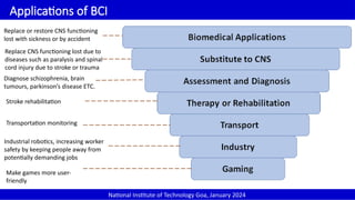
Applications of BCI
National Institute of Technology Goa, January 2024
Replace or restore CNS functioning
lost with sickness or by accident
Replace CNS functioning lost due to
diseases such as paralysis and spinal
cord injury due to stroke or trauma
Diagnose schizophrenia, brain
tumours, parkinson’s disease ETC.
Stroke rehabilitation
Transportation monitoring
Industrial robotics, increasing worker
safety by keeping people away from
potentially demanding jobs
Make games more user-
friendly
11.
National Institute ofTechnology, Patna , 22 March 2021
Motor Imagery
National Institute of Technology Goa, January 2024
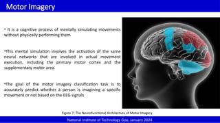
• It is a cognitive process of mentally simulating movements
without physically performing them
•This mental simulation involves the activation of the same
neural networks that are involved in actual movement
execution, including the primary motor cortex and the
supplementary motor area.
•The goal of the motor imagery classification task is to
accurately predict whether a person is imagining a specific
movement or not based on the EEG signals
Figure 7: The Neurofunctional Architecture of Motor Imagery
12.
National Institute ofTechnology, Patna , 22 March 2021
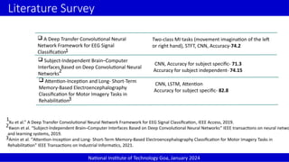
Literature Survey
Subject-Independent Brain–Computer
Interfaces Based on Deep Convolutional Neural
Networks
National Institute of Technology Goa, January 2024
A Deep Transfer Convolutional Neural
Network Framework for EEG Signal
Classification
Attention-Inception and Long- Short-Term
Memory-Based Electroencephalography
Classification for Motor Imagery Tasks in
Rehabilitation
1
Xu et al.” A Deep Transfer Convolutional Neural Network Framework for EEG Signal Classification, IEEE Access, 2019.
Kwon et al. “Subject-Independent Brain–Computer Interfaces Based on Deep Convolutional Neural Networks” IEEE transactions on neural netwo
and learning systems, 2019.
Amin et al. “Attention-Inception and Long- Short-Term Memory-Based Electroencephalography Classification for Motor Imagery Tasks in
Rehabilitation” IEEE Transactions on Industrial Informatics, 2021.
2
3
1
2
3
Two-class MI tasks (movement imagination of the left
or right hand), STFT, CNN, Accuracy-74.2
CNN, Accuracy for subject specific- 71.3
Accuracy for subject independent- 74.15
CNN, LSTM, Attention
Accuracy for subject specific- 82.8
13.
National Institute ofTechnology, Patna , 22 March 2021
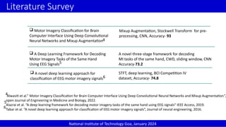
Literature Survey
National Institute of Technology Goa, January 2024
Motor Imagery Classification for Brain
Computer Interface Using Deep Convolutional
Neural Networks and Mixup Augmentation
A Deep Learning Framework for Decoding
Motor Imagery Tasks of the Same Hand
Using EEG Signals5
Alwasiti et al.” Motor Imagery Classification for Brain Computer Interface Using Deep Convolutional Neural Networks and Mixup Augmentation”,
open Journal of Engineering in Medicine and Biology, 2022.
Alazrai et al. “A deep learning framework for decoding motor imagery tasks of the same hand using EEG signals” IEEE Access, 2019.
Tabar et al. “A novel deep learning approach for classification of EEG motor imagery signals”, Journal of neural engineering, 2016.
6
5
6
Mixup Augmentation, Stockwell Transform for pre-
processing, CNN, Accuracy- 93
A novel three-stage framework for decoding
MI tasks of the same hand, CWD, sliding window, CNN
Accuracy-73.2
A novel deep learning approach for
classification of EEG motor imagery signals
STFT, deep learning, BCI Competition IV
dataset, Accuracy- 74.8
4
4
14.
National Institute ofTechnology, Patna , 22 March 2021
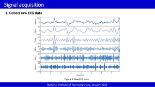
Signal acquisition
National Institute of Technology Goa, January 2024
1. Collect raw EEG data
Figure 8: Raw EEG data
15.
National Institute ofTechnology, Patna , 22 March 2021
Channel
National Institute of Technology Goa, January 2024
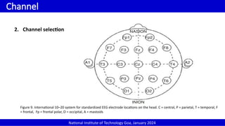
2. Channel selection
Figure 9. International 10–20 system for standardized EEG electrode locations on the head. C = central, P = parietal, T = temporal, F
= frontal, Fp = frontal polar, O = occipital, A = mastoids
16.
National Institute ofTechnology, Patna , 22 March 2021
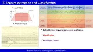
3. Feature extraction and Classification
National Institute of Technology Goa, September 2023
Apply filters
Artefact removal
Extract time or frequency component as a feature
Classification
Prosthetics Control
17.
National Institute ofTechnology, Patna , 22 March 2021
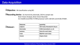
Data Acquisition
National Institute of Technology Goa, January 2024
Objective: MI classification using BCI
Recording device: 32 channel dry electrode, 250 Hz sample rate
Brain Vision Analyzer, Brain Vision Recorder
An Intel(R) Core(TM) i7-4790 processor with 3.60 GHz and 8 GB of RAM .
Dataset :
features quantity
subjects 10(7 male,3 female)(18- 28 yrs)
electrodes 3 EEG channels(c3, cz, c4)
sessions 2(one for each class)
trials 60(30 for each class)
classes 2(open,close)
18.
National Institute ofTechnology, Patna , 22 March 2021
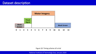
Dataset description
National Institute of Technology Goa, January 2024
0 1 2 3 4 5 6 7 8 9 10 11 12 13
Motor Imagery
Blank
Screen
Blank Screen
Visual
Signal
Figure 10: Timing scheme of a trial
19.
National Institute ofTechnology, Patna , 22 March 2021
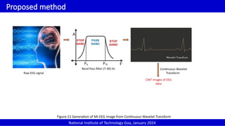
Proposed method
National Institute of Technology Goa, January 2024
Raw EEG signal
Band Pass filter (7-30) Hz
CWT images of EEG
data
Figure:11 Generation of MI-EEG image from Continuous Wavelet Transform
Continuous Wavelet
Transform
20.
National Institute ofTechnology, Patna , 22 March 2021
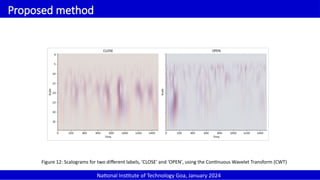
Proposed method
National Institute of Technology Goa, January 2024
Figure 12: Scalograms for two different labels, 'CLOSE' and 'OPEN', using the Continuous Wavelet Transform (CWT)
21.
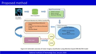
Proposed method
National Instituteof Technology Goa, January 2024
Figure 14: Schematic overview of motor imagery classification using Attention based CNN-BiLSTM model
22.
National Institute ofTechnology, Patna , 22 March 2021
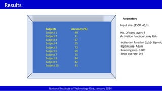
Results
National Institute of Technology Goa, January 2024
Subjects Accuracy (%)
Subject 1 90
Subject 2 71
Subject 3 67
Subject 4 55
Subject 5 73
Subject 6 69
Subject 7 75
Subject 8 84
Subject 9 82
Subject 10 61
Parameters
Input size- (1500, 40,3)
No. Of conv layers-4
Activation function Leaky Relu
Activation function (o/p)- Sigmoid
Optimizers- Adam
Learning rate- 0.001
Drop out rate- 0.4
23.
National Institute ofTechnology, Patna , 22 March 2021
References
National Institute of Technology Goa, January 2024
1. Rao, R.P.: Brain-computer interfacing: an introduction. Cambridge University Press (2013)
2. Kumar, S., Rajshekher, G., Prabhakar, S., et al.: Positron emission tomography in neurological diseases.
Neurology India 53(2), 149 (2005)
3. Bablani, A., Edla, D.R., Tripathi, D., Cheruku, R.: Survey on brain-computer interface: An emerging
computational intelligence paradigm. ACM Computing Surveys (CSUR) 52(1), 1–32 (2019).
4. F. Cincotti, D. Mattia, F. Aloise, S. Bufalari, G. Schalk, G. Oriolo, A. Cherubini, M. G. Marciani, F. Babiloni,
Non-invasive brain–computer interface system: towards its application as assistive technology, Brain
research bulletin 75 (6) (2008) 796–803.
5. E. M. Schmidt, Single neuron recording from motor cortex as a possible source of signals for control of
external devices, Annals of biomedical engineering 8 (4) (1980) 339–349.
6. N. Veena, N. Anitha, A review of non-invasive bci devices, Int. J. Biomed. Eng. Technol 34 (3) (2020) 205–233.


![EEG BASED MOTOR IMAGERY DECODING [Autosaved].pptx](https://cdn.slidesharecdn.com/ss_thumbnails/eegbasedmotorimagerydecodingautosaved-251208043154-f4c6e54a-thumbnail.jpg?width=640&height=640&fit=bounds)