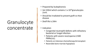
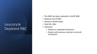
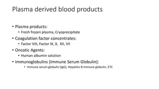
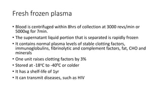
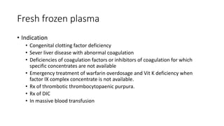
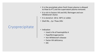
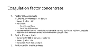
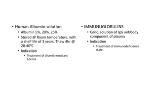
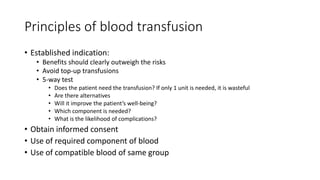
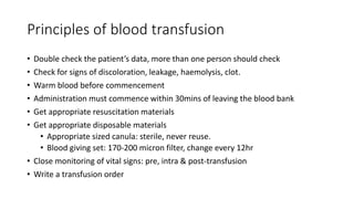
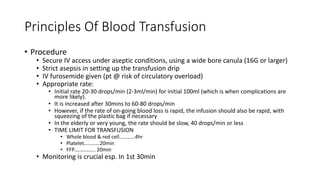
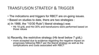
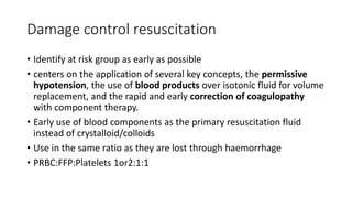
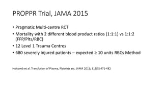
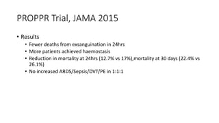
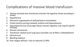
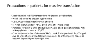
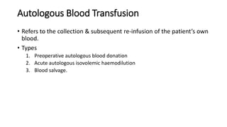
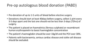
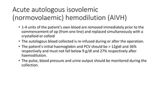
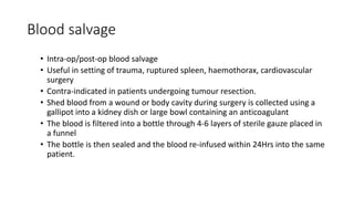
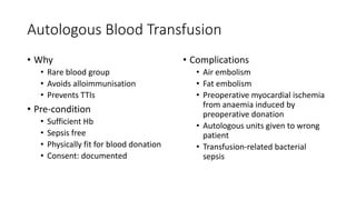
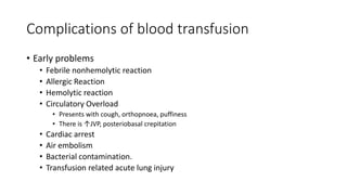
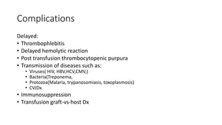
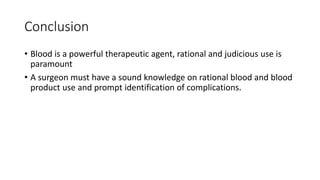
The document discusses the use of blood and blood products in surgery. It covers the indications and uses of various blood products like whole blood, packed red cells, platelet concentrates, plasma derivatives, and coagulation factor concentrates. It discusses principles of blood transfusion like compatibility testing, administration procedures, and complications of transfusion. It also covers topics like massive blood transfusion protocols, autologous blood transfusion, and challenges in blood transfusion.
The document discusses blood transfusion in the ICU, including:
- The history of blood transfusion and evolution of transfusion medicine.
- Controversy around transfusion triggers in the ICU, with studies showing increased risks with liberal transfusion strategies.
- Risks of blood transfusion including infections, TRALI, and immunomodulation effects.
- Specific transfusion guidelines and considerations for septic patients, bleeding patients, and massive transfusion situations.
1. Venous thromboembolism (VTE) is common in surgical patients and is a leading cause of preventable death.
2. The document provides guidelines for assessing VTE risk and recommendations for thromboprophylaxis for various types of surgeries.
3. The recommendations generally suggest early ambulation for low risk procedures and pharmacological thromboprophylaxis like low molecular weight heparin for higher risk procedures.
Pericarditis is an inflammation of the pericardium surrounding the heart that can be caused by infections, autoimmune diseases, or other etiologies. It is classified as acute, subacute, or chronic based on duration. The document defines pericarditis and its types, describes the pathophysiology involving fluid accumulation in the pericardial space, lists clinical manifestations such as chest pain and potential complications like cardiac tamponade, and outlines the diagnostic and management approaches including medications, imaging, and procedures.
Venous Thromboembolism refers to deep vein thrombosis and pulmonary embolism. The document discusses the pathophysiology and risk factors for venous thromboembolism. It covers normal vein function, causes of deep vein thrombosis, pulmonary embolism, post-thrombotic syndrome, and Virchow's triad of factors that increase thrombosis risk. It also discusses hereditary and acquired thrombophilias, or hypercoagulable states, that can increase thrombosis risk. Testing for thrombophilias can help determine duration of anticoagulation treatment after an initial thrombotic event.
Effect of restrictive versus liberal transfusion strategies on outcomes in pa...Mohd Saif Khan
Restrictive red cell transfusion policies are recommended as safe for most hospital patients with anaemia. Uncertainty exists for patients with cardiovascular disease, whose hearts may be more susceptible to limited coronary oxygen supply.
This document outlines guidelines for monitoring patients after cardiac surgery. It discusses common cardiac surgeries and their complications, immediate post-operative care including assessments, labs, hemodynamic management, respiratory function and more. The goal is to monitor for complications, ensure patient comfort, and begin early movement and discharge education to aid in recovery.
Blood transfusion, Nutrition and water & electrolyte balanceRamesh Parajuli
This document discusses blood products and transfusion. It defines blood products as therapeutic substances prepared from human blood, including whole blood, blood components, and plasma derivatives. It describes the major blood groups (ABO and Rh) and components found in whole blood. The effects of storage on whole blood and its components are outlined. Indications, contraindications, and administration of various blood products like packed red blood cells, platelet concentrates, and leukocyte-depleted red blood cells are summarized. Processing and preparation of these blood products is also briefly explained.
This document provides guidelines for the diagnosis and treatment of deep vein thrombosis (DVT). It recommends venous duplex scanning to diagnose DVT and further tests like venography if the scan is negative but clinical suspicion remains high. For treatment, it suggests anticoagulant drugs like low molecular weight heparin or warfarin depending on the patient's risk factors, pregnancy status, and whether they are hospitalized. It also provides recommendations for screening for thrombophilia and lengths of treatment with warfarin based on the type and risk factors associated with the DVT. Catheter-directed thrombolysis is considered for recent large DVT to help prevent post-thrombotic syndrome.
This document provides guidelines on the management of pericardial diseases from the ESC. It discusses the etiology, diagnosis, and treatment of various pericardial conditions including pericarditis, cardiac tamponade, constrictive pericarditis. For constrictive pericarditis specifically, it highlights two key pathophysiological characteristics - exaggerated ventricular interdependence and the halting of intrathoracic pressure changes on the ventricles by a thickened pericardium. Surgical pericardiectomy is the definitive treatment for constrictive pericarditis when medical management fails.
This highly energetic lecture presents the pathophysiology of S-T elevation myocardial infarction in an easy to understand style to help you best identify, triage and treat patients presenting with acute coronary syndromes. Using the latest research behind the AHA Guidelines changes, AHA National Faculty Rom Duckworth will help you better coordinate with you partners along the continuum of cardiac care. Emphasis is placed on risk factors, recognizing truly sick patients and coordinating care with hospital personnel.
Learning Objectives: Students will learn:
-The pathophysiology of S-T elevation myocardial infarction.
-The difference between STEMI, NSTEMI and unstable angina.
-Differing treatment methods and priorities for different cardiac syndromes.
-The function and importance of 12 lead ECG and prehospital diagnostic testing.
-The roles and responsibilities of EMS providers as the key element in “door-to-balloon” and “door-to-needle” time for STEMI patients.
www.romduck.com
www.RescueDigest.com
This document provides an evaluation process for thrombocytopenia in children and neonates. It involves taking a medical history, performing a physical exam and blood tests to determine the cause of the low platelet count. Common causes include infections, prematurity, drugs, underlying diseases or idiopathic thrombocytopenia. Treatment depends on the identified etiology but may include treating any underlying condition, transfusions to maintain adequate platelet levels, and addressing signs of bleeding if present. The goal is to diagnose the specific cause to guide management and prevent long-term complications.
This document discusses the use of venous blood gas (VBG) analysis as an alternative to arterial blood gas (ABG) analysis in emergency situations. It finds that VBG measurements of pCO2, pH, and bicarbonate correlate well with ABG measurements and are sufficient to guide treatment for conditions like diabetic ketoacidosis. It also finds that a VBG pCO2 level below 45 mmHg can reliably rule out clinically significant hypercarbia. The vast majority of patients can be managed using VBG alone. An ABG is only needed if the VBG results are discordant with the clinical presentation, such as in hemodynamically unstable patients.
The document discusses Patient Blood Management (PBM), which is a multidisciplinary approach to optimize care for patients who may need blood transfusions. It involves strategies before, during, and after surgery/procedures to minimize blood loss and transfusions. Key preoperative strategies include identifying and treating anemia, assessing bleeding risk, and considering preoperative autologous blood donation. Intraoperative strategies focus on techniques to reduce blood loss like cell salvage and tranexamic acid. Postoperative care emphasizes continued efforts to minimize blood loss and optimize physiology. The overall goals are to improve patient outcomes, reduce costs, and ensure an adequate blood supply.
Red cell transfusions in the critically ill compatibleBharath T
This document provides an overview of red cell transfusions in critically ill patients. It discusses the history of blood transfusions, reasons for anemia in ICU patients, physiological effects of red blood cells, technical aspects of blood compatibility testing and storage, hazards of transfusions, and evidence from studies on restrictive vs liberal transfusion thresholds. The TRICC study found no significant difference in mortality between restrictive and liberal transfusion strategies, though subgroups with lower illness severity saw benefits with restriction. Overall the document examines the risks and benefits of red cell transfusions in critical care.
The document provides an overview of blood conservation strategies in perioperative patients. It discusses why blood conservation is important to conserve limited blood resources and reduce risks of transfusion. Key strategies mentioned include preoperative patient optimization, use of antifibrinolytics like tranexamic acid, controlled hypotension, cell salvaging, normovolemic hemodilution, and autologous blood donation and transfusion to avoid allogeneic transfusions and their associated risks. The document emphasizes a multidisciplinary team approach and utilization of the latest drugs, techniques and technology to minimize blood loss and reduce need for allogeneic blood transfusions in surgical patients.
Post cardiac surgery monitoring & follow upRubayet Anwar
This document provides information on post-cardiac surgery monitoring and follow up. It discusses admission to the ICU, initial assessment, monitoring techniques, complications that can occur like bleeding and hemodynamic issues, and management strategies for those complications. The early focus is on stabilizing vital signs, addressing hypothermia, identifying potential issues like low cardiac output, and treating medical causes of bleeding through correcting coagulation abnormalities.
This document discusses the classification, presentation, diagnosis, and treatment of acute coronary syndrome (ACS). ACS results from an imbalance between myocardial oxygen supply and demand due to a thrombotic coronary artery. It is classified as ST-elevation myocardial infarction (STEMI), non-ST-elevation myocardial infarction (NSTEMI), or unstable angina (UA) based on electrocardiogram findings and cardiac biomarker levels. Initial treatment involves oxygen, nitroglycerin, aspirin, a P2Y12 inhibitor, and anticoagulation. STEMI patients should receive reperfusion via primary percutaneous coronary intervention or fibrinolysis if primary PCI cannot be performed in a timely manner.
This document discusses the management of hypertrophic cardiomyopathy (HCM). It covers the natural history of HCM, risk stratification including the role of implantable cardioverter defibrillators, pharmacological treatments, and invasive treatments such as alcohol septal ablation and surgical myectomy. Key points discussed include the use of beta blockers as first-line pharmacological therapy, guidelines for ICD implantation, the technique and outcomes of alcohol septal ablation versus surgical myectomy, and recommendations for experienced centers to perform these invasive procedures.
This document discusses acute coronary syndromes and ischemic heart disease. It begins with an overview of heart anatomy and physiology. It then defines acute coronary syndrome and myocardial infarction, describing signs and symptoms. Risk factors for ischemic heart disease are outlined. The document concludes with descriptions of nursing assessments, diagnoses, and interventions for patients with acute coronary syndromes or ischemic heart disease, focusing on pain management, improving perfusion, and reducing anxiety through education.
The document discusses advanced cardiac life support (ACLS) and basic life support (BLS) protocols for treating cardiac arrest and other medical emergencies. It outlines the key components of high-quality CPR in BLS, including chest compressions, rescue breathing, activation of emergency services, and use of an automated external defibrillator (AED). The document then details the basic life support sequence, appropriate use of shock versus CPR first, electrode placement for defibrillation, drugs not recommended for routine use, preferred methods for drug delivery, the potential role of precordial thump, and post-resuscitation care measures.
The document discusses hypertensive emergencies, which are acute, severe elevations in blood pressure that can cause target organ damage. It notes key risk factors and various potential causes. It outlines goals for lowering blood pressure during hypertensive emergencies, which depend on the specific target organ(s) affected and time since presentation. Common medications used for treatment are discussed along with their indications and special considerations. Treatment goals differ for conditions like pregnancy, stroke, and aortic dissection. The importance of determining whether target organ damage is present and tailoring treatment accordingly is emphasized.
This document discusses the management and treatment of patients with advanced heart failure who require admission to the intensive care unit (ICU). It defines advanced heart failure and provides criteria for determining which heart failure patients should be admitted to the ICU. It covers monitoring in the ICU, classification of heart failure, medical treatment including diuretics and inotropic drugs, and mechanical circulatory support options. The future of treatments like ventricular assist devices is also mentioned.
Fluid resuscitation is complex with choices between crystalloids and colloids. Initial resuscitation aims to restore normal blood pressure, heart rate and end-organ perfusion. Ongoing fluid needs are harder to determine as patients may appear "wet" but still be intravascularly depleted. Simple tests like passive leg raising can predict fluid responsiveness by assessing changes in blood pressure and flow. The optimal amount and type of fluid varies between patients and clinical contexts.
1. A 30-year-old male with fever and altered mental status was found to have a potassium level disturbance based on his ECG.
2. ECG changes due to electrolyte imbalances can vary between individuals and depend on other electrolyte levels as well.
3. However, certain consistent ECG features often indicate increased or decreased potassium, making ECG useful for identifying electrolyte issues if prior tracings are available for comparison.
This document discusses blood conservation techniques used in cardiac surgery to minimize blood transfusions. It begins with a brief history of blood transfusions in surgery and the efforts to reduce them due to risks. The scope of blood conservation is outlined as using techniques to minimize blood needed for treatment. It emphasizes a multidisciplinary team approach including various medical professionals and the patient/family. Phases of the surgery where conservation is used are described from pre-op patient screening to intra-op techniques like acute normovolemic hemodilution and post-op restrictive transfusion thresholds. Specific intra-op techniques are then detailed for each phase before, during, and after use of the cardiopulmonary bypass machine.
This document reviews STEMI (ST elevation myocardial infarction) recognition and treatment. It defines a STEMI as elevated ST segments on an ECG due to blocked coronary arteries. Imposters like left bundle branch block can mimic STEMIs. The anatomy, ECG interpretation rules, and signs of STEMI versus no STEMI are described. Treatment includes aspirin, nitroglycerin, oxygen, and morphine or dilaudid for pain management in the pre-hospital setting. Recognizing true STEMIs amid imposters like left bundle branch block is a critical skill for emergency responders.
Whole blood can be separated into components which allows for optimal survival of each constituent and transfusion of only the specific component needed by the patient. This avoids unnecessary transfusion and allows blood from one donor to treat several patients. Components include red blood cell concentrate, platelet concentrate, leukocyte-reduced products, plasma components like fresh frozen plasma and derivatives, and cellular components like granulocytes.
Blood and blood products were presented. Key points included:
1. Blood functions to transport vital substances throughout the body.
2. Blood typing and cross-matching must be done correctly to avoid transfusion reactions.
3. Several blood products exist including packed red blood cells, platelets, and plasma derivatives that are used to treat different conditions.
4. Blood transfusions can have complications and must only be done when necessary following all safety protocols.
This document provides an overview of blood component therapy. It discusses the composition of blood and history of blood transfusion. It describes the preparation of various blood components like red blood cells, platelets, plasma, and cryoprecipitate. It outlines the indications and guidelines for transfusion of these components. It also reviews trials on restrictive versus liberal transfusion strategies and discusses adverse effects and management of transfusion reactions.
Blood groups,blood components and blood transfusion By Dr Bimalesh Kumar GuptaDrbimalesh Gupta
This document provides an overview of blood groups, blood components, and blood transfusion. It defines key terms like blood, blood products, and blood transfusion. It describes the major blood groups like ABO and Rh, and the process of cross-matching. It discusses components of blood like red cells, platelets, fresh frozen plasma, and cryoprecipitate. It covers topics like blood donation, transfusion reactions, and alternatives to transfusion. Overall, the document provides a comprehensive overview of blood and transfusion medicine.
Transfusion of blood and blood products can be used to treat various conditions related to deficiencies in red blood cells, platelets, or clotting factors. There are several types of blood products including packed red blood cells, fresh whole blood, platelet concentrates, fresh frozen plasma, and cryoprecipitate. Massive transfusions involving 10 or more units of blood in 24 hours require special consideration and guidelines recommend maintaining a 1:1:1 ratio of plasma, platelets, and packed red blood cells. Acute normovolemic hemodilution involves removing blood pre-operatively and replacing volume with crystalloids or colloids to reduce transfusion needs during anticipated significant blood loss.
Surgery resident postgraduate presentation on the use of blood and products presented dept of surgery, Niger Delta University Teaching Hospital, Okolobiri, Bayelsa State, Nigeria
Blood and its components can be prepared from whole blood to provide targeted therapy for patients. Whole blood can be separated via centrifugation into components like red blood cells, platelets, and plasma. This separation allows specific transfusion of only the required elements and avoids unnecessary elements. Red blood cells, platelets, and plasma components are prepared and have defined indications, storage guidelines, and administration protocols to provide optimal clinical benefits to different patient conditions. Blood banking aims to safely and effectively utilize blood products based on individual patient needs.
Components Of Blood (For Transfusion)
• Each unit of blood is tested for evidence of hepatitis-b,
hepatitis-c, Human Immune deficiency Virus I & II.
• The blood is then processed into sub-components.
• Whole blood
• Packed cell volume
• Fresh frozen plasma
• Platelets
• Cryoprecipitate
This document provides information about blood transfusion, including:
- The composition and functions of blood, as well as total blood volume and components.
- Blood grouping, typing, and compatibility with the Rh factor.
- An overview of blood transfusion including indications, calculations for allowable blood loss, and blood products like packed red blood cells, plasma, platelets, and fresh frozen plasma.
- Criteria for blood donation, collection of blood for transfusion, and blood storage guidelines.
- Common anticoagulants used in blood storage like citrates and heparin.
Blood products topic is very important for Medical students as they have to know which blood product will be much beneficial to patients when they go into clinical practice. This PPT provides all of them.
Blood, Blood transfusion and Blood products bijay19
This document discusses blood and blood products. It begins by introducing blood, its components and functions. It then describes various blood properties and groups. It discusses different blood products like packed red blood cells, platelets, fresh frozen plasma and their uses. The document outlines indications and contraindications for transfusions. It notes complications of transfusions and massive transfusions. Finally, it briefly introduces blood substitutes.
This document discusses blood and blood products. It begins by introducing blood, its components and functions. It then describes various blood properties and groups. It discusses different blood products like packed red cells, platelets, fresh frozen plasma and cryoprecipitate. It details their composition, indications, contraindications and storage. The document also covers topics like blood transfusion, complications, massive transfusion, and blood substitutes. It emphasizes the importance of blood, cautions on judicious transfusion, and concludes by thanking the reader.
This document provides guidelines for blood transfusion practices. It discusses the history of blood transfusions from the early 1900s developments to modern practices. It outlines the components of blood that can be transfused including red blood cells, platelets, fresh frozen plasma, and cryoprecipitated anti-hemophilic factor. Thresholds and indications for transfusing each component are provided based on factors like hemoglobin level and platelet count. Proper procedures for blood transfusions including consent, preparation, and compatibility checking are also outlined.
It contains indications of blood and blood products and perioperative blood therapy that we usually follow in Aiims Patna ..its is most recent one made in April 2020
This document summarizes blood transfusion, including its components, indications, storage, and complications. It discusses red blood cell concentrates, plasma, platelet concentrates, and plasma derivatives that can be transfused. The objectives, triggers, and indications for transfusion are outlined. Details are provided on blood collection, storage, and shelf life of different components. Immediate complications discussed include hemolytic and non-hemolytic reactions like allergic, febrile, and anaphylactic reactions. Delayed complications mentioned are delayed hemolytic transfusion, post-transfusion purpura, graft-versus-host disease, and transfusion-transmitted infections.
This document discusses blood transfusion and blood products. It begins by defining blood transfusion as transferring blood or blood products between individuals. It then describes the main types of transfusion as homologous and autologous. The document goes on to provide a brief history of blood transfusion and discusses indications for transfusion such as acute blood loss or anemia. It also outlines donor criteria and collection/storage of blood and blood products like packed cells, plasma, platelets, and artificial substitutes. Complications of transfusion and massive transfusion are noted.
This document discusses whole blood, blood components, and blood derivatives. Whole blood contains all blood components, while blood components are parts separated from whole blood through centrifugation or apheresis. Blood derivatives are products made from fractionating plasma from multiple donors. The document describes various blood components like packed red blood cells, platelets, fresh frozen plasma and cryoprecipitate. It also discusses blood derivatives including albumin, coagulation factor concentrates, and immunoglobulins. It provides details on how these products are prepared, stored, and used to treat different conditions.
This document summarizes the components of blood and their clinical uses. It discusses whole blood as well as separated blood components including red blood cells, platelets, fresh frozen plasma, and cryoprecipitate. For each component, it describes what it contains, how it is prepared, appropriate clinical indications and transfusions guidelines, dosing and expected results, contraindications and precautions. It also provides instructions for proper storage of blood products prior to transfusion. The overall purpose is to explain the definitions and appropriate clinical use of different blood components.
This document provides an overview of blood components therapy, including their indications and guidelines for use. It discusses the various components that can be derived from whole blood, such as packed red blood cells, platelets, fresh frozen plasma, cryoprecipitate, and granulocytes. Storage conditions, shelf lives, and therapeutic doses are provided. The main reasons for transfusion in Africa are described as childhood malaria, hemoglobinopathies, obstetric bleeding, trauma, and certain surgical procedures. Contraindications and risks of transfusion-transmitted infections are also covered. The document emphasizes considering alternatives to transfusion and whether benefits outweigh risks in each clinical situation.
This document discusses blood components and their uses. It begins by explaining that effective blood transfusion now relies on separating whole blood into components. These components can meet most patient transfusion needs while minimizing risks. The document then discusses the various cellular and plasma components that can be derived from whole blood, including red blood cells, platelets, fresh frozen plasma, cryoprecipitate, and more specialized components. It provides details on the preparation methods, storage, and clinical indications for each component type.
Imaging plays an indispensable role in urology. Ultrasonography is a commonly used first-line imaging modality due to its noninvasive nature and ability to evaluate the kidneys, bladder, prostate, testes and surrounding structures. Transrectal ultrasound provides high resolution imaging of the prostate. While ultrasound has advantages, other modalities like CT, MRI and nuclear imaging provide additional clinical information. A thorough understanding of anatomy is important for accurate image interpretation in urologic imaging.
The document provides information on club foot (talipes equinovarus), including its incidence, classification, pathoanatomy, and management. It describes the Ponseti technique for nonoperative treatment of club foot, which involves serial casting and manipulation of the foot, often with Achilles tenotomy, followed by use of a foot abduction brace for several years to maintain correction and prevent recurrence of the deformity. Complications of nonoperative treatment include residual deformities like a rocker bottom foot or bean shaped foot, as well as potential fractures, pressure sores, failure to fully correct, or recurrence of the club foot deformity.
This document discusses the principles and management of malunion, which is the consolidation of a fracture in an abnormal position resulting in deformity or functional deficit. It outlines the causes of malunion, types of deformities, classification systems, evaluation methods including imaging, and surgical and non-surgical treatment options. Treatment is tailored based on the location and characteristics of the deformity, and may involve osteotomies, external fixation, or joint replacement to correct angular, rotational, translational or length discrepancies while avoiding complications. Close follow up is needed to monitor for issues like under/overcorrection and ensure proper healing.
This document outlines the residency training program for orthopedics in Nigeria. It discusses the admission requirements, registration process, educational objectives, and format of training. The training consists of basic medical sciences, junior residency, and senior residency phases.
The junior residency phase lasts a minimum of 30 months and involves rotations in orthopedics/trauma, general surgery, burns/plastic surgery, neurosurgery, and electives. The senior residency phase also lasts 30 months minimum and includes rotations in trauma/ICU, arthroplasty, arthroscopy/sports medicine, pediatric orthopedics, and an elective. Both phases aim to develop clinical, technical, teaching, and research competencies to produce skilled orth
Promoting Wellbeing - Applied Social Psychology - Psychology SuperNotesPsychoTech Services
A proprietary approach developed by bringing together the best of learning theories from Psychology, design principles from the world of visualization, and pedagogical methods from over a decade of training experience, that enables you to: Learn better, faster!
8 Surprising Reasons To Meditate 40 Minutes A Day That Can Change Your Life.pptxHolistified Wellness
We’re talking about Vedic Meditation, a form of meditation that has been around for at least 5,000 years. Back then, the people who lived in the Indus Valley, now known as India and Pakistan, practised meditation as a fundamental part of daily life. This knowledge that has given us yoga and Ayurveda, was known as Veda, hence the name Vedic. And though there are some written records, the practice has been passed down verbally from generation to generation.
The skin is the largest organ and its health plays a vital role among the other sense organs. The skin concerns like acne breakout, psoriasis, or anything similar along the lines, finding a qualified and experienced dermatologist becomes paramount.
Does Over-Masturbation Contribute to Chronic Prostatitis.pptxwalterHu5
In some case, your chronic prostatitis may be related to over-masturbation. Generally, natural medicine Diuretic and Anti-inflammatory Pill can help mee get a cure.
Osteoporosis - Definition , Evaluation and Management .pdfJim Jacob Roy
Osteoporosis is an increasing cause of morbidity among the elderly.
In this document , a brief outline of osteoporosis is given , including the risk factors of osteoporosis fractures , the indications for testing bone mineral density and the management of osteoporosis
Histololgy of Female Reproductive System.pptxAyeshaZaid1
Dive into an in-depth exploration of the histological structure of female reproductive system with this comprehensive lecture. Presented by Dr. Ayesha Irfan, Assistant Professor of Anatomy, this presentation covers the Gross anatomy and functional histology of the female reproductive organs. Ideal for students, educators, and anyone interested in medical science, this lecture provides clear explanations, detailed diagrams, and valuable insights into female reproductive system. Enhance your knowledge and understanding of this essential aspect of human biology.
Kosmoderma Academy, a leading institution in the field of dermatology and aesthetics, offers comprehensive courses in cosmetology and trichology. Our specialized courses on PRP (Hair), DR+Growth Factor, GFC, and Qr678 are designed to equip practitioners with advanced skills and knowledge to excel in hair restoration and growth treatments.
REGULATION FOR COMBINATION PRODUCTS AND MEDICAL DEVICES.pptx
blood products.pptx
1. The use of blood and blood
products in surgery
Dr PJ Shindang
2. Outline
• Introduction
• Indication and use of the blood products
• Principles of blood transfusion
• Massive blood transfusion.
• Autologous blood transfusion.
• Complications of blood transfusion
• Alternatives to blood transfusion
• Challenges
• Conclusion
• References
3. Introduction
• Blood transfusion refers to the intravenous transfer of compatible blood or blood
products from one individual to the same (autologous) or to another individual
(homologous/allogenic)
• WHOLE BLOOD: un-separated blood collected into an approved container containing
an anticoagulant-preservative solution.
• BLOOD PRODUCT: Any therapeutic substance prepared from human blood.
• Cellular derivatives: Packed red cell, Leucocytes depleted red cell, Platelet concentrate,
granulocyte concentrate
• Plasma derivatives: Fresh frozen plasma, Cryoprecipitate
• Coagulation factor concentrates: Factor VIII, Factor IX, X, XII, VII
• Oncotic Agents: Human albumin solution
• Immunoglobulins (Immune Serum Globulin): Immune serum globulin (IgG), Hepatitis B
immune globulin..ETC
4. Historical perspective
• 1665 – First recorded blood transfusion in England , R Lower revived a dog by
transfusing blood from another dog via a tied artery
• 1900 Karl Landsteiner discovers the first three human blood groups, A, B and O.
• 1914 Adolf Hustin discovers that sodium citrate can anticoagulate blood for
transfusion, allowing it to be stored and later transfused safely to patients on the
battlefield
• 1940 The Rh blood group is discovered when RBCs of monkeys were injected into
rabbits .
• 1985 The first HIV blood-screening test is licensed and implemented by blood
banks.
5. Blood collection and Storage
• Collection of blood should be done under strict asepsis form suitable donors
• Standard blood bag contains 450 +/- 45mls blood, with 60mls of anticoagulant preservative
• Stored at 2-6oC
• Anticoagulants include
• Heparin: 24 hours
• Acid-citrate-dextrose (ACD) : 21 days (obsolete)
• Citrate-phosphate-dextrose (CPD): 28 days
• Citrate-phosphate-dextrose-adenine(CPDA): 35days
• SAGM: 42days
• Storage
• Whole blood & packed cells: 2-60C
• Platelet concentrate: 20-240C (room temperature)
• FFP & cryoprecipitate (frozen): -18 to -400C.
• Shelf-life:
• Platelets- 5days
• FFP/Cryoppt-1yr
• Factor concentrates- 2yrs
• Albumin- 4yrs
6. Effects of blood storage
• CELLS
• RBC:
• Swell, lose K+ to plasma.
• 1% is lost for each day of storage
• 2,3-DPG levels fall after 1 week
• WBC: Survives for 30-90 min in the recipient's blood. WBC’s are not
viable after 24h of storage.
• Platelets: there are no viable platelets after 24h. However, non-viable
platelets remain for 2 weeks.
7. Effect of storage
• Electrolytes
• The plasma potassium rises at the rate of
1mmol/day.
• The sodium concentration of the plasma
is increased because of the sodium
citrate in the CPD anticoagulant.
• Calcium: There is no ionized calcium.
Ionized calcium displaces sodium in
disodium citrate, forming unionized
calcium citrate.
• pH:- falls from about 7.2 at the time of
collection to about 6.8 at 20 days.
• Plasma Hb levels rise during storage due to
leakage of Hb from cells. At 20 days the level
is about 0.2 g/L.
• The ammonia concentration also rises.
8. Effect of storage
• Clotting factors
• Factor Vlll (AHF) declines rapidly and activity falls by 40% after 24h of storage, There is little
activity after 7 days.
• Factor V declines rapidly after 24h and there is very little activity after 7 days.
• Factor IX declines rapidly after 7 days and there is no activity after 14 days.
• Factor X loses its activity after 7 days.
• Factor Vll declines only after 14 days.
• Fibrinogen and factor II are stable for 21 days.
9. Whole blood
• It contains all the blood components
• It’s use is now limited except in hospitals and areas where facilities for
producing blood fractions are unavailable.
• Indications
• To restore blood volume after acute loss of > 25% blood volume
• Exchange blood transfusion: hyperbilirubinemia, priapism.
• Extracorporeal circulation: haemo-dialysis, heart-lung machine
• Autologous blood transfusion
• Where blood component therapy is unavailable.
• Massive blood transfusion
10. Packed Red
cell
Preparation
• Whole blood is centrifuged
at 3000 revs/min (or 5000 x
g) for 5 min.
• The plasma removed to give
a Hct of 0.55-0.75 (PCV 55-
75%).
• One unit raises the Hb by
approximately 1g/dl in a
70kg adult.
• Stored like whole blood, with
shelf life of 42days.
• Indications
• Acute blood loss (after
crystalloid fluid resuscitation)
• Symptomatic chronic anemia:
• Leukemia
• aplastic anemia
• Malignancies
• CRF
• Pre-op transfusion (before
emergency surgery. If elective
surgery, use other appropriate
means)
• Severe burns with risk of
hyperkalemia
• The elderly
• cachetic patients.
11. Platelet
concentrate
• It is the precipitate after platelet rich plasma is
centrifuged at 3000rev/min (or 5000 x g) for 5 min
• Platelet-rich plasma is the supernatant plasma after
whole blood is centrifuged at 1000/min or2000x g for
3 min.
• Each unit has a volume of 50-60ml, containing 5.5x10⁹
Platelet.
• One unit of platelet concentrate raises the platelet
count by 5-10 x 109/L in an adult.
• Platelets are stored at room temperature (20-240C)
under constant agitation to prevent clumping
• Shelf life of 3-5days
• Transfused at a rate of 0.1unit/kg
12. Platelet
concentrate
Indications
• Management of severe or life-threatening
thrombocytopaenia
• Thrombocytopenia caused by massive blood loss and
replacement with platelet-poor products.
• Qualitative platelet disorders.
• Chemotherapy Induced marrow suppression
• Massive blood transfusion
13. Granulocyte
concentrate
• Prepared by leukopharesis
• Vol 220ml which contains 1 x 10¹⁰granulocytes
/unit.
• Should be irradiated to prevent graft-vs-Host
disease
• Shelf life is 24Hr
• Indication
• Congenital neutrophil defects with refractory
bacterial or fungal infection.
• Patients with severe neutropenia (<500
PMNs/uL).
• Patients on Intensive chemotherapy & transplant
• Reversible bone marrow hypoplasia
14. Leucocyte
Depleted RBC
• The WBC has been reduced to <5x10⁶ WBC
• Reduces risk of CMV
• Stored as whole blood.
• Shell life: 24hr
• Indication
• Patient on repeated transfusion.
• Patient with previous reaction to red cell
transfusion
16. Fresh frozen plasma
• Blood is centrifuged within 8hrs of collection at 3000 revs/min or
5000xg for 7min.
• The supernatant liquid portion that is separated is rapidly frozen
• It contains normal plasma levels of stable clotting factors,
immunoglobulins, fibrinolytic and complement factors, fat, CHO and
minerals
• One unit raises clotting factors by 3%
• Stored at -18oC to -400C or colder
• It has a shelf-life of 1yr
• It can transmit diseases, such as HIV
17. Fresh frozen plasma
• Indication
• Congenital clotting factor deficiency
• Sever liver disease with abnormal coagulation
• Deficiencies of coagulation factors or inhibitors of coagulation for which
specific concentrates are not available
• Emergency treatment of warfarin overdosage and Vit K deficiency when
factor IX complex concentrate is not available.
• Rx of thrombotic thrombocytopaenic purpura.
• Rx of DIC
• In massive blood transfusion
18. Cryoprecipitate
• It is the precipitate when fresh frozen plasma is allowed
to thaw to 4°C and the supernatant plasma removed.
• It is rich in Factors VIII and XIII, fibrinogen and von
Willebrand's factor.
• It is stored at -18 to -40oC or colder.
• Shell life….1yr. Thaw 24hr
• Indication
• Used in Rx of haemophilia A
• Hypofibrinogenaemia
• Von Willebrand's disease
• Factor XIII deficiency
• DIC
19. Coagulation factor concentrate
1. Factor VIII concentrate
• Contains 250 IU of factor VIII per vial
• Stored @ +2 to +60C
• Indication:
• Rx of Hemophilia A
• Rx of Von Willebrand dx
• Recombinant factor VIII and IX are available but are very expensive. However, they are
free from diseases transmitted by blood derived concentrates
2. Factor IX concentrate
• Contains 350-600 IU per vial of factor IX.
• Stored @ +2 to +60C.
• Indication: Rx of Hemophilia B.
3. Antithrombin III concentrate
20. • Human Albumin solution
• Albumin 5%, 20%, 25%
• Stored @ Room temperature, with
a shelf life of 3 years. Thaw 4hr @
20-400C
• Indication
• Treatment of diuretic-resistant
Edema
• IMMUNUGLOBULINS
• Conc. solution of IgG antibody
component of plasma
• Indication
• Treatment of immunodeficiency
state
21. Principles of blood transfusion
• Established indication:
• Benefits should clearly outweigh the risks
• Avoid top-up transfusions
• 5-way test
• Does the patient need the transfusion? If only 1 unit is needed, it is wasteful
• Are there alternatives
• Will it improve the patient’s well-being?
• Which component is needed?
• What is the likelihood of complications?
• Obtain informed consent
• Use of required component of blood
• Use of compatible blood of same group
22. Principles of blood transfusion
• Double check the patient’s data, more than one person should check
• Check for signs of discoloration, leakage, haemolysis, clot.
• Warm blood before commencement
• Administration must commence within 30mins of leaving the blood bank
• Get appropriate resuscitation materials
• Get appropriate disposable materials
• Appropriate sized canula: sterile, never reuse.
• Blood giving set: 170-200 micron filter, change every 12hr
• Close monitoring of vital signs: pre, intra & post-transfusion
• Write a transfusion order
23. Principles Of Blood Transfusion
• Procedure
• Secure IV access under aseptic conditions, using a wide bore canula (16G or larger)
• Strict asepsis in setting up the transfusion drip
• IV furosemide given (pt @ risk of circulatory overload)
• Appropriate rate:
• Initial rate 20-30 drops/min (2-3ml/min) for initial 100ml (which is when complications are
more likely).
• It is increased after 30mins to 60-80 drops/min
• However, if the rate of on-going blood loss is rapid, the infusion should also be rapid, with
squeezing of the plastic bag if necessary
• In the elderly or very young, the rate should be slow, 40 drops/min or less
• TIME LIMIT FOR TRANSFUSION
• Whole blood & red cell…………4hr
• Platelet…………20min
• FFP……………. 20min
• Monitoring is crucial esp. In 1st 30min
24. TRANSFUSION STRATEGY & TRIGGER
• The indications and triggers for RBCT are on-going issues.
• Based on studies to date, there are two strategies :
a) In 1988, the “10/30 Rule”( liberal strategy) was
• Hb 10 g/dL and Hct 30% and transfusions were performed based on
those values
b) Recently, the restrictive strategy (Hb level below 7 g/dL)
• more accepted due to evidence regarding the negative impact on
prognoses following RBCT per the liberal strategy as well as the
complications and costs associated with RBCT
25. Damage control resuscitation
• Identify at risk group as early as possible
• centers on the application of several key concepts, the permissive
hypotension, the use of blood products over isotonic fluid for volume
replacement, and the rapid and early correction of coagulopathy
with component therapy.
• Early use of blood components as the primary resuscitation fluid
instead of crystalloid/colloids
• Use in the same ratio as they are lost through haemorrhage
• PRBC:FFP:Platelets 1or2:1:1
26. PROPPR Trial, JAMA 2015
• Pragmatic Multi-centre RCT
• Mortality with 2 different blood product ratios (1:1:1) vs 1:1:2
(FFP/Plts/RBC)
• 12 Level 1 Trauma Centres
• 680 severely injured patients – expected ≥ 10 units RBCs Method
Holcomb et al. Transfusion of Plasma, Platelets etc. JAMA 2015; 313(5):471-482
27. PROPPR Trial, JAMA 2015
• Results
• Fewer deaths from exsanguination in 24hrs
• More patients achieved haemostasis
• Reduction in mortality at 24hrs (12.7% vs 17%),mortality at 30 days (22.4% vs
26.1%)
• No increased ARDS/Sepsis/DVT/PE in 1:1:1
28. Massive blood
transfusion
• In adults.
• Transfusion of half of a patient’s blood volume in 4
hours.
• Administration of 10 or more packed red blood cell
within 24hrs (adult blood volume is approximately 70
mL/kg).
• Transfusion of >4 RBC units in 1 h with anticipation of
continued need for blood product support.
• In children
• Transfusion of more than 40 mL blood/kg in 24 hrs
(blood volume of children older than neonates is
approximately 80 mL/kg).
29. Complications of massive blood transfusion
1. Volume overload over-transfusion (monitor Hb regularly, titrate according to
needs)
2. Hypothermia
3. Dilutional coagulopathy of clotting factors and platelets
4. Citrate toxicity causing metabolic acidosis and hypocalcaemia
5. Hyperkalaemia (use of younger blood, monitor regularly, may require specific
therapy)
6. Disease transmission
7. Transfusion related acute lung injury (consider use of filters, leukodepletion)
8. Clerical error.
9. Bleeding diathesis
10. Poor oxygen delivery—due to reduced 2,3 DPG
30. Precautions in patients for massive transfusion
• Adequate care in documentation etc- to prevent clerical errors
• Warm the blood- to prevent hypothermia
• Calcium gluconate: After every 1L of blood
• FFP: For every 6 units of RBCs, give 6 units of FFP (1:1 ratio)
• Platelets: for every 6 units of RBCs (& FFP), give one 6-pack of platelets. Aim
to keep platelet counts > 100,000
• Cryoprecipitate: After 1st 6 units of RBCs, check fibrinogen level. If <100mg/dl,
give 20 units of cryoprecipitate (which contains 2g of fibrinogen). Repeat as
needed, depending on fibrinogen level
31. Autologous Blood Transfusion
• Refers to the collection & subsequent re-infusion of the patient’s own
blood.
• Types
1. Preoperative autologous blood donation
2. Acute autologous isovolemic haemodilution
3. Blood salvage.
32. Pre-op autologous blood donation (PABD)
• Pre-donation of up to 1-5 units of blood before elective surgery
• Donations should start at least 40days before surgery, collect 1 pint every
3-5 days apart and the last one should not be less than 3-days (72hrs) of
surgery.
• The patient is placed on haematinics (ferrous sulphate) or recombinant
human erythropoietin to boost haemoglobin concentration
• The patient's haemoglobin should be over 10g/dl and the PCV over 30%.
• Patients with bacteraemia, serious cardiac disease and sickle cell disease
should be excluded.
33. Acute autologous isovolemic
(normovolaemic) hemodilution (AIVH)
• 1-4 units of the patient's own blood are removed immediately prior to the
commencement of op (from one line) and replaced simultaneously with a
crystalloid or colloid
• The autologous blood collected is re-infused during or after the operation.
• The patient's initial haemoglobin and PCV should be > 12gldl and 36%
respectively and must not fall below 9 g/dl and 27% respectively after
haemodilution.
• The pulse, blood pressure and urine output should be monitored during the
collection.
34. Blood salvage
• Intra-op/post-op blood salvage
• Useful in setting of trauma, ruptured spleen, haemothorax, cardiovascular
surgery
• Contra-indicated in patients undergoing tumour resection.
• Shed blood from a wound or body cavity during surgery is collected using a
gallipot into a kidney dish or large bowl containing an anticoagulant
• The blood is filtered into a bottle through 4-6 layers of sterile gauze placed in
a funnel
• The bottle is then sealed and the blood re-infused within 24Hrs into the same
patient.
35. Autologous Blood Transfusion
• Why
• Rare blood group
• Avoids alloimmunisation
• Prevents TTIs
• Pre-condition
• Sufficient Hb
• Sepsis free
• Physically fit for blood donation
• Consent: documented
• Complications
• Air embolism
• Fat embolism
• Preoperative myocardial ischemia
from anaemia induced by
preoperative donation
• Autologous units given to wrong
patient
• Transfusion-related bacterial
sepsis
36. Complications of blood transfusion
• Early problems
• Febrile nonhemolytic reaction
• Allergic Reaction
• Hemolytic reaction
• Circulatory Overload
• Presents with cough, orthopnoea, puffiness
• There is ↑JVP, posteriobasal crepitation
• Cardiac arrest
• Air embolism
• Bacterial contamination.
• Transfusion related acute lung injury
38. ALTERNATIVES TO BLOOD TRANSFUSION
• RED CELL SUBSTITUTES
• Per fluorocarbon
• Porphyrin
• Recombinant haemoglobin: Diaspirin Cross-linked Hemoglobin, Polymerized stroma-free Hemoglobin.
• PLASMA SUBSTITUTES
• Crystalloids
• Colloids
• Stable plasma protein solution
• Albumin solution
• Dextran
• Synthetic gelatin colloids (hemaccel, gelofusine)
• Hydroxyethyl starch preparations (hetastarch, pentastarch)
• Platelet substitute: Pegylated Recombinant Human Megakaryocyte Growth and Development
Factor (PEG-rHuMGDF)
• Others
• Erythropoietin
• Desmopressin: in mild factor 8 deficiency
39. CHALLENGES OF BLOOD TRANSFUSION
• Shortage of voluntary blood donors
• TTIs:
• Parasitic: malaria, T. Cruzi
• Viruses: HBV, HCV, HIV, CMV, HTLV 1 & 2, parvovirus
• Prions
• Bacteria
• Ineffective blood transfusion: anaemic paid donor
• Absence of a coordinated blood transfusion service
• Weak regulatory mechanisms
• Poor transport and communication networks
• Limited awareness
• Infrastructural inadequacy
• Storage problems (erratic power supply)
• Lack of facilities for preparation of blood products
• Low level of community participation
40. Conclusion
• Blood is a powerful therapeutic agent, rational and judicious use is
paramount
• A surgeon must have a sound knowledge on rational blood and blood
product use and prompt identification of complications.
41. References
• Badoe E. A; principles and practice of surgery, 5th edition
• Handbook of transfusion medicine by McClelland B
• Advances in blood transfusion. American Society of Haematology
• Update on Blood transfusions and Blood substitutes by Miller R.N.
IARS Review course lectures
• Courtney M. T; Sabiston Textbook of surgery 6th edition.
1. resulting in increased affinity of Hb for O2, with less O2 delivery to the tissues. 2,3-DPG recovery takes place within 24hrs after transfusion
and by 75% after 5 days.
Hb concentration alone is not enough indicator of need for blood transfusion, but should be based on Hx, PE, Inv (indication should be symptomatic anemia).
. One
unit of platelet concentrate has a volume of approximately
50 mL. Platelet preparations are capable of transmitting infectious
diseases and can account for allergic reactions similar to
those caused by red blood cell transfusion. A therapeutic level
of platelets is in the range of 50,000 to 100,000/μL, but is very
dependent on the clinical situation. Recent evidence suggests
that earlier use of platelets may improve outcomes in bleeding
patients.
The daily dose of granulocytes in adults and children is 1.5 to 3 × 10⁸/kg of body weight
(i.e., chronic granulomatous disease
failing to respond to appropriate antimicrobial therapy for more than 24 to 48 hours may be considered for granulocyte transfusion.
FFP is an effective volume expander because of the plasma proteins that it contains
Contain about half of Factor VIII & Fibrinogen in donated whole blood
Factor VIII: 80-100 IU/pack.
Fibrinogen 150-300iu/pack
(name, blood group, hospital number, ward) against the blood to be transfused (date of collection, expiry date, blood group of donor)
‘Critical bleeding’ may be defined as major haemorrhage that is life threatening and likely to result in the need for massive transfusion.
(3.0ml for every 1.0ml of blood collected) or colloid (1ml for every 1ml of blood collected) through another line to maintain the circulating blood volume.