This document provides details on bioassay methods for several compounds including vasopressin, digitalis, d-tubocurarine, histamine, and 5-hydroxytryptamine (5-HT).
It describes two common bioassay methods for vasopressin - the first uses rats to measure changes in blood pressure, the second uses rats and measures anti-diuretic activity. For digitalis, it outlines guinea pig and pigeon bioassays measuring the lethal dose. The d-tubocurarine bioassay uses rabbits to measure head drop or isolated frog muscle to measure contraction reduction. Histamine is assayed using guinea pig ileum or other tissues and measuring contraction. Finally, 5
Bioassay of Digitalis, d-tubocurarine , OxytocinHeena Parveen
This document summarizes several bioassay methods for determining the potency of digitalis, oxytocin, and d-tubocurarine (d-tb) extracts, including guinea pig, cat, and pigeon methods for digitalis; depression of blood pressure in chickens, contraction of rat uterus, and measurement of milk ejection pressure in lactating rats for oxytocin; and rabbit head drop and frog rectus abdominis muscle preparation methods for d-tb. Standard preparations and procedures for administering test and standard extracts and measuring responses are described for each method.
Bioassy of insulin according to Indian pharmacopoeiaSONALPANDE5
This document summarizes several methods for bioassaying insulin, including preparation of standard and test solutions, rabbit, mouse, rat diaphragm, and rat epididymal fat pad methods. It also describes the radioimmunoassay method, which uses radiolabeled insulin and antibodies to determine insulin concentration in test samples by comparing to standard curves.
This document summarizes different bioassay methods used to test the potency of insulin samples, including the rabbit method, mouse method, rat diaphragm method, and epididymal fat pad of rat method. The rabbit method involves measuring changes in blood sugar levels after administering standardized and test insulin doses to groups of rabbits. The mouse method compares the percentage of convulsions produced by standardized and test insulin doses in mice. The rat diaphragm and epididymal fat pad methods examine the effects of insulin on glucose uptake in rat tissue samples.
This document describes the bioassay method for determining the potency of digitalis extracts using guinea pigs and pigeons. The potency of a test sample is compared to a standard digitalis preparation by determining the lethal dose needed to arrest the heart. For guinea pigs, the extract is administered via cannulated jugular vein until heart arrest, and the lethal dose is calculated. For pigeons, the lethal dose per kg that causes heart arrest or emesis is determined and used to calculate the potency relative to the standard. Proper preparation of extracts, use of minimum 6 animals, and determination of average lethal doses are described.
Insulin bioassay is the measurement of insulin potency by biological activity measurement using a standard biological system. An often used technique is to inject insulin samples into animals, usually rats or rabbits, and then track their blood sugar levels to see how well insulin lowers blood sugar. An alternate strategy is to measure glucose absorption or metabolic reactions using cultured cells sensitive to insulin, including adipocytes or muscle cells. By giving important information on insulin potency, purity, and stability, these tests guarantee the quality of insulin products intended for therapeutic use. Bioassays continue to be necessary for insulin preparation quality control and regulatory clearance even with the development of analytical methods.
This document describes experiments to study the effects of various drugs on isolated frog heart, blood pressure, and heart rate in dogs. The experiments involve setting up perfused frog heart preparations and measuring the chronotropic and inotropic effects of drugs. The effects of drugs are also studied on normal and calcium-depleted hypodynamic frog hearts. In addition, the effects of sympathomimetic and parasympathomimetic drugs on mean blood pressure and heart rate are measured in anesthetized dogs using a cannulation technique.
This document provides an overview of bioassay procedures. It defines bioassay as the comparative assessment of the potency of a test compound to a standard compound using a living biological system. The basic bioassay procedure involves preparing tissues, attaching them to an organ bath, constructing dose-response curves for standard and test compounds, and calculating the potency of the test compound based on its curve's position relative to the standard. Sources of error include biological variation between tissues and methodological errors in experimental design or implementation.
Bioassay of insulin & Bioassay of VasopressinHeena Parveen
This document summarizes bioassay methods for measuring the potency of insulin and vasopressin, including:
- Rabbit and mouse methods that measure the hypoglycemic or convulsive effects of test samples compared to standards.
- Rat diaphragm and fat pad methods measuring insulin's effects on glucose uptake and fat metabolism.
- Radioimmunoassay, a sensitive technique using radiolabeled antigens to quantify insulin concentrations via competitive binding.
- A suggested method for vasopressin involving cannulation of rats to measure changes in blood pressure after administration of test samples versus a reference standard.
This document describes several bioassay methods for measuring the potency of insulin samples, including rabbit, mouse, rat diaphragm, and rat epididymal fat pad methods. For the rabbit method, insulin samples and a standard are injected subcutaneously in rabbits and blood sugar levels are measured over time, comparing the hypoglycemic effect between samples. The mouse method compares the percentage of mice experiencing convulsions after insulin injection between samples and a standard. The rat diaphragm and epididymal fat pad methods measure glucose uptake in tissue samples incubated with insulin to determine insulin-like activity.
Respiratory and reproduction pharmacology ManjuJhakhar
This document summarizes animal models used in respiratory and reproductive pharmacology. For respiratory pharmacology, it describes in vitro tests including histamine receptor binding and effects on isolated organs. It also describes various in vivo tests in anesthetized guinea pigs to assess bronchospasmolytic activity. For reproduction pharmacology, it discusses models to evaluate inhibition of fertility and gonadotropin secretion, including suppressing organ weights in rats treated with steroids.
Expt 9- To evaluate the effect of insulin in rabbits at different intervalsMirzaAnwarBaig1
This study evaluated the effect of insulin on rabbits at different time intervals. Rabbits were given 20 units of insulin prepared with saline, HCl, phenol, and glycerin. Blood glucose levels were measured before insulin injection and over the next 5 hours. The results showed that insulin decreased blood glucose levels over time, with the maximum decline occurring at 2 hours, demonstrating insulin's hypoglycemic effect. This confirms that insulin regulates blood glucose levels by absorbing glucose from the blood into cells.
This document describes the method for determining the unknown concentration of histamine using guinea pig ileum. Guinea pig ileum is highly sensitive to histamine due to the presence of H1 receptors. The assay involves setting up guinea pig ileum in an organ bath with tyrode solution and measuring the contractile response to additions of histamine of known and unknown concentrations. The responses are used to construct a dose-response curve and calculate the concentration of the unknown histamine sample. Proper experimental controls and techniques are required to obtain accurate and reproducible results from this bioassay method.
This document describes the bioassay of digitoxin, which is extracted from the plant Digitalis lanata and used to treat conditions like congestive heart failure. It discusses two common bioassay methods - the guinea pig method and pigeon method. Both methods involve injecting extracts of the test and standard digitoxin samples intravenously into guinea pigs or pigeons and determining the lethal dose that causes heart arrest. The potency of the test sample is then calculated based on its lethal dose compared to that of the standard.
This document summarizes several bioassay methods for estimating the potency of adrenaline (epinephrine) samples, including methods using anesthetized dogs, spinal cats, rabbit duodenums, rat uteruses, and isolated rabbit hearts. The potency of a test adrenaline sample is estimated by comparing its physiological effects, such as increases in blood pressure or force of heart contraction, to those of a standard adrenaline preparation with a known potency. The document describes the experimental procedures and physiological readouts for each bioassay method.
Invivo screening methods for anti inflammatory agentsSravani Ganti
This document describes various in vivo screening methods used to test potential anti-inflammatory agents. It discusses acute, subacute, and chronic inflammation phases and associated screening methods. Methods described include carrageenan-induced paw edema in rats, croton-oil induced ear edema in mice, oxazolone induced ear edema in mice, UV erythema in guinea pigs, pleurisy test, granuloma pouch technique, and vascular permeability test. Each method involves inducing inflammation and measuring the ability of test compounds to reduce inflammatory responses like edema formation compared to control groups.
This document describes the bioassay of digitalis, a plant extract used to treat heart conditions. It discusses two main types of bioassays - quantal assays which involve an all-or-none response, and graded assays where the response increases proportionally with dose. The digitalis bioassay uses either guinea pigs or pigeons to compare the potency of a test sample to a standard preparation by determining the lethal dose needed to stop the animal's heart. The potency of the test sample is calculated based on the lethal dose relative to the standard preparation.
Antiallergic activity by mast cell stabilization assayHimikaRathi
This document describes procedures for evaluating the antiallergic and mast cell stabilizing activity of drugs. It involves using guinea pigs to test if drugs can prevent bronchoconstriction induced by histamine aerosol exposure. It also involves collecting mast cells from rat peritoneal fluid and observing if drugs can prevent degranulation when exposed to egg albumin. The procedures provide a way to test if drugs can stabilize mast cell membranes and prevent the release of inflammatory mediators associated with allergic responses.
In-vitro and in-vivo methods of diuretics & antihypertensive final.pptxAishwaryaPatil697206
This ppt contains in-vitro and in-vivo preclinical methods of diuretics and antihypertensive drugs. It contains classification as well as mechanism of action.
This document summarizes a study comparing a new antivenom produced by ICP for Papuan taipan snake bites to the existing CSL antivenom. In a phase I clinical trial, 18 patients receiving bites from Papuan taipans were randomly assigned to receive either antivenom. Results showed no significant differences in safety or effectiveness between the antivenoms as measured by resolution of coagulopathy and development of neurotoxicity. Based on equivalent performance, the new antivenom was deemed suitable for further evaluation in a larger phase II clinical trial.
The document describes assays for determining the potency of tetanus antitoxin, streptokinase, and urokinase. The assays involve comparing the ability of the test sample to neutralize tetanus toxin or activate plasminogen, relative to international reference standards. This is done by measuring the dose needed to protect mice from tetanus paralysis or the clot lysis time in mixtures of the sample versus standard. The potency of the test sample is calculated based on its dose-response or clot lysis time relative to the standard.
This document describes three bioassay methods for measuring the potency of oxytocin: 1) by measuring decreases in blood pressure in chickens, 2) by measuring contractions of isolated rat uteruses, and 3) by measuring increases in milk ejection pressure in lactating rats. The potency of oxytocin is determined by comparing the biological activity of test samples to a standard preparation of oxytocin using one of these three assay methods. Detailed procedures are provided for each of the three bioassay methods.
Expt. 4 Study of anti ulcer activity of a drug using pylorus ligand (SHAY) ra...VISHALJADHAV100
This document describes an experiment to study the anti-ulcer activity of drugs like ranitidine and cimetidine using a pylorus ligation rat model. Rats were divided into control, cimetidine, and ranitidine groups. The pylorus of rats were ligated for 4 hours to induce ulcers. Drugs were administered before ligation. After ligation, gastric contents were analyzed for volume, pH, and acidity. Stomachs were examined for ulcers. Cimetidine and ranitidine showed reduced gastric acid secretion and fewer ulcers compared to controls, demonstrating their antiulcer activity.
Nervous tissue is found in the brain, spinal cord, and nerves and is responsible for communication. There are two main cell types that make up nervous tissue: neurons and neuroglia. Neurons are excitable cells that are responsible for sending and receiving messages by converting stimuli into electrical signals called nerve action potentials, which they conduct to other neurons, muscles, or glands. Neuroglia provide support and nutrients to neurons.
The document provides an overview of the human body's organization and systems. It discusses the different levels of structural complexity from atoms and molecules to cells, tissues, organs, and systems. The major body systems are described including musculoskeletal, digestive, urinary, reproductive, cardiovascular, nervous, endocrine, respiratory, integumentary, and their functions in maintaining homeostasis. Homeostasis involves control systems that use negative feedback to regulate internal variables like temperature, pH, and glucose levels. Imbalances can develop if control is poor and threaten health.
The lymphatic system consists of lymph, lymphatic vessels, lymph nodes, and lymphatic tissues. Lymph is formed from interstitial fluid that has filtered from blood capillaries. It is transported unidirectionally through lymphatic vessels by skeletal muscle and respiratory pumping action. Lymph vessels branch and join, eventually forming two main ducts that drain into veins in the neck. Lymph passes through lymph nodes, which filter lymph and activate immune cells. Together with organs like the spleen and thymus, the lymphatic system helps maintain fluid balance, absorb fats, and fight infection.
The document provides detailed information about the structure and functions of the cell membrane and transport processes across it. It discusses the following key points in 3 sentences:
The cell membrane is a selectively permeable lipid bilayer that regulates what passes in and out of the cell. It contains integral proteins like channels and carriers that facilitate the movement of substances through passive diffusion or active transport using cellular energy. The document thoroughly explains the structure of the membrane and bilayer, as well as different transport mechanisms like simple diffusion, facilitated diffusion, osmosis, and active transport involving primary and secondary carriers.
1) Drugs act by interacting with discrete target molecules like enzymes, ion channels, transporters, and receptors located inside or on the surface of cells.
2) The main types of drug actions are stimulation, depression, irritation, replacement, cytotoxic effects, and modification of immune status. Drugs can stimulate, depress, or irritate specialized cells.
3) The major mechanisms of drug action involve altering the activity of enzymes, opening or closing ion channels, inhibiting transporters, and activating or blocking cell surface receptors. This leads to changes in second messenger signaling and gene expression that ultimately cause a pharmacological response.
Cellular junctions connect neighboring cells and allow for cellular adhesion and communication. There are four main types of cellular junctions: tight junctions, gap junctions, focal junctions, and adherence junctions. Each type serves a different function in cellular adhesion and communication.
Bradykinins are inflammatory mediators that are 9-amino acid peptides formed through the proteolytic cleavage of kininogens by the enzyme kallikrein. They activate G protein-coupled receptors to cause powerful vasodilation, increased capillary permeability inducing edema, and stimulation of inflammation and pain. Bradykinins also cause cardiac stimulation through indirect and direct effects like tachycardia and increased cardiac output as well as coronary vasodilation. While they have pharmacological actions like vasodilation and increased vascular permeability, there are currently no therapeutic uses of kinin analogues though increased bradykinin levels may be involved in the cough and therapeutic efficiency produced by ACE inhibitors.
This document discusses insecticide and opioid poisoning and treatment. It covers the following main points:
1) It describes the main groups of insecticides (organophosphorus, carbamates, chlorinated, naphthalene) and their modes of action as acetylcholinesterase inhibitors or through other mechanisms.
2) The symptoms, diagnosis, and treatment of poisoning from each group is outlined, including use of atropine as an antidote for organophosphorus and carbamates.
3) It also discusses opioids like morphine, their pharmacological effects, and treatment of overdose with opioid antagonists like naloxone, naltrexone, and nalmef
This document discusses biosimilars and their manufacturing and regulation. It defines biosimilars as biological products that are similar but not identical to already approved biologics. Their manufacturing involves analyzing the reference product and replicating its structure through living cell cultures. Biosimilars undergo clinical trials to demonstrate similarity in safety and efficacy. Regulatory approval requires demonstrating comparability to the reference product. Issues include potential differences in efficacy and immunogenicity compared to the reference product.
This document provides information on cell culture techniques. It discusses:
1. Primary cell culture involves directly culturing cells separated from tissues using enzymatic or mechanical methods. Adherent cells attach to surfaces while suspension cells remain floating.
2. Secondary cell cultures are produced when primary cells are subcultured. Cell lines can be either finite, with limited life spans, or continuous, capable of indefinite growth.
3. Cell culture media must provide nutrients and an appropriate environment for cell growth. Serum-containing and serum-free media are commonly used. Equipment like incubators, storage units, and cryogenic tanks are also needed to maintain cell cultures.
Dr Sarita Sharma is an Associate Professor at MMCP, MMDU. She holds a doctorate and teaches at MMCP, which is part of MMDU. This brief document provides the name, title, and place of employment of Dr Sarita Sharma.
1) Opioid poisoning can be treated with naloxone or naltrexone, which are opioid receptor antagonists that reverse the effects of opioids like morphine.
2) Heavy metal poisoning is treated with chelating agents like dimercaprol, dimercaptosuccinic acid, or penicillamine that bind to heavy metals like mercury and lead and help remove them from the body.
3) Specific agents are used for different heavy metals - dimercaprol for arsenic or mercury, disodium edetate for lead, desferrioxamine for iron overload in conditions like thalassemia, and penicillamine for copper in Wilson's disease.
Pharmacotherapeutics of Asthma discusses the treatment of asthma through pharmacotherapy. It begins with an introduction to asthma, defining it as a chronic inflammatory disease involving reversible airway obstruction. It then covers the pathophysiology of asthma including bronchoconstriction, inflammation, and the types of asthma. The document discusses the goals of asthma treatment which are to provide acute relief and reverse inflammation. It provides an overview of the major drug classes used to treat asthma, including bronchodilators, leukotriene antagonists, mast cell stabilizers, corticosteroids, and anti-IgE antibodies. The mechanisms of action, uses, and side effects of common asthma medications like salbutamol, theophyll
Lipid derived autacoids like eicosanoids and platelet activating factor (PAF) are biologically active derivatives of essential fatty acids that act as local hormones. Eicosanoids include prostaglandins and leukotrienes, which are derived from arachidonic acid and perform diverse functions throughout the body like regulation of blood pressure, platelet aggregation, and uterine contraction. PAF also activates platelets, recruits white blood cells, and causes vasodilation. These lipid mediators play important roles in processes like inflammation and anaphylaxis.
NSAIDs have analgesic, antipyretic and anti-inflammatory properties. They work by inhibiting cyclooxygenase (COX) enzymes and reducing prostaglandin production. NSAIDs are classified as nonselective COX inhibitors like aspirin, preferential COX-2 inhibitors like nimesulide, or selective COX-2 inhibitors like celecoxib. Common adverse effects include gastrointestinal irritation. NSAIDs are used to treat pain, fever, and inflammation conditions like arthritis.
This document discusses inflammation and repair. It provides details on innate immunity, the mechanisms of acute inflammation including vascular events and cellular events, and the process of phagocytosis. Specifically, it notes that acute inflammation involves alteration in vascular permeability and blood flow, migration of white blood cells, and typically resolves within 2 weeks. Chronic inflammation lasts longer and involves lymphocytes, plasma cells and macrophages.
This document discusses the pathophysiology of various hematological disorders including erythrocyte, leukocyte, and platelet disorders. It covers anemias caused by impaired red blood cell production or increased destruction. It also discusses leukopenia, leukocytosis, leukemia, polycythemia, and thrombocytopenia. The causes, signs, symptoms, and treatments of these disorders are described in detail over multiple paragraphs.
Cerebrospinal fluid (CSF) is produced in the brain by modified ependymal cells in the choroid plexus and surrounding blood vessels. It acts as a cushion and protects the brain, removes waste, and maintains homeostasis. CSF contains more sodium than potassium and some lymphocytes. The blood-brain barrier regulates substances entering the brain and consists of tight junctions between endothelial cells and surrounding astrocytes.
This document provides an overview of wound healing, its functions, stages, mechanisms, factors affecting it, and complications.
A wound is a break in the integrity of the skin or tissues, which may be associated with disruption of the structure and function.
Healing is the body’s response to injury in an attempt to restore normal structure and functions.
Healing can occur in two ways: Regeneration and Repair
There are 4 phases of wound healing: hemostasis, inflammation, proliferation, and remodeling. This document also describes the mechanism of wound healing. Factors that affect healing include infection, uncontrolled diabetes, poor nutrition, age, anemia, the presence of foreign bodies, etc.
Complications of wound healing like infection, hyperpigmentation of scar, contractures, and keloid formation.
How to Manage Reception Report in Odoo 17Celine George
A business may deal with both sales and purchases occasionally. They buy things from vendors and then sell them to their customers. Such dealings can be confusing at times. Because multiple clients may inquire about the same product at the same time, after purchasing those products, customers must be assigned to them. Odoo has a tool called Reception Report that can be used to complete this assignment. By enabling this, a reception report comes automatically after confirming a receipt, from which we can assign products to orders.
🔥🔥🔥🔥🔥🔥🔥🔥🔥
إضغ بين إيديكم من أقوى الملازم التي صممتها
ملزمة تشريح الجهاز الهيكلي (نظري 3)
💀💀💀💀💀💀💀💀💀💀
تتميز هذهِ الملزمة بعِدة مُميزات :
1- مُترجمة ترجمة تُناسب جميع المستويات
2- تحتوي على 78 رسم توضيحي لكل كلمة موجودة بالملزمة (لكل كلمة !!!!)
#فهم_ماكو_درخ
3- دقة الكتابة والصور عالية جداً جداً جداً
4- هُنالك بعض المعلومات تم توضيحها بشكل تفصيلي جداً (تُعتبر لدى الطالب أو الطالبة بإنها معلومات مُبهمة ومع ذلك تم توضيح هذهِ المعلومات المُبهمة بشكل تفصيلي جداً
5- الملزمة تشرح نفسها ب نفسها بس تكلك تعال اقراني
6- تحتوي الملزمة في اول سلايد على خارطة تتضمن جميع تفرُعات معلومات الجهاز الهيكلي المذكورة في هذهِ الملزمة
واخيراً هذهِ الملزمة حلالٌ عليكم وإتمنى منكم إن تدعولي بالخير والصحة والعافية فقط
كل التوفيق زملائي وزميلاتي ، زميلكم محمد الذهبي 💊💊
🔥🔥🔥🔥🔥🔥🔥🔥🔥
Leveraging Generative AI to Drive Nonprofit InnovationTechSoup
In this webinar, participants learned how to utilize Generative AI to streamline operations and elevate member engagement. Amazon Web Service experts provided a customer specific use cases and dived into low/no-code tools that are quick and easy to deploy through Amazon Web Service (AWS.)
Gender and Mental Health - Counselling and Family Therapy Applications and In...PsychoTech Services
A proprietary approach developed by bringing together the best of learning theories from Psychology, design principles from the world of visualization, and pedagogical methods from over a decade of training experience, that enables you to: Learn better, faster!
A Free 200-Page eBook ~ Brain and Mind Exercise.pptxOH TEIK BIN
(A Free eBook comprising 3 Sets of Presentation of a selection of Puzzles, Brain Teasers and Thinking Problems to exercise both the mind and the Right and Left Brain. To help keep the mind and brain fit and healthy. Good for both the young and old alike.
Answers are given for all the puzzles and problems.)
With Metta,
Bro. Oh Teik Bin 🙏🤓🤔🥰
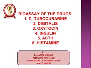
1. BIOASSAY OF THE DRUGS:
1. D- TUBOCURARINE
2. DIGITALIS
3. OXYTOCIN
4. INSULIN
5. ACTH
6. HISTAMINE
BY:
Dr. SARITA SHARMA
ASSOCIATE PROFESSOR
DEPARTMENT OF PHARMACOLOGY
MMCP, MMDU
3. 1. Rabbit Head-drop Method :
Principle: d-Tubocararine hydrochloride is
injected into the marginal vein of a rabbit’s ear
till the rabbit’s neck muscles are relaxed such
that the animal cannot hold its head up. The
total amount of test sample required to produce
the endpoint is compared with the total amount
of the standard sample required to produce
similar endpoint.
Selection of Rabbits: Rabbits weighing 2 kg.
are used. Animals should be free from disease,
obtained from a healthy colony and should be
accustomed with the experimental procedure.
4. Experimental Procedure:
o Rabbit is placed in a holder with its head protruding
outside.
o The head should be freely movable.
o Minimum 8 rabbits are used.
Rabbit head drop method for the bioassay of d-tubocurarine.
(i) : i.v. inj. of d-tubocurarine. (ii) : Head drop after injection.
5. o They are divided into two groups each
containing 4 rabbits.
o First group will receive standard sample and
the second group will receive the sample
under test.
o d-Tubocurarine solution is injected at a
constant speed by infusion apparatus through
the marginal vein.
o Injection should be given at a rate of 0.4
ml/min and should take about 10 min. Dose
0.012% w/v in saline.
6. o Infusion is continued till the rabbit will not be
in a position to hold its head erect or there
will be no response by focusing light on the
eyes.
o Rabbits recover immediately from the effect
of curarization.
o During the experiment there is a possibility of
respiratory embarrassment which is treated
by injecting neostigmine, methyl sulphate
(0.05 mg.) and atropine sulphate immediately
through the marginal ear vein.
7. o Cross-over test is carried out to minimize
biological error due to animal variation.
o Those rabbits which received the standard
sample on the first day will be given test
sample on the second day of experiment and
vice versa.
o Mean dose which produces head drop of the
test sample is compared with the mean dose
of standard preparation.
8. Principle: Potency of the test sample is
compared with that of the standard preparation by
determining the action on the cardiac muscle.
Standard Preparation and Units: The standard
preparation is a mixture of dried and powdered
digitalis leaves (1 unit = 76 mg.)
Preparation of Extracts: Exact amount of the
powder is extracted with dehydrated alcohol in a
continuous extraction apparatus for six hours. The
final extract should contain 10 ml. (5 ml. alcohol +
5 ml. water) per 10 g. of digitalis powder. It should
be stored in between 5 oC and –5 oC.
9. o Minimum 6 pigeons are used for testing each
sample.
o The weight of the heaviest pigeon should not
exceed twice the weight of the lightest pigeon.
o Food is withheld 16-28 hours before the
experiment.
o Pigeons are divided on the basis of their sex,
weight and breed, into two groups.
10. o They are anaesthetized with anesthetic ether.
o One side of the wing is dissected and the alar
vein is cannulated by means of a venous
cannula. Dilutions are made with normal
saline.
o The test sample and standard sample is
infused through cannula.
11. o In pigeons, stoppage of heart is associated with
a characteristic vomiting response called
‘emesis’.
o The milk from the crop sac of pigeons is being
ejected out. This may be taken as the end point
response of digitalis.
o The lethal dose per kg. of body weight is
determined for each pigeon.
o The potency of the test sample is determined
by dividing the mean lethal dose of standard by
the mean lethal dose of the test sample.
12. Principle: The potency of oxytocin is
determined by comparing its activity with that
of the Standard Preparation of oxytocin under
the conditions of a suitable method of assay.
Standard Preparation: The Standard
Preparation is the 4th International Standard
for Oxytocin, established in 1978, consisting
of freeze-dried synthetic oxytocin peptide with
human albumin and citric acid (supplied in
ampoules containing 12.5 Units)
13. o Anaesthetize a young healthy adult cockerel weighing
1.2 to 2.3 kg with an anesthetic that will maintain a
prolonged and constant high blood pressure.
o Expose the gluteus primus muscle in one thigh and cut
and retract it to reveal the popliteal artery and crural
vein.
o Cannulate the popliteal artery and record the blood
pressure.
o Cannulate the crural or brachial vein.
14. o Prepare standard solution with saline. Inject
0.1-0.5 ml.
o Inject 2 doses of standard solution into
cannulated vein and record blood pressure.
o Dose should cause decrease in B.P.
o Interval between 2 injections is 3-10 mins or
depends on rate at which B.P. comes to
normal.
o Dilute test sample with saline same as
standard one.
15. o Ratio between standard and test should be
equal.
o If animal become insensitive due to repetitive
doses so another animal is taken.
o Measure all responses and result is calculated
by standard statistical method.