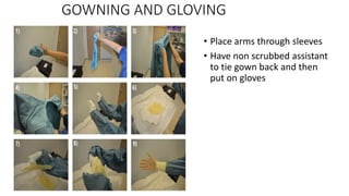
Basic Surgical Skills
Dr Rajeev Kumar Pandit
FCPS Resident ( Surgery)
Manmohan Memorial Medical College,
Swayambhu, Nepal
Learning Objectives
• TO UNDERSTAND:-
• The principles of patient positioning and operating room safety
• The Principles of skin and abdominal incision
• The Principles of wound closure
• The Principles of bowel and vascular anastomosis
• TO BE AWARE OF:-
• The principles of drain usages
• The principles of diathermy.
Introduction
• The successful outcomes in surgery depend upon the knowledge,
skills and judgement.
• Along with technical, non technical skills like communication,
empathy and teamwork are important.
PATIENT POSITIONING AND SAFETY ON THE
OPERATING TABLE
• The safety of the patient in the operating theatre is paramount at all
times, and is a key responsibility of the surgeon, regardless of grade,
experience or seniority. For all cases it is the surgeon’s responsibility
to make sure the patient is placed on the table to maximize exposure
for the procedure itself and to ensure risks of injury are avoided.
TRANSFER TO AND FROM THE OPERATING TABLE
• The transfer of the anesthetized patient is a critical moment
where there are significant risks of fall, injuries. Staff should
all receive regular training in manual handling. Patients at
additional risk include the obese, elderly and emaciated.
These groups require additional care and specialized
equipment.
POSITIONING ON THE TABLE
• Surgeons responsibility for
• Safety
• Adequate exposure for procedure
• Placement of diathermy
• Placement of operating lights
• These should be done before scrub
PATIENT PREPARATION
• Identification of patient
• History and physical examination of patient
• Hair of surgical site are better clipped or depilated than
shaving and it should be done just prior to operation.
SURGICAL SCRUBBING
• Hand washing is important measure for infection prevention.
• Nails are area of greatest contamination
• Remove all jewelries
• Use soap, brush and running water
• Scrub hands, elbow up to arms
• Turn off tap with elbow
• Dry with sterile towels
• Hold hand and forearm away from body and above elbow until
putting a sterile gown and glove
GOWNING AND GLOVING
• Place arms through sleeves
• Have non scrubbed assistant
to tie gown back and then
put on gloves
SKIN PREPARATION
• Before operation wash surgical site and surroundings with soap and
water
• Prepare skin with antiseptic solution from centre to periphery in
circular manner.
• Antiseptics-chlorhexidine, iodophores, alcohol
• Solution should remain wet on skin for atleast 2 minutes.
• The most important principle is progress from clean to dirty
• Areas of high microbial count like axilla, groin, vagina, perineum, anus
are prepped last with separate sponge
DRAPPING
• Cover all part of body except the operative field and area necessary
for anesthesia.
SKIN INCISION
• Skin incision to be made with scalpel with blade being placed vertical
and down to skin and then drawn toward the area of desire direction.
• The incision is facilitated by tension being applied across the line of
incision with fingers.
Different types of surgical blade
FACTORS CONSIDERED WHEN PLANNING A SKIN
INCISION
1. Skin tension line ( Langer’s line) represents dermal collagen
orientation and results in better scar.
2. Anatomical structures-avoid bony prominence, crossing skin
creases, consider nerve and vessels
3. Cosmetic factors-
4. Adequate access for procedure-
ABDOMINAL INCISION
• It should be planned to access the relevant organs, surface landmarks ,
pain control and cosmetic outcomes.
• The incision should be carried deeper through the subcutaneous tissue.
• The muscle layers should be divided or split and peritoneum displayed.
• Peritoneum should be held with 2 forceps and gently incised to ensure
there is no damage to the underlying organs.
• The length of suture material should be at least four times the length of
wound to minimize risk of wound dehiscence and later incisional hernia.
Picture of skin incision
SUTURE MATERIALS
• Characters of suture materials
1. Physical structures- Monofilament or Multifilament
2. Strength- depends upon the constiuent materials, its thickness and how it
handled in the tissue.
3. Tensile strength-Elastic or Plastic
4. Absorbability – absorbable or non absorbable
5. Biological behavior- natural/synthetic
Barbed sutures – novel suture materials, eradicate need for knot tying.
SIZES OF SUTURE MATERIALS
2— Thick. For pedicle ligation.
1—
1—zero.
2—zero. For bowel suturing.
3—zero.
4—zero.
5—zero. For vascular anastomosis.
6—zero. ”
7—zero.
8—zero.
9—zero. For ophthalmic surgery
TYPES OF SUTURE MATERIALS
• According to source
• Natural – catgut, silk
• Synthetic – vicryl, prolene
• According to absorbability
• Absorbable – catgut, vicryl
• Non absorbable- prolene, silk
• According to number of filament
• Monofilament – prolene, silk
• Multifilament/Braided – catgut, vicryl
• According to relation with needle
• Atraumatic –eyeless needle
• Traumatic-needle with eye
NEEDLE
• Needles can be atraumatic or traumatic with eye
• It has main three parts
• Shank
• Body
• Point
• It should be hold approximately one third to one half of the way back from
the rear of needle.
KNOTTING TECHNIQUE
The knot must be tied firmly, but without strangulating the tissues.
The knot must be unable to slip or unravel.
The knot must be as small as possible to minimise the amount of foreign material.
The knot must be tightened without exerting any tension or pressure on the tissues being ligated, i.e. the
knot should be bedded down carefully, only exerting pressure against counter-pressure from the index
finger or thumb.
During tying, the suture material must not be ‘sawed’ as this weakens the thread.
The suture material must be laid square during tying, otherwise tension applied during tightening may
cause breakage or fracture of the thread.
When tying an instrument knot, the thread should only be grasped at the free end, as gripping the thread
with artery forceps or needle holders can damage the material and again result in breakage or fracture.
TYPES OF KNOT
SUTURE TECHNIQUE
1. Continuous suturing
2. Interrupted simple suturing
3. Mattress suturing
Vertical
Horizontal
4. Subcuticular suturing
Alternative to sutures
• Skin adhesive strip - Tegaderm or Bioclusive
• Tissue glue - n-butyl-2-cyanoacrylate monomer solution
• Staples
WOUND CLOSURE
• Types of wound healing
Primary intention
Clean wound
Secondary intention
Healthy granulation tissue
Overexuberant granulation tissue
Infected sloughy wound
Black eschar
Tertiary intention
Delayed closure
Skin grafting
PRINCIPLE OF ANASTOMOSIS
Intestinal anastomoses
Ensure good blood supply to both bowel ends before and after
formation of anastomosis
Ensure the anastomosis is under no tension
Avoid risk to mesenteric vessels by clamps or sutures
Use atraumatic bowel clamps to minimize contamination
Interrupted and continuous single-layer suture techniques are
adequate and safe
Stapling devices are an alternative when speed is required or
access is a major factor
Vascular anastomosis
• Non-absorbable monofilament suture material should be used, e.g.
polypropylene
• A smooth intimal suture line is essential
• Knots require multiple throws in order to ensure security
• The suture must pass from within outwards on the downflow aspect
of the anastomosis
DRAINS
• Drains are inserted to allow fluid or air that might
collect at an operation site or in a wound to drain
freely to the surface.
• Three basic principles apply in the use of drains:
1 Open drains that utilize the principle of gravity
2 Semi-open drains that work on the principle of the
capillary effect
3 Closed drain systems that utilize suction.
PRINCIPLE OF DRAIN REMOVAL
1. Drains put in to cover perioperative bleeding may usually be
removed after 24 hours, e.g. thyroidectomy.
2. Drains put in to drain serous collections usually can be removed
after 5 days, e.g. mastectomy.
3. Drains put in because of infection should be left until the infection
is subsiding or the drainage is minimal.
4. Drains put in to cover colorectal anastomoses should be removed at
about 5–7 days. However, it should be stressed that in no way does
a drain prevent any intestinal leakage, but merely may assist any
such leakage to drain externally rather than to produce life-
threatening peritonitis.
THE PRINCIPLE OF DIATHERMY: ELECTROCAUTERY
• When an electrical current passes through a conductor, some of its
energy appears as heat. The heat produced depends on:
1. the intensity of the current;
2. the wave form of the current;
3. the electrical property of the tissues through which the current
passes;
4. the relative sizes of the two electrodes.
• There are two basic types of diathermy system in use, monopolar
diathermy and bipolar diathermy
EFFECT OF CAUTERY
• Diathermy can be used for three purposes:
1 Coagulation: the sealing of blood vessels.
2 Fulguration: the destructive coagulation of tissues with
charring.
3 Cutting: used to divide tissues during bloodless surgery.
COMPLICATION OF DIATHERMY
• Electrocution
• Explosion
• Burn
• Channeling
• Interference with pacemaker
• Reference :-
• Bailey and love’s short practice of surgery 27th edition
• Farquharson’s textbook of operative surgery
• SRB surgical operation textbook.
Thank you

Basic surgical skills

  • 1.
    Basic Surgical Skills DrRajeev Kumar Pandit FCPS Resident ( Surgery) Manmohan Memorial Medical College, Swayambhu, Nepal
  • 2.
    Learning Objectives • TOUNDERSTAND:- • The principles of patient positioning and operating room safety • The Principles of skin and abdominal incision • The Principles of wound closure • The Principles of bowel and vascular anastomosis • TO BE AWARE OF:- • The principles of drain usages • The principles of diathermy.
  • 3.
    Introduction • The successfuloutcomes in surgery depend upon the knowledge, skills and judgement. • Along with technical, non technical skills like communication, empathy and teamwork are important.
  • 6.
    PATIENT POSITIONING ANDSAFETY ON THE OPERATING TABLE • The safety of the patient in the operating theatre is paramount at all times, and is a key responsibility of the surgeon, regardless of grade, experience or seniority. For all cases it is the surgeon’s responsibility to make sure the patient is placed on the table to maximize exposure for the procedure itself and to ensure risks of injury are avoided.
  • 7.
    TRANSFER TO ANDFROM THE OPERATING TABLE • The transfer of the anesthetized patient is a critical moment where there are significant risks of fall, injuries. Staff should all receive regular training in manual handling. Patients at additional risk include the obese, elderly and emaciated. These groups require additional care and specialized equipment.
  • 8.
    POSITIONING ON THETABLE • Surgeons responsibility for • Safety • Adequate exposure for procedure • Placement of diathermy • Placement of operating lights • These should be done before scrub
  • 9.
    PATIENT PREPARATION • Identificationof patient • History and physical examination of patient • Hair of surgical site are better clipped or depilated than shaving and it should be done just prior to operation.
  • 10.
    SURGICAL SCRUBBING • Handwashing is important measure for infection prevention. • Nails are area of greatest contamination • Remove all jewelries • Use soap, brush and running water • Scrub hands, elbow up to arms • Turn off tap with elbow • Dry with sterile towels • Hold hand and forearm away from body and above elbow until putting a sterile gown and glove
  • 13.
    GOWNING AND GLOVING •Place arms through sleeves • Have non scrubbed assistant to tie gown back and then put on gloves
  • 14.
    SKIN PREPARATION • Beforeoperation wash surgical site and surroundings with soap and water • Prepare skin with antiseptic solution from centre to periphery in circular manner. • Antiseptics-chlorhexidine, iodophores, alcohol • Solution should remain wet on skin for atleast 2 minutes. • The most important principle is progress from clean to dirty • Areas of high microbial count like axilla, groin, vagina, perineum, anus are prepped last with separate sponge
  • 15.
    DRAPPING • Cover allpart of body except the operative field and area necessary for anesthesia.
  • 16.
    SKIN INCISION • Skinincision to be made with scalpel with blade being placed vertical and down to skin and then drawn toward the area of desire direction. • The incision is facilitated by tension being applied across the line of incision with fingers.
  • 17.
    Different types ofsurgical blade
  • 18.
    FACTORS CONSIDERED WHENPLANNING A SKIN INCISION 1. Skin tension line ( Langer’s line) represents dermal collagen orientation and results in better scar. 2. Anatomical structures-avoid bony prominence, crossing skin creases, consider nerve and vessels 3. Cosmetic factors- 4. Adequate access for procedure-
  • 20.
    ABDOMINAL INCISION • Itshould be planned to access the relevant organs, surface landmarks , pain control and cosmetic outcomes. • The incision should be carried deeper through the subcutaneous tissue. • The muscle layers should be divided or split and peritoneum displayed. • Peritoneum should be held with 2 forceps and gently incised to ensure there is no damage to the underlying organs. • The length of suture material should be at least four times the length of wound to minimize risk of wound dehiscence and later incisional hernia.
  • 21.
  • 22.
    SUTURE MATERIALS • Charactersof suture materials 1. Physical structures- Monofilament or Multifilament 2. Strength- depends upon the constiuent materials, its thickness and how it handled in the tissue. 3. Tensile strength-Elastic or Plastic 4. Absorbability – absorbable or non absorbable 5. Biological behavior- natural/synthetic Barbed sutures – novel suture materials, eradicate need for knot tying.
  • 23.
    SIZES OF SUTUREMATERIALS 2— Thick. For pedicle ligation. 1— 1—zero. 2—zero. For bowel suturing. 3—zero. 4—zero. 5—zero. For vascular anastomosis. 6—zero. ” 7—zero. 8—zero. 9—zero. For ophthalmic surgery
  • 24.
    TYPES OF SUTUREMATERIALS • According to source • Natural – catgut, silk • Synthetic – vicryl, prolene • According to absorbability • Absorbable – catgut, vicryl • Non absorbable- prolene, silk • According to number of filament • Monofilament – prolene, silk • Multifilament/Braided – catgut, vicryl • According to relation with needle • Atraumatic –eyeless needle • Traumatic-needle with eye
  • 26.
    NEEDLE • Needles canbe atraumatic or traumatic with eye • It has main three parts • Shank • Body • Point • It should be hold approximately one third to one half of the way back from the rear of needle.
  • 28.
    KNOTTING TECHNIQUE The knotmust be tied firmly, but without strangulating the tissues. The knot must be unable to slip or unravel. The knot must be as small as possible to minimise the amount of foreign material. The knot must be tightened without exerting any tension or pressure on the tissues being ligated, i.e. the knot should be bedded down carefully, only exerting pressure against counter-pressure from the index finger or thumb. During tying, the suture material must not be ‘sawed’ as this weakens the thread. The suture material must be laid square during tying, otherwise tension applied during tightening may cause breakage or fracture of the thread. When tying an instrument knot, the thread should only be grasped at the free end, as gripping the thread with artery forceps or needle holders can damage the material and again result in breakage or fracture.
  • 29.
  • 30.
    SUTURE TECHNIQUE 1. Continuoussuturing 2. Interrupted simple suturing 3. Mattress suturing Vertical Horizontal 4. Subcuticular suturing
  • 31.
    Alternative to sutures •Skin adhesive strip - Tegaderm or Bioclusive • Tissue glue - n-butyl-2-cyanoacrylate monomer solution • Staples
  • 32.
    WOUND CLOSURE • Typesof wound healing Primary intention Clean wound Secondary intention Healthy granulation tissue Overexuberant granulation tissue Infected sloughy wound Black eschar Tertiary intention Delayed closure Skin grafting
  • 33.
    PRINCIPLE OF ANASTOMOSIS Intestinalanastomoses Ensure good blood supply to both bowel ends before and after formation of anastomosis Ensure the anastomosis is under no tension Avoid risk to mesenteric vessels by clamps or sutures Use atraumatic bowel clamps to minimize contamination Interrupted and continuous single-layer suture techniques are adequate and safe Stapling devices are an alternative when speed is required or access is a major factor
  • 34.
    Vascular anastomosis • Non-absorbablemonofilament suture material should be used, e.g. polypropylene • A smooth intimal suture line is essential • Knots require multiple throws in order to ensure security • The suture must pass from within outwards on the downflow aspect of the anastomosis
  • 35.
    DRAINS • Drains areinserted to allow fluid or air that might collect at an operation site or in a wound to drain freely to the surface. • Three basic principles apply in the use of drains: 1 Open drains that utilize the principle of gravity 2 Semi-open drains that work on the principle of the capillary effect 3 Closed drain systems that utilize suction.
  • 36.
    PRINCIPLE OF DRAINREMOVAL 1. Drains put in to cover perioperative bleeding may usually be removed after 24 hours, e.g. thyroidectomy. 2. Drains put in to drain serous collections usually can be removed after 5 days, e.g. mastectomy. 3. Drains put in because of infection should be left until the infection is subsiding or the drainage is minimal. 4. Drains put in to cover colorectal anastomoses should be removed at about 5–7 days. However, it should be stressed that in no way does a drain prevent any intestinal leakage, but merely may assist any such leakage to drain externally rather than to produce life- threatening peritonitis.
  • 37.
    THE PRINCIPLE OFDIATHERMY: ELECTROCAUTERY • When an electrical current passes through a conductor, some of its energy appears as heat. The heat produced depends on: 1. the intensity of the current; 2. the wave form of the current; 3. the electrical property of the tissues through which the current passes; 4. the relative sizes of the two electrodes. • There are two basic types of diathermy system in use, monopolar diathermy and bipolar diathermy
  • 38.
    EFFECT OF CAUTERY •Diathermy can be used for three purposes: 1 Coagulation: the sealing of blood vessels. 2 Fulguration: the destructive coagulation of tissues with charring. 3 Cutting: used to divide tissues during bloodless surgery.
  • 39.
    COMPLICATION OF DIATHERMY •Electrocution • Explosion • Burn • Channeling • Interference with pacemaker
  • 40.
    • Reference :- •Bailey and love’s short practice of surgery 27th edition • Farquharson’s textbook of operative surgery • SRB surgical operation textbook. Thank you