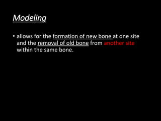
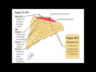
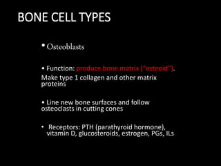
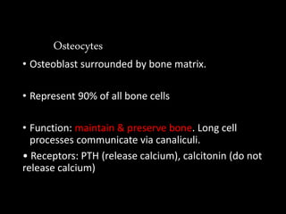
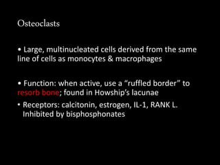
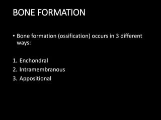
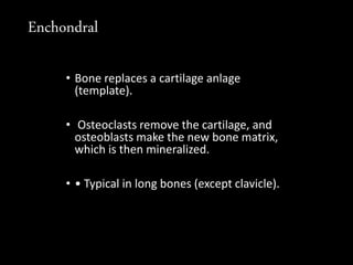
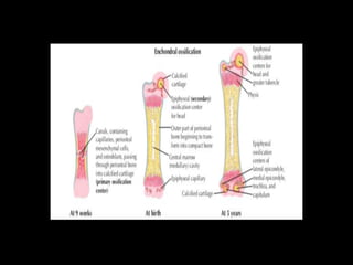
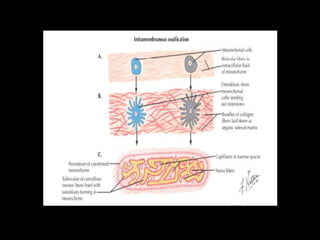
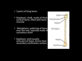
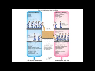
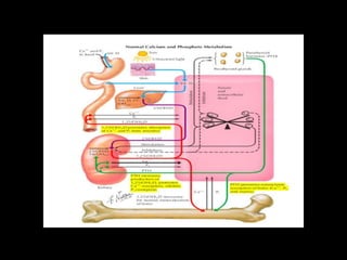
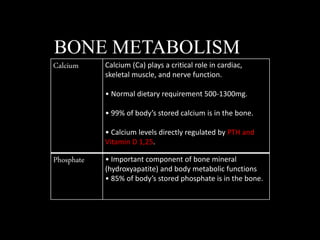
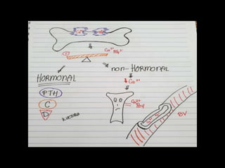
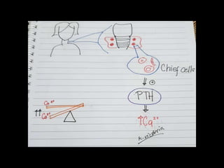
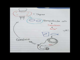
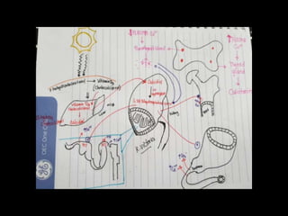
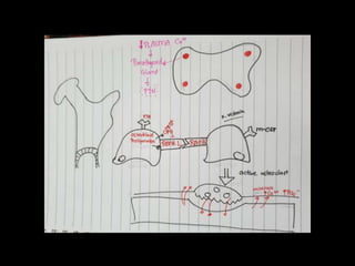
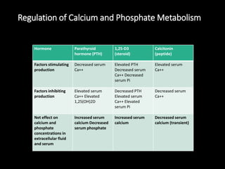
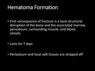
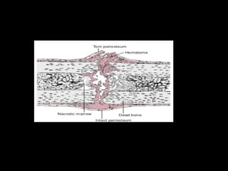
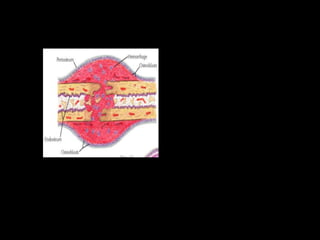
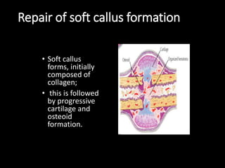
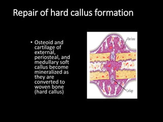
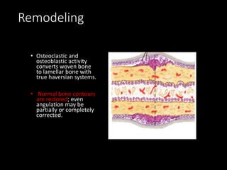
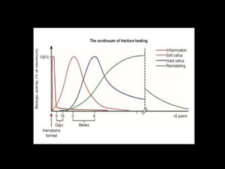
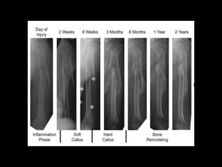
ORTHOrocks
Keiko Gwendoline A. Victoria RN, MD
Department of Orthopaedic
Region II Trauma and Medical
Center
Bayombong, Nueva Vizcaya
Bony skeleton
• remarkable organ that serves both a
• Structural function
• Reservoir function
Modeling
• allows for the formation of new bone at one site
and the removal of old bone from another site
within the same bone.
Remodeling
• Much of the cellular activity in a bone consists of
removal and replacement at the same site
Cells
• Osteoblasts, osteocytes, osteoclasts
BONE CELL TYPES
•Osteoblasts
• Function: produce bone matrix (“osteoid”).
Make type 1 collagen and other matrix
proteins
• Line new bone surfaces and follow
osteoclasts in cutting cones
• Receptors: PTH (parathyroid hormone),
vitamin D, glucosteroids, estrogen, PGs, ILs
Osteocytes
• Osteoblast surrounded by bone matrix.
• Represent 90% of all bone cells
• Function: maintain & preserve bone. Long cell
processes communicate via canaliculi.
• Receptors: PTH (release calcium), calcitonin (do not
release calcium)
Osteoclasts
• Large, multinucleated cells derived from the same
line of cells as monocytes & macrophages
• Function: when active, use a “ruffled border” to
resorb bone; found in Howship’s lacunae
• Receptors: calcitonin, estrogen, IL-1, RANK L.
Inhibited by bisphosphonates
BONE FORMATION
• Bone formation (ossification) occurs in 3 different
ways:
1. Enchondral
2. Intramembranous
3. Appositional
Enchondral
• Bone replaces a cartilage anlage
(template).
• Osteoclasts remove the cartilage, and
osteoblasts make the new bone matrix,
which is then mineralized.
• • Typical in long bones (except clavicle).
Intramembranous
• • Bone develops directly from mesenchymal cells
without a cartilage anlage. • Mesenchymal cells
differentiate into osteoblasts, which produce bone.
• • Examples: flat bones (e.g., the cranium) and
clavicle
MICROSCOPIC BONE TYPES
• Woven
Lamellar
• Mature bone
• highly organized with stress orientation
STRUCTURAL BONE TYPES
• Proteoglycans
• Macromolecules made up of a
hyaluronic backbone w/ multiple
glycosaminoglycans
• Glycosaminoglycans (GAG): made of
core protein w/ chondroitin & keratin
branches
Gives bone compressive strength
CORTICAL (COMPACT BONE) CANCELLOUS BONE
Cortical (compact)
• Strong, dense bone,
makes up 80% of the
skeleton
• Composed of multiple
osteons (haversian
systems) with intervening
interstitial lamellae
Cancellous (spongy/trabecular)
• Crossed lattice structure, makes up
20% of the skeleton
• High bone turnover rate.
BONE FORMS
• Long bones
•Form by enchondral ossification (except
clavicle):
primary (in shaft) and secondary growth
centers
• 3 parts of long bone:
• Diaphysis: shaft, made of thick
cortical bone, filled with bone
marrow
• Metaphysis: widening of bone
near the end, typically made of
cancellous bone
• Epiphysis: end (usually
articular) of bone, forms from
secondary ossification centers
BONE COMPOSITION
• Bone is composed of multiple
components:
• 1. Organic phase (“matrix:” proteins,
macromolecules, cells)
• 2. Inorganic phase (minerals, e.g., Ca)
• 3. Water
Inorganic phase
• Approximately 60% of bone weight
• Calcium hydroxyapatite Ca10(PO4)6(OH)
Primary mineral in bone. Adds compressive strength.
• Osteocalcium phosphate
“Brushite” is a secondary/minor mineral in bone.
Organic phase
• 90% of organic phase
• Type 1 collagen gives tensile strength
• Mineralization occurs at ends (hole zones) and
along sides (pores) of the collagen fibers.
Collagen types and locations
Noncollagen proteins
• e.g osteocalcin
• #1 indicator of increased bone turnover
• Osteonectin and osteopontin
Water
• Approximately 5% of bone weight (varies with age
and location)
• Periosteum surrounds the bone, is thicker in
children, and responsible for the growing diameter
(width) of long bones.
• video
BONE METABOLISM
Calcium Calcium (Ca) plays a critical role in cardiac,
skeletal muscle, and nerve function.
• Normal dietary requirement 500-1300mg.
• 99% of body’s stored calcium is in the bone.
• Calcium levels directly regulated by PTH and
Vitamin D 1,25.
Phosphate • Important component of bone mineral
(hydroxyapatite) and body metabolic functions
• 85% of body’s stored phosphate is in the bone.
Regulation of Calcium and Phosphate Metabolism
Hormone Parathyroid
hormone (PTH)
1,25-D3
(steroid)
Calcitonin
(peptide)
Factors stimulating
production
Decreased serum
Ca++
Elevated PTH
Decreased serum
Ca++ Decreased
serum Pi
Elevated serum
Ca++
Factors inhibiting
production
Elevated serum
Ca++ Elevated
1,25(OH)2D
Decreased PTH
Elevated serum
Ca++ Elevated
serum Pi
Decreased serum
Ca++
Net effect on
calcium and
phosphate
concentrations in
extracellular fluid
and serum
Increased serum
calcium Decreased
serum phosphate
Increased serum
calcium
Decreased serum
calcium (transient)
• STAGES OF ENCHONDRAL F RACTURE REPAIR
•Hematoma Formation
•Soft callus
•Hard callus
•Remodeling
Hematoma Formation
• First consequence of fracture is a local structural
disruption of the bone and the associated marrow,
periosteum, surrounding muscle, and blood
vessels.
• Lasts for 7 days
• Periosteum and local soft tissues are stripped off
Repair of soft callus formation
• Soft callus
forms, initially
composed of
collagen;
• this is followed
by progressive
cartilage and
osteoid
formation.
Repair of hard callus formation
• Osteoid and
cartilage of
external,
periosteal, and
medullary soft
callus become
mineralized as
they are
converted to
woven bone
(hard callus)
Remodeling
• Osteoclastic and
osteoblastic activity
converts woven bone
to lamellar bone with
true haversian systems.
• Normal bone contours
are restored; even
angulation may be
partially or completely
corrected.
Basic science in Orthopaedic

Basic science in Orthopaedic

  • 1.
    ORTHOrocks Keiko Gwendoline A.Victoria RN, MD Department of Orthopaedic Region II Trauma and Medical Center Bayombong, Nueva Vizcaya
  • 2.
    Bony skeleton • remarkableorgan that serves both a • Structural function • Reservoir function
  • 4.
    Modeling • allows forthe formation of new bone at one site and the removal of old bone from another site within the same bone.
  • 5.
    Remodeling • Much ofthe cellular activity in a bone consists of removal and replacement at the same site
  • 6.
  • 8.
    BONE CELL TYPES •Osteoblasts •Function: produce bone matrix (“osteoid”). Make type 1 collagen and other matrix proteins • Line new bone surfaces and follow osteoclasts in cutting cones • Receptors: PTH (parathyroid hormone), vitamin D, glucosteroids, estrogen, PGs, ILs
  • 9.
    Osteocytes • Osteoblast surroundedby bone matrix. • Represent 90% of all bone cells • Function: maintain & preserve bone. Long cell processes communicate via canaliculi. • Receptors: PTH (release calcium), calcitonin (do not release calcium)
  • 10.
    Osteoclasts • Large, multinucleatedcells derived from the same line of cells as monocytes & macrophages • Function: when active, use a “ruffled border” to resorb bone; found in Howship’s lacunae • Receptors: calcitonin, estrogen, IL-1, RANK L. Inhibited by bisphosphonates
  • 11.
    BONE FORMATION • Boneformation (ossification) occurs in 3 different ways: 1. Enchondral 2. Intramembranous 3. Appositional
  • 12.
    Enchondral • Bone replacesa cartilage anlage (template). • Osteoclasts remove the cartilage, and osteoblasts make the new bone matrix, which is then mineralized. • • Typical in long bones (except clavicle).
  • 14.
    Intramembranous • • Bonedevelops directly from mesenchymal cells without a cartilage anlage. • Mesenchymal cells differentiate into osteoblasts, which produce bone. • • Examples: flat bones (e.g., the cranium) and clavicle
  • 16.
  • 17.
  • 18.
    Lamellar • Mature bone •highly organized with stress orientation
  • 19.
  • 20.
    • Proteoglycans • Macromoleculesmade up of a hyaluronic backbone w/ multiple glycosaminoglycans • Glycosaminoglycans (GAG): made of core protein w/ chondroitin & keratin branches Gives bone compressive strength
  • 21.
    CORTICAL (COMPACT BONE)CANCELLOUS BONE
  • 22.
    Cortical (compact) • Strong,dense bone, makes up 80% of the skeleton • Composed of multiple osteons (haversian systems) with intervening interstitial lamellae
  • 24.
    Cancellous (spongy/trabecular) • Crossedlattice structure, makes up 20% of the skeleton • High bone turnover rate.
  • 25.
    BONE FORMS • Longbones •Form by enchondral ossification (except clavicle): primary (in shaft) and secondary growth centers
  • 26.
    • 3 partsof long bone: • Diaphysis: shaft, made of thick cortical bone, filled with bone marrow • Metaphysis: widening of bone near the end, typically made of cancellous bone • Epiphysis: end (usually articular) of bone, forms from secondary ossification centers
  • 28.
  • 30.
    • Bone iscomposed of multiple components: • 1. Organic phase (“matrix:” proteins, macromolecules, cells) • 2. Inorganic phase (minerals, e.g., Ca) • 3. Water
  • 31.
    Inorganic phase • Approximately60% of bone weight • Calcium hydroxyapatite Ca10(PO4)6(OH) Primary mineral in bone. Adds compressive strength. • Osteocalcium phosphate “Brushite” is a secondary/minor mineral in bone.
  • 32.
    Organic phase • 90%of organic phase • Type 1 collagen gives tensile strength • Mineralization occurs at ends (hole zones) and along sides (pores) of the collagen fibers.
  • 33.
  • 34.
    Noncollagen proteins • e.gosteocalcin • #1 indicator of increased bone turnover • Osteonectin and osteopontin
  • 35.
    Water • Approximately 5%of bone weight (varies with age and location) • Periosteum surrounds the bone, is thicker in children, and responsible for the growing diameter (width) of long bones.
  • 39.
  • 40.
    BONE METABOLISM Calcium Calcium(Ca) plays a critical role in cardiac, skeletal muscle, and nerve function. • Normal dietary requirement 500-1300mg. • 99% of body’s stored calcium is in the bone. • Calcium levels directly regulated by PTH and Vitamin D 1,25. Phosphate • Important component of bone mineral (hydroxyapatite) and body metabolic functions • 85% of body’s stored phosphate is in the bone.
  • 46.
    Regulation of Calciumand Phosphate Metabolism Hormone Parathyroid hormone (PTH) 1,25-D3 (steroid) Calcitonin (peptide) Factors stimulating production Decreased serum Ca++ Elevated PTH Decreased serum Ca++ Decreased serum Pi Elevated serum Ca++ Factors inhibiting production Elevated serum Ca++ Elevated 1,25(OH)2D Decreased PTH Elevated serum Ca++ Elevated serum Pi Decreased serum Ca++ Net effect on calcium and phosphate concentrations in extracellular fluid and serum Increased serum calcium Decreased serum phosphate Increased serum calcium Decreased serum calcium (transient)
  • 47.
    • STAGES OFENCHONDRAL F RACTURE REPAIR
  • 48.
  • 49.
    Hematoma Formation • Firstconsequence of fracture is a local structural disruption of the bone and the associated marrow, periosteum, surrounding muscle, and blood vessels. • Lasts for 7 days • Periosteum and local soft tissues are stripped off
  • 52.
    Repair of softcallus formation • Soft callus forms, initially composed of collagen; • this is followed by progressive cartilage and osteoid formation.
  • 53.
    Repair of hardcallus formation • Osteoid and cartilage of external, periosteal, and medullary soft callus become mineralized as they are converted to woven bone (hard callus)
  • 54.
    Remodeling • Osteoclastic and osteoblasticactivity converts woven bone to lamellar bone with true haversian systems. • Normal bone contours are restored; even angulation may be partially or completely corrected.