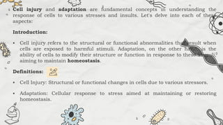
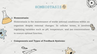
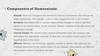
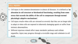
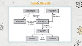
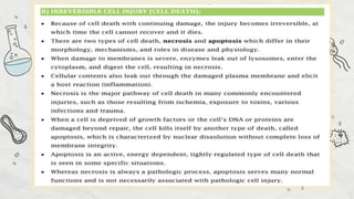
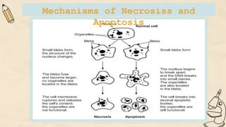
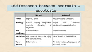
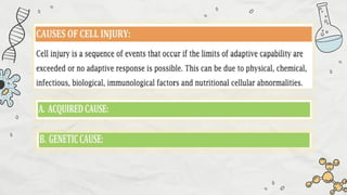
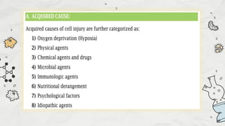
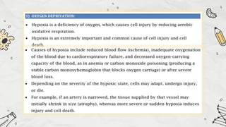
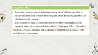
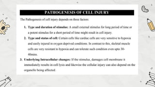
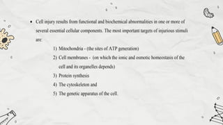
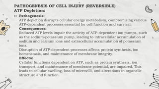
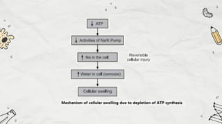
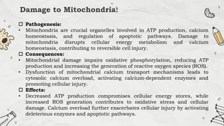
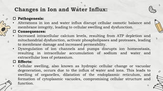
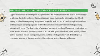
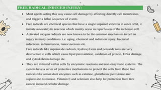
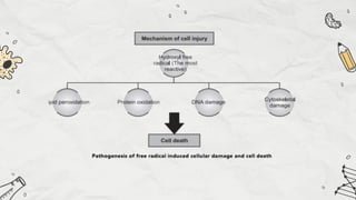
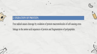
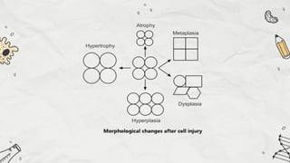
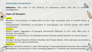
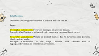
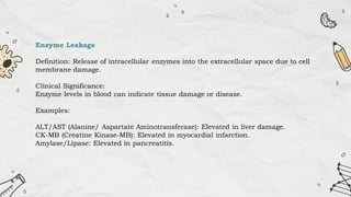
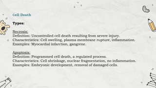
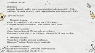
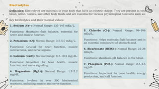
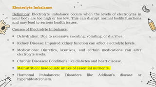
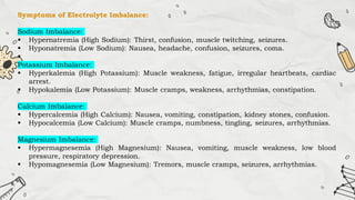
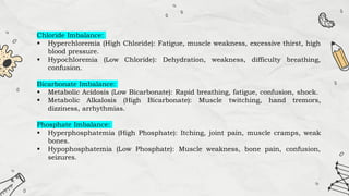
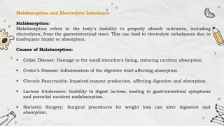
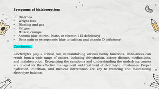
BASIC
PRINCIPLES OF
CELL INJURY
Cell injury and adaptation are fundamental concepts in understanding the
response of cells to various stresses and insults. Let's delve into each of these
aspects:
Introduction:
 Cell injury refers to the structural or functional abnormalities that result when
cells are exposed to harmful stimuli. Adaptation, on the other hand, is the
ability of cells to modify their structure or function in response to these stimuli,
aiming to maintain homeostasis.
Definitions:
 Cell Injury: Structural or functional changes in cells due to various stressors.
 Adaptation: Cellular response to stress aimed at maintaining or restoring
homeostasis.
 Homeostasis:
• Homeostasis is the maintenance of stable internal conditions within an
organism despite external changes. In cellular terms, it involves
regulating variables such as pH, temperature, and ion concentrations
to ensure optimal function.
 Components and Types of Feedback Systems:
HOMEOSTASIS
• Stimuli: These are changes in the internal or external environment that disrupt the
body's equilibrium. For example, a rise in body temperature due to hot weather.
• Receptor: Specialized cells or sensory organs detect changes in stimuli and send
signals to the control center. In our example, sensory receptors in the skin detect the
temperature change.
• Control Center: The control center receives information from the receptors and
determines the appropriate response. In this case, the control center could be the
hypothalamus in the brain.
• Effector: Effectors are structures, typically muscles or glands, that carry out the
response instructed by the control center. In our example, sweat glands are effectors
that produce sweat to cool down the body.
Components of Homeostasis:
Feedback systems are crucial in maintaining homeostasis. There are two
main types:
 Negative Feedback: Works to counteract deviations from the set point,
restoring homeostasis (e.g., blood glucose rise).
 Positive Feedback: Amplifies deviations from the set point, potentially
leading to a cascade of events (e.g., blood clotting).
Types of Homeostasis:
Negative Feedback - Blood Glucose Rise:
Stimulus: Increased blood glucose levels after a meal.
Receptor: Specialized cells in the pancreas called beta cells sense the elevated
blood glucose levels.
Control Center: The control center in this scenario is the pancreas. It receives
input from the beta cells and initiates a response.
Effector: The effector in this case is the release of insulin by the pancreas into the
bloodstream.
Response: Insulin facilitates the uptake of glucose by cells, promotes its
conversion into glycogen for storage, and enhances glucose utilization in
tissues, thus lowering blood glucose levels.
Example: After consuming a meal rich in carbohydrates, blood glucose levels rise.
Beta cells in the pancreas detect this increase and release insulin. Insulin then
prompts cells throughout the body to take up glucose from the bloodstream,
reducing blood glucose levels back to the normal range.
Positive Feedback - Blood Coagulation:
Stimulus: Injury leading to blood vessel damage.
Receptor: Platelets and damaged endothelial cells in the blood vessel wall detect
the injury.
Control Center: The liver synthesizes and releases clotting factors into the
bloodstream in response to signals from the damaged tissue.
Effector: Platelets aggregate at the site of injury, and clotting factors catalyze the
formation of fibrin, a protein mesh that stabilizes the clot.
Response: Clot formation reinforces the initial platelet plug, preventing further
blood loss at the injury site.
Example: When you sustain a cut, damaged blood vessel walls expose collagen
fibers and tissue factors. Platelets adhere to the exposed collagen and release
chemicals that attract more platelets. As platelets aggregate, they release
additional clotting factors, leading to the formation of a blood clot. This clotting
cascade continues until the clot is stabilized and bleeding stops.
CELL INJURY
Mechanisms of Necrosiss and
Apoptosis
Differences between necrosis &
apoptosis
 Pathogenesis:
ATP depletion disrupts cellular energy metabolism, compromising various
ATP-dependent processes essential for cell function and survival.
Consequences:
Reduced ATP levels impair the activity of ATP-dependent ion pumps, such
as the sodium-potassium pump, leading to intracellular accumulation of
sodium and calcium ions and extracellular accumulation of potassium
ions.
Disruption of ATP-dependent processes affects protein synthesis, ion
homeostasis, and maintenance of membrane integrity.
Effects:
Cellular functions dependent on ATP, such as protein synthesis, ion
transport, and maintenance of membrane potential, are impaired. This
leads to cellular swelling, loss of microvilli, and alterations in organelle
structure and function.
PATHOGENESIS OF CELL INJURY (REVERSIBLE)
ATP Depletion:
Damage to Mitochondria:
 Pathogenesis:
• Mitochondria are crucial organelles involved in ATP production, calcium
homeostasis, and regulation of apoptotic pathways. Damage to
mitochondria disrupts cellular energy metabolism and calcium
homeostasis, contributing to reversible cell injury.
 Consequences:
• Mitochondrial damage impairs oxidative phosphorylation, reducing ATP
production and increasing the generation of reactive oxygen species (ROS).
• Dysfunction of mitochondrial calcium transport mechanisms leads to
cytosolic calcium overload, activating calcium-dependent enzymes and
promoting cellular injury.
 Effects:
• Decreased ATP production compromises cellular energy stores, while
increased ROS generation contributes to oxidative stress and cellular
damage. Calcium overload further exacerbates cellular injury by activating
deleterious enzymes and apoptotic pathways.
Changes in Ion and Water Influx:
 Pathogenesis:
• Alterations in ion and water influx disrupt cellular osmotic balance and
membrane integrity, leading to cellular swelling and dysfunction.
 Consequences:
• Increased intracellular calcium levels, resulting from ATP depletion and
mitochondrial dysfunction, activate phospholipases and proteases, leading
to membrane damage and increased permeability.
• Dysregulation of ion channels and pumps disrupts ion homeostasis,
resulting in intracellular accumulation of sodium and water and
extracellular loss of potassium.
 Effects:
• Cellular swelling, also known as hydropic cellular change or vacuolar
degeneration, occurs due to the influx of water and ions. This leads to
swelling of organelles, dilatation of the endoplasmic reticulum, and
formation of cytoplasmic vacuoles, compromising cellular structure and
function.
In summary, reversible cell injury involves multiple interconnected
mechanisms, including ATP depletion, damage to mitochondria, and
alterations in ion and water influx. These mechanisms contribute to
cellular dysfunction and structural changes, which can be reversed if the
injurious stimuli are removed promptly. Understanding these mechanisms
is essential for elucidating the pathogenesis of reversible cell injury and
developing strategies for intervention and prevention.
IRREVERSIBLE CELL INJURY
Mitochondrial Damage:
Pathogenesis:
Mitochondria play a crucial role in cellular energy production, calcium
homeostasis, and apoptosis. Damage to mitochondria disrupts ATP
synthesis, increases reactive oxygen species (ROS) production, and
promotes mitochondrial permeability transition, triggering apoptosis.
Consequences:
ATP depletion compromises cellular energy stores, leading to dysfunction of
ATP-dependent processes. Increased ROS production causes oxidative
damage to cellular components, exacerbating cellular injury. Mitochondrial
permeability transition pore opening results in release of pro-apoptotic
factors, initiating apoptotic cell death.
Membrane Damage:
Pathogenesis:
Membrane damage disrupts cellular integrity and function, leading to
leakage of cellular contents and loss of osmotic balance.
Consequences:
Loss of membrane integrity allows influx of ions and water into the cell,
leading to cellular swelling and dysfunction. Leakage of intracellular
contents, such as enzymes and ions, into the extracellular space
activates inflammatory responses and contributes to tissue damage.
Cytoskeletal Damage:
Pathogenesis:
The cytoskeleton provides structural support, maintains cell shape, and
mediates intracellular transport and signaling. Damage to the cytoskeleton
disrupts these essential functions.
Consequences:
Cytoskeletal damage compromises cellular integrity and stability, leading to
loss of cell shape and structural integrity. Disruption of intracellular
transport impairs cellular functions and contributes to cell dysfunction and
death.
Nuclear Damage:
• Pyknosis: Condensation of chromatin within the nucleus, resulting in
shrinkage and increased basophilia.
• Karyorrhexis: Fragmentation of the nucleus into smaller, irregularly shaped
fragments.
• Karyolysis: Dissolution of nuclear chromatin, resulting in loss of nuclear
staining and structure.
Consequences:
Nuclear damage disrupts DNA integrity and gene expression, impairing
essential cellular processes such as replication and transcription.
Fragmentation and dissolution of the nucleus are characteristic features of
irreversible cell injury and apoptosis.
Lysosomal Damage:
Pathogenesis:
Lysosomes contain hydrolytic enzymes involved in intracellular digestion
and recycling of cellular components. Damage to lysosomal membranes
leads to leakage of these enzymes into the cytoplasm.
Consequences:
Release of lysosomal enzymes into the cytoplasm results in autodigestion
of cellular organelles and structures, leading to further cellular damage
and dysfunction.
Cell Death and Phagocytosis:
Pathogenesis: Irreversible cell injury culminates in cell death, which can
occur via necrosis or apoptosis. Necrosis is characterized by cellular swelling,
rupture of the cell membrane, and release of cellular contents, eliciting an
inflammatory response. Apoptosis, or programmed cell death, is a regulated
process involving activation of caspases and fragmentation of the cell into
apoptotic bodies.
Consequences: Cell death eliminates damaged cells and prevents further
tissue damage. Phagocytic cells, such as macrophages, engulf and remove
apoptotic cells and cellular debris, contributing to tissue repair and
resolution of inflammation.
In summary, irreversible cell injury involves extensive damage to
cellular structures and functions, leading to irreversible changes such
as mitochondrial dysfunction, membrane damage, cytoskeletal
disruption, nuclear alterations, lysosomal damage, and ultimately, cell
death. Understanding these mechanisms is crucial for elucidating the
pathogenesis of irreversible cell injury and developing strategies for
intervention and treatment.
Morphology of cell injury – Adaptive changes
(Atrophy, Hypertrophy, hyperplasia,
Metaplasia, Dysplasia)
Cell injury can lead to various adaptive changes as cells strive to
maintain homeostasis in response to environmental stressors.
These adaptive changes include atrophy, hypertrophy,
hyperplasia, metaplasia, and dysplasia.
1. Atrophy
Definition: Atrophy is a decrease in cell size and functional capacity due to a reduction in
cell substance.
Types:
Physiological Atrophy: Normal process of aging (e.g., thymus gland atrophy in children).
Pathological Atrophy: Due to inadequate nutrition, disuse, diminished blood supply, or
denervation.
Causes:
Disuse: Muscle atrophy from prolonged immobilization; Denervation: Loss of nerve supply
leading to muscle atrophy; Ischemia: Reduced blood supply to tissues (e.g., cerebral
atrophy due to atherosclerosis); Malnutrition: Inadequate nutrition leading to general
wasting (e.g., cachexia in chronic diseases); Loss of endocrine stimulation: Hormonal
changes (e.g., endometrial atrophy post-menopause).
Mechanism:
- Decreased protein synthesis and increased protein degradation, primarily through the
ubiquitin-proteasome pathway.
- Autophagy, where cell components are degraded by lysosomes.
Examples:
- Muscle atrophy in a limb immobilized by a cast.
- Brain atrophy in Alzheimer’s disease.
2. Hypertrophy
Definition: Hypertrophy is an increase in cell size and functional capacity without an
increase in cell number.
Types:
-Physiological Hypertrophy: Due to increased functional demand or hormonal
stimulation (e.g., skeletal muscle hypertrophy with exercise).
-Pathological Hypertrophy: Due to abnormal stress or disease conditions (e.g.,
cardiac hypertrophy due to hypertension).
Causes:
- Increased workload: E.g., weightlifting leading to skeletal muscle hypertrophy.
- Hormonal stimulation: E.g., uterine hypertrophy during pregnancy.
Mechanism:
- Increased protein synthesis and organelle number within cells.
- Activation of growth factors and signal transduction pathways (e.g., IGF-1 in muscle
cells).
Examples:
- Left ventricular hypertrophy in response to chronic hypertension.
- Skeletal muscle hypertrophy in response to resistance training.
Definition: Hyperplasia is an increase in the number of cells in an organ or tissue, leading
to increased mass.
Types:
- **Physiological Hyperplasia**: Hormonal (e.g., breast enlargement during pregnancy) or
compensatory (e.g., liver regeneration after partial hepatectomy).
- **Pathological Hyperplasia**: Due to excessive hormonal stimulation or growth factors
(e.g., benign prostatic hyperplasia).
Causes:
- **Hormonal**: E.g., estrogen-induced endometrial hyperplasia.
- **Compensatory**: Following tissue damage or partial organ removal.
- **Chronic irritation**: E.g., callus formation on skin.
Mechanism:
- Increased cellular proliferation driven by growth factors and hormones.
- Activation of stem cells and increased mitotic activity.
Examples:
- Endometrial hyperplasia due to prolonged estrogen stimulation.
- Compensatory liver hyperplasia after partial resection.
4. Metaplasia
**Definition**: Metaplasia is a reversible change in which one differentiated cell type is
replaced by another cell type.
**Types**:
- **Squamous Metaplasia**: Replacement of glandular epithelium by squamous
epithelium (e.g., in the respiratory tract due to smoking).
- **Columnar Metaplasia**: Replacement of squamous epithelium by columnar
epithelium (e.g., Barrett's esophagus).
**Causes**:
- **Chronic irritation or inflammation**: Leading to replacement by a cell type better
suited to withstand the stress.
- **Vitamin A deficiency**: Leading to squamous metaplasia in various epithelial
tissues.
**Mechanism**:
- Reprogramming of stem cells in the affected tissue.
- Cytokines, growth factors, and extracellular matrix components drive the
differentiation of stem cells to the new cell type.
**Examples**:
- Squamous metaplasia of bronchial epithelium in smokers.
5. Dysplasia
**Definition**: Dysplasia is an abnormal proliferation of cells that is characterized by
changes in cell size, shape, and organization. It is often considered a precancerous
condition.
**Types**:
- **Mild, Moderate, Severe Dysplasia**: Based on the degree of abnormal cellular and
architectural features.
- **Carcinoma in situ**: Severe dysplasia involving the full thickness of the epithelium but
not invading the basement membrane.
**Causes**:
- **Chronic irritation or inflammation**: E.g., cervical dysplasia due to human
papillomavirus (HPV) infection.
- **Genetic mutations**: Leading to disruption in normal cell regulatory mechanisms.
**Mechanism**:
- Genetic and epigenetic changes leading to loss of normal control of cellular growth and
differentiation.
- Accumulation of mutations in oncogenes and tumor suppressor genes.
**Examples**:
- Cervical dysplasia detected by Pap smear.
- Dysplasia in the bronchial epithelium of smokers.
Cell Swelling
Definition: Cell swelling, also known as hydropic swelling or oncosis, occurs
when cells take in excess water due to an inability to maintain ionic and fluid
homeostasis.
Mechanism:
Injury: Physical, chemical, or biological factors damage the cell membrane or
disrupt cellular metabolism.
Ionic Imbalance: Damage to ion pumps (e.g., Na+/K+ ATPase) leads to an
accumulation of sodium inside the cell.
Osmotic Imbalance: Water follows sodium into the cell, leading to swelling.
Examples:
Hypoxic Injury: In conditions like ischemia (restricted blood flow), cells lack
oxygen, leading to ATP depletion and impaired ion pump function.
Toxic Injury: Exposure to toxins such as bacterial toxins or drugs can damage
cellular membranes.
Intracellular Accumulation
Definition: This refers to the build-up of substances within cells due to metabolic
derangements.
Types and Examples:
Lipids:
Steatosis: Accumulation of triglycerides in liver cells, commonly seen in alcohol abuse or
obesity.
Atherosclerosis: Cholesterol accumulates in macrophages and smooth muscle cells within
arterial walls.
Proteins:
Mallory Bodies: Aggregates of damaged intermediate filaments in liver cells, often seen in
alcoholic liver disease.
Amyloidosis: Accumulation of misfolded proteins forming amyloid deposits in various tissues.
Glycogen:
Glycogen Storage Diseases: Genetic disorders like Pompe disease, where glycogen accumulates
in tissues due to enzyme deficiencies.
Pigments:
Melanin: Increased melanin in skin cells leading to hyperpigmentation disorders like melasma.
Hemosiderin: Accumulation of iron-containing pigment in conditions like hemochromatosis.
Calcification
Definition: Pathological deposition of calcium salts in tissues.
Types:
Dystrophic Calcification:Occurs in damaged or necrotic tissues.
Example: Calcification in atherosclerotic plaques or damaged heart valves.
Metastatic Calcification:Occurs in normal tissues due to hypercalcemia (elevated
blood calcium levels).
Example: Calcification in the lungs, kidneys, and stomach due to
hyperparathyroidism or chronic kidney disease.
Enzyme Leakage
Definition: Release of intracellular enzymes into the extracellular space due to cell
membrane damage.
Clinical Significance:
Enzyme levels in blood can indicate tissue damage or disease.
Examples:
ALT/AST (Alanine/ Aspartate Aminotransferase): Elevated in liver damage.
CK-MB (Creatine Kinase-MB): Elevated in myocardial infarction.
Amylase/Lipase: Elevated in pancreatitis.
Cell Death
Types:
Necrosis:
Definition: Uncontrolled cell death resulting from severe injury.
Characteristics: Cell swelling, plasma membrane rupture, inflammation.
Examples: Myocardial infarction, gangrene.
Apoptosis:
Definition: Programmed cell death, a regulated process.
Characteristics: Cell shrinkage, nuclear fragmentation, no inflammation.
Examples: Embryonic development, removal of damaged cells.
Acidosis & Alkalosis
Definition:
Acidosis: Excessive acidity in the blood and other body tissues (pH < 7.35).
Alkalosis: Excessive alkalinity in the blood and other body tissues (pH > 7.45).
Types and Causes:
• Metabolic Acidosis:
Cause: Increased acid production or loss of bicarbonate.
Examples: Diabetic ketoacidosis, lactic acidosis, renal failure.
• Respiratory Acidosis:
Cause: Accumulation of CO2 due to hypoventilation.
Examples: Chronic obstructive pulmonary disease (COPD), drug overdose.
• Metabolic Alkalosis:
Cause: Loss of acid or excessive bicarbonate.
Examples: Vomiting, diuretic use, hypokalemia.
• Respiratory Alkalosis:
Cause: Excessive loss of CO2 due to hyperventilation.
Examples: Anxiety, high altitude, fever.
Electrolytes
Definition: Electrolytes are minerals in your body that have an electric charge. They are present in your
blood, urine, tissues, and other body fluids and are essential for various physiological functions such as:
Key Electrolytes and Their Normal Values:
1. Sodium (Na+): Normal Range: 135-145 mEq/L.
Functions: Maintains fluid balance, essential for
nerve and muscle function.
2. Potassium (K+): Normal Range: 3.5-5.0 mEq/L.
Functions: Crucial for heart function, muscle
contractions, and nerve signals.
3. Calcium (Ca2+): Normal Range: 8.5-10.2 mg/dL.
Functions: Important for bone health, muscle
function, and nerve signaling.
4. Magnesium (Mg2+): Normal Range: 1.7-2.2
mg/dL.
Functions: Involved in over 300 biochemical
reactions, including muscle and nerve function.
5. Chloride (Cl-): Normal Range: 96-106
mEq/L.
Functions: Helps maintain fluid balance and is
an essential component of stomach acid.
6. Bicarbonate (HCO3-): Normal Range: 22-28
mEq/L.
Functions: Maintains pH balance in the blood.
7. Phosphate (PO4-): Normal Range: 2.5-4.5
mg/dL.
Functions: Important for bone health, energy
production, and cell function.
Electrolyte Imbalance
Definition: Electrolyte imbalance occurs when the levels of electrolytes in
your body are too high or too low. This can disrupt normal bodily functions
and may lead to serious health issues.
Causes of Electrolyte Imbalance:
 Dehydration: Due to excessive sweating, vomiting, or diarrhea.
 Kidney Disease: Impaired kidney function can affect electrolyte levels.
 Medications: Diuretics, laxatives, and certain medications can alter
electrolyte levels.
 Chronic Diseases: Conditions like diabetes and heart disease.
 Malnutrition: Inadequate intake of essential nutrients.
 Hormonal Imbalances: Disorders like Addison’s disease or
hyperaldosteronism.
Symptoms of Electrolyte Imbalance:
Sodium Imbalance:
 Hypernatremia (High Sodium): Thirst, confusion, muscle twitching, seizures.
 Hyponatremia (Low Sodium): Nausea, headache, confusion, seizures, coma.

Potassium Imbalance:
 Hyperkalemia (High Potassium): Muscle weakness, fatigue, irregular heartbeats, cardiac
arrest.
 Hypokalemia (Low Potassium): Muscle cramps, weakness, arrhythmias, constipation.
Calcium Imbalance:
 Hypercalcemia (High Calcium): Nausea, vomiting, constipation, kidney stones, confusion.
 Hypocalcemia (Low Calcium): Muscle cramps, numbness, tingling, seizures, arrhythmias.
Magnesium Imbalance:
 Hypermagnesemia (High Magnesium): Nausea, vomiting, muscle weakness, low blood
pressure, respiratory depression.
 Hypomagnesemia (Low Magnesium): Tremors, muscle cramps, seizures, arrhythmias.
Chloride Imbalance:
 Hyperchloremia (High Chloride): Fatigue, muscle weakness, excessive thirst, high
blood pressure.
 Hypochloremia (Low Chloride): Dehydration, weakness, difficulty breathing,
confusion.
Bicarbonate Imbalance:
 Metabolic Acidosis (Low Bicarbonate): Rapid breathing, fatigue, confusion, shock.
 Metabolic Alkalosis (High Bicarbonate): Muscle twitching, hand tremors,
dizziness, arrhythmias.
Phosphate Imbalance:
 Hyperphosphatemia (High Phosphate): Itching, joint pain, muscle cramps, weak
bones.
 Hypophosphatemia (Low Phosphate): Muscle weakness, bone pain, confusion,
seizures.
Malabsorption and Electrolyte Imbalance
Malabsorption:
Malabsorption refers to the body's inability to properly absorb nutrients, including
electrolytes, from the gastrointestinal tract. This can lead to electrolyte imbalances due to
inadequate intake or absorption.
Causes of Malabsorption:
 Celiac Disease: Damage to the small intestine's lining, reducing nutrient absorption.
 Crohn’s Disease: Inflammation of the digestive tract affecting absorption.
 Chronic Pancreatitis: Impaired enzyme production, affecting digestion and absorption.
 Lactose Intolerance: Inability to digest lactose, leading to gastrointestinal symptoms
and potential nutrient malabsorption.
 Bariatric Surgery: Surgical procedures for weight loss can alter digestion and
absorption.
Symptoms of Malabsorption:
• Diarrhea
• Weight loss
• Bloating and gas
• Fatigue
• Muscle cramps.
• Anemia (due to iron, folate, or vitamin B12 deficiency)
• Bone pain or osteoporosis (due to calcium and vitamin D deficiency)
Conclusion:
Electrolytes play a critical role in maintaining various bodily functions. Imbalances can
result from a wide range of causes, including dehydration, kidney disease, medications,
and malabsorption. Recognizing the symptoms and understanding the underlying causes
are crucial for the effective management and treatment of electrolyte imbalances. Proper
hydration, nutrition, and medical intervention are key to restoring and maintaining
electrolyte balance.
THANK YOU

BASIC PRINCIPLES OF CELL INJURY & ADAPTATION

  • 1.
  • 2.
    Cell injury andadaptation are fundamental concepts in understanding the response of cells to various stresses and insults. Let's delve into each of these aspects: Introduction:  Cell injury refers to the structural or functional abnormalities that result when cells are exposed to harmful stimuli. Adaptation, on the other hand, is the ability of cells to modify their structure or function in response to these stimuli, aiming to maintain homeostasis. Definitions:  Cell Injury: Structural or functional changes in cells due to various stressors.  Adaptation: Cellular response to stress aimed at maintaining or restoring homeostasis.
  • 3.
     Homeostasis: • Homeostasisis the maintenance of stable internal conditions within an organism despite external changes. In cellular terms, it involves regulating variables such as pH, temperature, and ion concentrations to ensure optimal function.  Components and Types of Feedback Systems: HOMEOSTASIS
  • 4.
    • Stimuli: Theseare changes in the internal or external environment that disrupt the body's equilibrium. For example, a rise in body temperature due to hot weather. • Receptor: Specialized cells or sensory organs detect changes in stimuli and send signals to the control center. In our example, sensory receptors in the skin detect the temperature change. • Control Center: The control center receives information from the receptors and determines the appropriate response. In this case, the control center could be the hypothalamus in the brain. • Effector: Effectors are structures, typically muscles or glands, that carry out the response instructed by the control center. In our example, sweat glands are effectors that produce sweat to cool down the body. Components of Homeostasis:
  • 5.
    Feedback systems arecrucial in maintaining homeostasis. There are two main types:  Negative Feedback: Works to counteract deviations from the set point, restoring homeostasis (e.g., blood glucose rise).  Positive Feedback: Amplifies deviations from the set point, potentially leading to a cascade of events (e.g., blood clotting). Types of Homeostasis:
  • 6.
    Negative Feedback -Blood Glucose Rise: Stimulus: Increased blood glucose levels after a meal. Receptor: Specialized cells in the pancreas called beta cells sense the elevated blood glucose levels. Control Center: The control center in this scenario is the pancreas. It receives input from the beta cells and initiates a response. Effector: The effector in this case is the release of insulin by the pancreas into the bloodstream. Response: Insulin facilitates the uptake of glucose by cells, promotes its conversion into glycogen for storage, and enhances glucose utilization in tissues, thus lowering blood glucose levels. Example: After consuming a meal rich in carbohydrates, blood glucose levels rise. Beta cells in the pancreas detect this increase and release insulin. Insulin then prompts cells throughout the body to take up glucose from the bloodstream, reducing blood glucose levels back to the normal range.
  • 7.
    Positive Feedback -Blood Coagulation: Stimulus: Injury leading to blood vessel damage. Receptor: Platelets and damaged endothelial cells in the blood vessel wall detect the injury. Control Center: The liver synthesizes and releases clotting factors into the bloodstream in response to signals from the damaged tissue. Effector: Platelets aggregate at the site of injury, and clotting factors catalyze the formation of fibrin, a protein mesh that stabilizes the clot. Response: Clot formation reinforces the initial platelet plug, preventing further blood loss at the injury site. Example: When you sustain a cut, damaged blood vessel walls expose collagen fibers and tissue factors. Platelets adhere to the exposed collagen and release chemicals that attract more platelets. As platelets aggregate, they release additional clotting factors, leading to the formation of a blood clot. This clotting cascade continues until the clot is stabilized and bleeding stops.
  • 10.
  • 12.
  • 13.
  • 25.
     Pathogenesis: ATP depletiondisrupts cellular energy metabolism, compromising various ATP-dependent processes essential for cell function and survival. Consequences: Reduced ATP levels impair the activity of ATP-dependent ion pumps, such as the sodium-potassium pump, leading to intracellular accumulation of sodium and calcium ions and extracellular accumulation of potassium ions. Disruption of ATP-dependent processes affects protein synthesis, ion homeostasis, and maintenance of membrane integrity. Effects: Cellular functions dependent on ATP, such as protein synthesis, ion transport, and maintenance of membrane potential, are impaired. This leads to cellular swelling, loss of microvilli, and alterations in organelle structure and function. PATHOGENESIS OF CELL INJURY (REVERSIBLE) ATP Depletion:
  • 27.
    Damage to Mitochondria: Pathogenesis: • Mitochondria are crucial organelles involved in ATP production, calcium homeostasis, and regulation of apoptotic pathways. Damage to mitochondria disrupts cellular energy metabolism and calcium homeostasis, contributing to reversible cell injury.  Consequences: • Mitochondrial damage impairs oxidative phosphorylation, reducing ATP production and increasing the generation of reactive oxygen species (ROS). • Dysfunction of mitochondrial calcium transport mechanisms leads to cytosolic calcium overload, activating calcium-dependent enzymes and promoting cellular injury.  Effects: • Decreased ATP production compromises cellular energy stores, while increased ROS generation contributes to oxidative stress and cellular damage. Calcium overload further exacerbates cellular injury by activating deleterious enzymes and apoptotic pathways.
  • 28.
    Changes in Ionand Water Influx:  Pathogenesis: • Alterations in ion and water influx disrupt cellular osmotic balance and membrane integrity, leading to cellular swelling and dysfunction.  Consequences: • Increased intracellular calcium levels, resulting from ATP depletion and mitochondrial dysfunction, activate phospholipases and proteases, leading to membrane damage and increased permeability. • Dysregulation of ion channels and pumps disrupts ion homeostasis, resulting in intracellular accumulation of sodium and water and extracellular loss of potassium.  Effects: • Cellular swelling, also known as hydropic cellular change or vacuolar degeneration, occurs due to the influx of water and ions. This leads to swelling of organelles, dilatation of the endoplasmic reticulum, and formation of cytoplasmic vacuoles, compromising cellular structure and function.
  • 29.
    In summary, reversiblecell injury involves multiple interconnected mechanisms, including ATP depletion, damage to mitochondria, and alterations in ion and water influx. These mechanisms contribute to cellular dysfunction and structural changes, which can be reversed if the injurious stimuli are removed promptly. Understanding these mechanisms is essential for elucidating the pathogenesis of reversible cell injury and developing strategies for intervention and prevention.
  • 32.
  • 33.
    Mitochondrial Damage: Pathogenesis: Mitochondria playa crucial role in cellular energy production, calcium homeostasis, and apoptosis. Damage to mitochondria disrupts ATP synthesis, increases reactive oxygen species (ROS) production, and promotes mitochondrial permeability transition, triggering apoptosis. Consequences: ATP depletion compromises cellular energy stores, leading to dysfunction of ATP-dependent processes. Increased ROS production causes oxidative damage to cellular components, exacerbating cellular injury. Mitochondrial permeability transition pore opening results in release of pro-apoptotic factors, initiating apoptotic cell death.
  • 34.
    Membrane Damage: Pathogenesis: Membrane damagedisrupts cellular integrity and function, leading to leakage of cellular contents and loss of osmotic balance. Consequences: Loss of membrane integrity allows influx of ions and water into the cell, leading to cellular swelling and dysfunction. Leakage of intracellular contents, such as enzymes and ions, into the extracellular space activates inflammatory responses and contributes to tissue damage.
  • 35.
    Cytoskeletal Damage: Pathogenesis: The cytoskeletonprovides structural support, maintains cell shape, and mediates intracellular transport and signaling. Damage to the cytoskeleton disrupts these essential functions. Consequences: Cytoskeletal damage compromises cellular integrity and stability, leading to loss of cell shape and structural integrity. Disruption of intracellular transport impairs cellular functions and contributes to cell dysfunction and death.
  • 36.
    Nuclear Damage: • Pyknosis:Condensation of chromatin within the nucleus, resulting in shrinkage and increased basophilia. • Karyorrhexis: Fragmentation of the nucleus into smaller, irregularly shaped fragments. • Karyolysis: Dissolution of nuclear chromatin, resulting in loss of nuclear staining and structure. Consequences: Nuclear damage disrupts DNA integrity and gene expression, impairing essential cellular processes such as replication and transcription. Fragmentation and dissolution of the nucleus are characteristic features of irreversible cell injury and apoptosis.
  • 37.
    Lysosomal Damage: Pathogenesis: Lysosomes containhydrolytic enzymes involved in intracellular digestion and recycling of cellular components. Damage to lysosomal membranes leads to leakage of these enzymes into the cytoplasm. Consequences: Release of lysosomal enzymes into the cytoplasm results in autodigestion of cellular organelles and structures, leading to further cellular damage and dysfunction.
  • 38.
    Cell Death andPhagocytosis: Pathogenesis: Irreversible cell injury culminates in cell death, which can occur via necrosis or apoptosis. Necrosis is characterized by cellular swelling, rupture of the cell membrane, and release of cellular contents, eliciting an inflammatory response. Apoptosis, or programmed cell death, is a regulated process involving activation of caspases and fragmentation of the cell into apoptotic bodies. Consequences: Cell death eliminates damaged cells and prevents further tissue damage. Phagocytic cells, such as macrophages, engulf and remove apoptotic cells and cellular debris, contributing to tissue repair and resolution of inflammation.
  • 39.
    In summary, irreversiblecell injury involves extensive damage to cellular structures and functions, leading to irreversible changes such as mitochondrial dysfunction, membrane damage, cytoskeletal disruption, nuclear alterations, lysosomal damage, and ultimately, cell death. Understanding these mechanisms is crucial for elucidating the pathogenesis of irreversible cell injury and developing strategies for intervention and treatment.
  • 48.
    Morphology of cellinjury – Adaptive changes (Atrophy, Hypertrophy, hyperplasia, Metaplasia, Dysplasia)
  • 49.
    Cell injury canlead to various adaptive changes as cells strive to maintain homeostasis in response to environmental stressors. These adaptive changes include atrophy, hypertrophy, hyperplasia, metaplasia, and dysplasia.
  • 50.
    1. Atrophy Definition: Atrophyis a decrease in cell size and functional capacity due to a reduction in cell substance. Types: Physiological Atrophy: Normal process of aging (e.g., thymus gland atrophy in children). Pathological Atrophy: Due to inadequate nutrition, disuse, diminished blood supply, or denervation. Causes: Disuse: Muscle atrophy from prolonged immobilization; Denervation: Loss of nerve supply leading to muscle atrophy; Ischemia: Reduced blood supply to tissues (e.g., cerebral atrophy due to atherosclerosis); Malnutrition: Inadequate nutrition leading to general wasting (e.g., cachexia in chronic diseases); Loss of endocrine stimulation: Hormonal changes (e.g., endometrial atrophy post-menopause). Mechanism: - Decreased protein synthesis and increased protein degradation, primarily through the ubiquitin-proteasome pathway. - Autophagy, where cell components are degraded by lysosomes. Examples: - Muscle atrophy in a limb immobilized by a cast. - Brain atrophy in Alzheimer’s disease.
  • 51.
    2. Hypertrophy Definition: Hypertrophyis an increase in cell size and functional capacity without an increase in cell number. Types: -Physiological Hypertrophy: Due to increased functional demand or hormonal stimulation (e.g., skeletal muscle hypertrophy with exercise). -Pathological Hypertrophy: Due to abnormal stress or disease conditions (e.g., cardiac hypertrophy due to hypertension). Causes: - Increased workload: E.g., weightlifting leading to skeletal muscle hypertrophy. - Hormonal stimulation: E.g., uterine hypertrophy during pregnancy. Mechanism: - Increased protein synthesis and organelle number within cells. - Activation of growth factors and signal transduction pathways (e.g., IGF-1 in muscle cells). Examples: - Left ventricular hypertrophy in response to chronic hypertension. - Skeletal muscle hypertrophy in response to resistance training.
  • 52.
    Definition: Hyperplasia isan increase in the number of cells in an organ or tissue, leading to increased mass. Types: - **Physiological Hyperplasia**: Hormonal (e.g., breast enlargement during pregnancy) or compensatory (e.g., liver regeneration after partial hepatectomy). - **Pathological Hyperplasia**: Due to excessive hormonal stimulation or growth factors (e.g., benign prostatic hyperplasia). Causes: - **Hormonal**: E.g., estrogen-induced endometrial hyperplasia. - **Compensatory**: Following tissue damage or partial organ removal. - **Chronic irritation**: E.g., callus formation on skin. Mechanism: - Increased cellular proliferation driven by growth factors and hormones. - Activation of stem cells and increased mitotic activity. Examples: - Endometrial hyperplasia due to prolonged estrogen stimulation. - Compensatory liver hyperplasia after partial resection.
  • 53.
    4. Metaplasia **Definition**: Metaplasiais a reversible change in which one differentiated cell type is replaced by another cell type. **Types**: - **Squamous Metaplasia**: Replacement of glandular epithelium by squamous epithelium (e.g., in the respiratory tract due to smoking). - **Columnar Metaplasia**: Replacement of squamous epithelium by columnar epithelium (e.g., Barrett's esophagus). **Causes**: - **Chronic irritation or inflammation**: Leading to replacement by a cell type better suited to withstand the stress. - **Vitamin A deficiency**: Leading to squamous metaplasia in various epithelial tissues. **Mechanism**: - Reprogramming of stem cells in the affected tissue. - Cytokines, growth factors, and extracellular matrix components drive the differentiation of stem cells to the new cell type. **Examples**: - Squamous metaplasia of bronchial epithelium in smokers.
  • 54.
    5. Dysplasia **Definition**: Dysplasiais an abnormal proliferation of cells that is characterized by changes in cell size, shape, and organization. It is often considered a precancerous condition. **Types**: - **Mild, Moderate, Severe Dysplasia**: Based on the degree of abnormal cellular and architectural features. - **Carcinoma in situ**: Severe dysplasia involving the full thickness of the epithelium but not invading the basement membrane. **Causes**: - **Chronic irritation or inflammation**: E.g., cervical dysplasia due to human papillomavirus (HPV) infection. - **Genetic mutations**: Leading to disruption in normal cell regulatory mechanisms. **Mechanism**: - Genetic and epigenetic changes leading to loss of normal control of cellular growth and differentiation. - Accumulation of mutations in oncogenes and tumor suppressor genes. **Examples**: - Cervical dysplasia detected by Pap smear. - Dysplasia in the bronchial epithelium of smokers.
  • 56.
    Cell Swelling Definition: Cellswelling, also known as hydropic swelling or oncosis, occurs when cells take in excess water due to an inability to maintain ionic and fluid homeostasis. Mechanism: Injury: Physical, chemical, or biological factors damage the cell membrane or disrupt cellular metabolism. Ionic Imbalance: Damage to ion pumps (e.g., Na+/K+ ATPase) leads to an accumulation of sodium inside the cell. Osmotic Imbalance: Water follows sodium into the cell, leading to swelling. Examples: Hypoxic Injury: In conditions like ischemia (restricted blood flow), cells lack oxygen, leading to ATP depletion and impaired ion pump function. Toxic Injury: Exposure to toxins such as bacterial toxins or drugs can damage cellular membranes.
  • 57.
    Intracellular Accumulation Definition: Thisrefers to the build-up of substances within cells due to metabolic derangements. Types and Examples: Lipids: Steatosis: Accumulation of triglycerides in liver cells, commonly seen in alcohol abuse or obesity. Atherosclerosis: Cholesterol accumulates in macrophages and smooth muscle cells within arterial walls. Proteins: Mallory Bodies: Aggregates of damaged intermediate filaments in liver cells, often seen in alcoholic liver disease. Amyloidosis: Accumulation of misfolded proteins forming amyloid deposits in various tissues. Glycogen: Glycogen Storage Diseases: Genetic disorders like Pompe disease, where glycogen accumulates in tissues due to enzyme deficiencies. Pigments: Melanin: Increased melanin in skin cells leading to hyperpigmentation disorders like melasma. Hemosiderin: Accumulation of iron-containing pigment in conditions like hemochromatosis.
  • 58.
    Calcification Definition: Pathological depositionof calcium salts in tissues. Types: Dystrophic Calcification:Occurs in damaged or necrotic tissues. Example: Calcification in atherosclerotic plaques or damaged heart valves. Metastatic Calcification:Occurs in normal tissues due to hypercalcemia (elevated blood calcium levels). Example: Calcification in the lungs, kidneys, and stomach due to hyperparathyroidism or chronic kidney disease.
  • 59.
    Enzyme Leakage Definition: Releaseof intracellular enzymes into the extracellular space due to cell membrane damage. Clinical Significance: Enzyme levels in blood can indicate tissue damage or disease. Examples: ALT/AST (Alanine/ Aspartate Aminotransferase): Elevated in liver damage. CK-MB (Creatine Kinase-MB): Elevated in myocardial infarction. Amylase/Lipase: Elevated in pancreatitis.
  • 60.
    Cell Death Types: Necrosis: Definition: Uncontrolledcell death resulting from severe injury. Characteristics: Cell swelling, plasma membrane rupture, inflammation. Examples: Myocardial infarction, gangrene. Apoptosis: Definition: Programmed cell death, a regulated process. Characteristics: Cell shrinkage, nuclear fragmentation, no inflammation. Examples: Embryonic development, removal of damaged cells.
  • 61.
    Acidosis & Alkalosis Definition: Acidosis:Excessive acidity in the blood and other body tissues (pH < 7.35). Alkalosis: Excessive alkalinity in the blood and other body tissues (pH > 7.45). Types and Causes: • Metabolic Acidosis: Cause: Increased acid production or loss of bicarbonate. Examples: Diabetic ketoacidosis, lactic acidosis, renal failure. • Respiratory Acidosis: Cause: Accumulation of CO2 due to hypoventilation. Examples: Chronic obstructive pulmonary disease (COPD), drug overdose. • Metabolic Alkalosis: Cause: Loss of acid or excessive bicarbonate. Examples: Vomiting, diuretic use, hypokalemia. • Respiratory Alkalosis: Cause: Excessive loss of CO2 due to hyperventilation. Examples: Anxiety, high altitude, fever.
  • 62.
    Electrolytes Definition: Electrolytes areminerals in your body that have an electric charge. They are present in your blood, urine, tissues, and other body fluids and are essential for various physiological functions such as: Key Electrolytes and Their Normal Values: 1. Sodium (Na+): Normal Range: 135-145 mEq/L. Functions: Maintains fluid balance, essential for nerve and muscle function. 2. Potassium (K+): Normal Range: 3.5-5.0 mEq/L. Functions: Crucial for heart function, muscle contractions, and nerve signals. 3. Calcium (Ca2+): Normal Range: 8.5-10.2 mg/dL. Functions: Important for bone health, muscle function, and nerve signaling. 4. Magnesium (Mg2+): Normal Range: 1.7-2.2 mg/dL. Functions: Involved in over 300 biochemical reactions, including muscle and nerve function. 5. Chloride (Cl-): Normal Range: 96-106 mEq/L. Functions: Helps maintain fluid balance and is an essential component of stomach acid. 6. Bicarbonate (HCO3-): Normal Range: 22-28 mEq/L. Functions: Maintains pH balance in the blood. 7. Phosphate (PO4-): Normal Range: 2.5-4.5 mg/dL. Functions: Important for bone health, energy production, and cell function.
  • 63.
    Electrolyte Imbalance Definition: Electrolyteimbalance occurs when the levels of electrolytes in your body are too high or too low. This can disrupt normal bodily functions and may lead to serious health issues. Causes of Electrolyte Imbalance:  Dehydration: Due to excessive sweating, vomiting, or diarrhea.  Kidney Disease: Impaired kidney function can affect electrolyte levels.  Medications: Diuretics, laxatives, and certain medications can alter electrolyte levels.  Chronic Diseases: Conditions like diabetes and heart disease.  Malnutrition: Inadequate intake of essential nutrients.  Hormonal Imbalances: Disorders like Addison’s disease or hyperaldosteronism.
  • 64.
    Symptoms of ElectrolyteImbalance: Sodium Imbalance:  Hypernatremia (High Sodium): Thirst, confusion, muscle twitching, seizures.  Hyponatremia (Low Sodium): Nausea, headache, confusion, seizures, coma.  Potassium Imbalance:  Hyperkalemia (High Potassium): Muscle weakness, fatigue, irregular heartbeats, cardiac arrest.  Hypokalemia (Low Potassium): Muscle cramps, weakness, arrhythmias, constipation. Calcium Imbalance:  Hypercalcemia (High Calcium): Nausea, vomiting, constipation, kidney stones, confusion.  Hypocalcemia (Low Calcium): Muscle cramps, numbness, tingling, seizures, arrhythmias. Magnesium Imbalance:  Hypermagnesemia (High Magnesium): Nausea, vomiting, muscle weakness, low blood pressure, respiratory depression.  Hypomagnesemia (Low Magnesium): Tremors, muscle cramps, seizures, arrhythmias.
  • 65.
    Chloride Imbalance:  Hyperchloremia(High Chloride): Fatigue, muscle weakness, excessive thirst, high blood pressure.  Hypochloremia (Low Chloride): Dehydration, weakness, difficulty breathing, confusion. Bicarbonate Imbalance:  Metabolic Acidosis (Low Bicarbonate): Rapid breathing, fatigue, confusion, shock.  Metabolic Alkalosis (High Bicarbonate): Muscle twitching, hand tremors, dizziness, arrhythmias. Phosphate Imbalance:  Hyperphosphatemia (High Phosphate): Itching, joint pain, muscle cramps, weak bones.  Hypophosphatemia (Low Phosphate): Muscle weakness, bone pain, confusion, seizures.
  • 66.
    Malabsorption and ElectrolyteImbalance Malabsorption: Malabsorption refers to the body's inability to properly absorb nutrients, including electrolytes, from the gastrointestinal tract. This can lead to electrolyte imbalances due to inadequate intake or absorption. Causes of Malabsorption:  Celiac Disease: Damage to the small intestine's lining, reducing nutrient absorption.  Crohn’s Disease: Inflammation of the digestive tract affecting absorption.  Chronic Pancreatitis: Impaired enzyme production, affecting digestion and absorption.  Lactose Intolerance: Inability to digest lactose, leading to gastrointestinal symptoms and potential nutrient malabsorption.  Bariatric Surgery: Surgical procedures for weight loss can alter digestion and absorption.
  • 67.
    Symptoms of Malabsorption: •Diarrhea • Weight loss • Bloating and gas • Fatigue • Muscle cramps. • Anemia (due to iron, folate, or vitamin B12 deficiency) • Bone pain or osteoporosis (due to calcium and vitamin D deficiency) Conclusion: Electrolytes play a critical role in maintaining various bodily functions. Imbalances can result from a wide range of causes, including dehydration, kidney disease, medications, and malabsorption. Recognizing the symptoms and understanding the underlying causes are crucial for the effective management and treatment of electrolyte imbalances. Proper hydration, nutrition, and medical intervention are key to restoring and maintaining electrolyte balance.
  • 68.