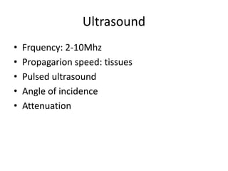
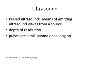
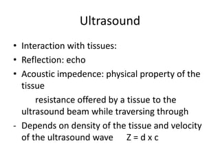
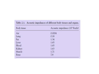
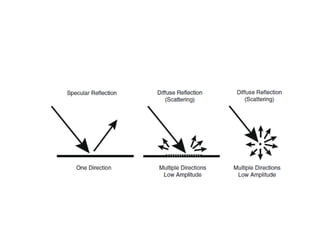
Ultrasound imaging works by transmitting high frequency sound waves into the body and receiving echoes from tissue to form an image. Ultrasound waves are generated by compressing and releasing tissue with a transducer. As the waves propagate through different tissues, they may be absorbed, refracted, reflected, scattered, or transmitted. The echo signals are used to generate an image on the screen by modulating brightness or motion. Image quality can be affected by artifacts from assumptions made during image formation not matching reality or by speckle from interference of signals from small scatterers within tissues.
Ultrasound Physics Made easy - By Dr Chandni WadhwaniChandni Wadhwani
History of ultrasound, Principle of Ultrasound.
Ultrasound wave and its interactions
Ultrasound machine and its parts, Image display, Artifacts and their clinical importance
what is Doppler ultrasound, Elastography and Recent advances in field of ultrasound.
Safety issues in ultrasound.
Ultrasound uses high frequency sound waves to create images of internal organs and structures. It has several medical applications such as visualizing soft tissues, assessing blood vessels, and guiding procedures. The document discusses how ultrasound works, including how sound waves are produced and reflected to form images, and factors that affect image quality such as frequency, attenuation, and gain. Ultrasound is a valuable medical imaging tool when used by an operator with the proper knowledge and skills to acquire and interpret the images.
This document discusses different types of ultrasound transducers and systems. It describes linear array, sector, and vector array transducers. It also discusses mechanical transducers, electronically steered systems, and phased array transducers. Finally, it outlines several specialized ultrasound transducers including those used for small parts, endocavity, transesophageal, transluminal, and intracardiac applications.
This document discusses the physical principles of ultrasound used in medical imaging. It defines key terms like frequency, wavelength, attenuation and resolution. It describes how piezoelectric transducers convert electrical pulses to ultrasound pulses and echoes. It explains how sector and linear array transducers work and the different display modes. It also discusses artifacts and the safety of diagnostic medical ultrasound.
Ultrasound uses high frequency sound waves to image internal structures. It works by sending sound waves into the body which bounce off tissues and organs, creating echoes. The echoes are detected and used to produce images on screen. Key physics principles include velocity, wavelength, frequency and amplitude of the sound waves. How the waves interact with different tissues through reflection, transmission, scattering and attenuation impacts image quality. Resolution, beamforming and processing power determine how well an ultrasound system can distinguish between tissues. Doppler and colour Doppler utilize the Doppler effect to evaluate blood flow velocity and direction to provide functional information.
Ultrasonography uses high frequency sound waves to produce images of internal organs and structures. Sound waves are transmitted into the body using a transducer, which converts electrical signals to sound and vice versa. Reflections from tissues are detected and used to construct images showing anatomical structures. Key physics principles include velocity, frequency, wavelength, and reflection based on acoustic impedance differences between tissues. Proper transducer design and focused beams are important for optimizing image quality and resolution.
Ultrasound uses longitudinal waves to produce diagnostic images. It transmits sound pulses and receives echoes to determine depth and structures within the body. The document discusses key aspects of ultrasound including its history, components like transducers, and interactions with tissue like reflection, refraction and absorption that allow ultrasound imaging. Transducers convert electrical pulses to sound waves and back using piezoelectric crystals. Factors like frequency, focal length and beam properties affect image resolution and depth.
Ultrasound uses high frequency sound waves that are transmitted into the body. The echoes that bounce back are used to form images of tissues and organs. Higher frequencies provide better resolution but penetrate less deeply. Ultrasound is used for diagnostic medical imaging and to guide procedures by providing real-time visualization of internal structures. It has advantages of being noninvasive, having no known health effects, and being relatively inexpensive compared to other imaging modalities.
Introduction to ultarsound machine and physicsmanishyadav513
Dr. Manish Yadav gave a presentation on introducing ultrasound machines and their basic physics. He described the main parts of an ultrasound machine including the display, CPU, transducer probes, keyboard, and storage devices. He explained how machines are used including different presets, features, and modes. The physics of ultrasound was discussed including sound wave properties like velocity, frequency, wavelength, and amplitude. Key interactions between ultrasound and tissue like transmission, reflection, refraction, scattering, and attenuation were also covered.
This document discusses various topics related to ultrasound imaging including goals, early pioneers, transducer types, Doppler instrumentation and physics, harmonic imaging, spatial compounding, extended field of view, fusion imaging, 3D and 4D ultrasound, and contrast enhanced ultrasound. It provides details on transducer selection, control settings, tissue harmonic imaging principles, spatial compounding benefits, fusion imaging steps, and contrast agent interactions.
The document discusses various topics related to ultrasound and knobology. It begins with an introduction to ultrasound, covering the properties of ultrasound including frequency, wavelength, velocity and attenuation. It then discusses the principles of ultrasound imaging using the pulse-echo technique. The document covers ultrasound tissue interaction through reflection, refraction, absorption and scattering. It also discusses ultrasound instrumentation components including transducers, imaging modes like B-mode and special imaging techniques like harmonic imaging. Finally, it provides a brief introduction to knobology.
Training Material inherited form Philips Basics of Ultrasonography. Covers the fundamentals of Ultrasound Waveform, Piezoelectric Effect, Phased Echo Concept, Goal of Ultrasound, Ultrasound Image Construction process, Types of Resolution, Probe Internals, The Doppler Effect, Spectrum Waveform and concept, Color Doppler, Components of Ultrasound.
This document discusses ultrasound physics and principles. It covers the characteristics of sound waves including their need for a medium, compression and rarefaction, and propagation. It describes ultrasound wave properties like range, velocity in different media, and how velocity relates to compressibility, density, and intensity. Transducers are discussed including their piezoelectric crystal, electrode, and backing block components. Modes of ultrasound like continuous wave and pulse wave are summarized. Key interactions of ultrasound with matter like reflection, refraction, and absorption are covered. Principles of Doppler ultrasound for blood flow measurement are outlined.
Ultrasound uses high frequency sound waves to generate images of the inside of the body without using ionizing radiation. It works by transmitting sound wave pulses into the body from a probe, detecting the echoes returning from tissue boundaries, and processing and displaying the images on a screen. Ultrasound gel is used between the probe and skin to allow for tight contact and transmission of the waves. Doppler ultrasound can detect the speed and direction of moving structures like blood cells by measuring the change in frequency of returning echoes. The images can display flow information in color or measure concentration and velocity.
1. Ultrasound uses high frequency sound waves and their echoes to produce medical images of the inside of the body. 2. The ultrasound machine transmits sound pulses into the body using a probe, which detects the echoes reflected back from tissues and organs. 3. By measuring the time it takes for the echoes to return, the machine can calculate distances to internal structures and display a 2D image on the screen based on the intensities of the echoes.
- Ultrasound uses high frequency sound waves between 1-20 MHz to create images of the inside of the body. Higher frequencies provide more detail while lower frequencies allow viewing of deeper structures.
- The transducer transmits sound waves into the body which reflect off boundaries between tissues and organs. The reflections are converted into a real-time image on a monitor showing the location and characteristics of internal structures.
- Common ultrasound modes include 2D brightness mode (B-mode) which shows a cross-sectional slice, motion mode (M-mode) for viewing heart walls, and Doppler modes for assessing blood flow. Proper patient positioning and use of ultrasound gel are required to obtain quality images.
This document provides an overview of ultrasound physics, transducers, and transducer jelly. It discusses the characteristics of sound waves including their generation through mechanical vibration and their transmission through solids, liquids, and gases. The history of ultrasound and piezoelectricity is summarized. Key ultrasound concepts like wavelength, frequency, propagation velocity, amplitude, and absorption are defined. The components and function of ultrasound transducers including the piezoelectric crystal and backing block are described. Finally, the properties and ingredients of transducer jelly used to couple the transducer to the skin are outlined.
Vascular ultrasound uses sound waves to image blood vessels. It combines real-time imaging (B-mode) with Doppler to show anatomy and blood flow. Ultrasound is generated by piezoelectric crystals in the transducer that convert electrical signals to sound waves. Reflected sound waves are converted back to electrical signals to form images. Factors like frequency, amplitude, and wavelength determine image quality and depth of penetration. Ultrasound provides information on vessel structure and blood flow velocity through Doppler modes.
Doppler ultrasound utilizes the Doppler effect to detect moving objects like blood flow inside the body. It works by transmitting ultrasound pulses into the body and detecting the change in frequency (Doppler shift) of echoes reflected from moving structures like blood cells. There are different Doppler ultrasound modes - continuous wave measures presence and direction of flow while pulsed wave provides depth information. Spectral Doppler displays flow information as a waveform while color Doppler images flow in color overlaid on anatomy. Optimization involves adjusting settings like Doppler angle, sample volume size, and velocity scale to improve sensitivity and accuracy.
The document discusses ultrasound technology including its history, basic principles, imaging modes, transducer types, and diagnostic applications. It provides details on how ultrasound works by sending sound waves into the body and analyzing the echoes. Key points covered include pulse echo imaging, Doppler imaging, resolution, propagation of ultrasound in tissue, and common ultrasound machines and transducer types.
This document discusses the principles of Doppler ultrasound. It begins with a brief history of Doppler and how the Doppler effect was discovered. It then covers the basic physics of Doppler ultrasound including the Doppler equation. The remainder of the document discusses specific Doppler parameters and how to optimize the Doppler examination including:
- Adjusting spectral and color Doppler parameters
- Normal arterial and venous flow patterns
- Changes in flow related to stenosis
This document provides an overview of ultrasound physics basics. It discusses how ultrasound uses sound waves between 10-20 MHz to generate images. Sound waves are longitudinal waves that travel through materials at different speeds depending on compressibility and density. Ultrasound imaging works by transmitting pulses into the body and receiving echoes, with transducers converting between electrical and sound signals. Factors like frequency, beam characteristics, and tissue interactions impact the resulting images and potential artifacts. Understanding ultrasound physics principles is important for optimizing scans and interpreting images.
This document discusses various types of artifacts that can occur in ultrasound imaging. It defines an artifact as anything in an ultrasound image that does not accurately represent the anatomical structures present. Common causes of artifacts include errors in assumptions about how ultrasound beams travel through the body and interact with tissues. The document categorizes artifacts and provides examples associated with beam characteristics, multiple echoes, velocity errors, and attenuation errors. It emphasizes the importance of recognizing artifacts to aid in diagnosis and improve image quality.
This document provides an overview of ultrasound probe types, imaging modes, and basic controls. It discusses the different types of probes and basic ultrasound imaging modes including B-mode, M-mode, color flow mode, and Doppler mode. For each mode, it lists the main controls and knob functions, and provides guidance on optimizing settings like frequency, depth, gain, and pulse repetition frequency. The document serves as a basic guide to ultrasound machine controls and settings for different imaging applications.
Ultrasound is produced by piezoelectric crystals in transducers that convert electrical pulses into sound waves and received echoes into electrical signals. Transducers operate in shock, burst, or continuous excitation modes. The piezoelectric crystals resonate at specific frequencies determined by their thickness and composition. Damping materials in transducers shorten pulse duration to improve image resolution by reducing echo overlap. Transducers use the pulse-echo principle to transmit sound pulses into the body and receive returning echoes to create ultrasound images.
Ultrasound Basics, Troubleshooting And Outline Of Uses In Anaesthesia is a presentation that covers:
1) The history, physics, and interactions of ultrasound waves including how ultrasound machines work and form images.
2) Current and potential applications of ultrasound in anesthesiology such as regional anesthesia, vascular access, airway assessment, and focused cardiac ultrasound.
3) An overview of ultrasound uses including lung, gastric, and cardiac ultrasound as well as emerging technologies.
This document provides an overview of the history and physics of ultrasound machines. It discusses how ultrasound works, including how sound waves are produced and received, how images are formed, and factors that affect image quality. The key components of an ultrasound machine are described, including the transducer probe, central processing unit, display, and storage devices. Different ultrasound imaging modes like A-mode, B-mode, and M-mode are introduced along with common medical applications of ultrasound imaging.
This document provides an overview of ultrasound imaging systems. It discusses how ultrasound uses high frequency sound waves to visualize internal organs and tissues. Key points include:
- Ultrasound uses sound waves above the range of human hearing (above 20 kHz) for medical imaging. It provides 2D, 3D, and 4D images of anatomy.
- The physics of ultrasound involves the longitudinal transmission of sound waves through tissues at different speeds depending on density and elasticity. Reflections at tissue boundaries create echoes that form images.
- Ultrasound transducers use piezoelectric materials like quartz or PZT to transmit sound and detect reflections. Array transducers with multiple elements beamform the ultrasound for
This document discusses the basic physics and settings of endoscopic ultrasound (EUS) systems. It covers the properties of ultrasound waves, how they propagate and are affected in different media like tissues. It describes transducer characteristics, imaging principles including resolution, scanning, Doppler and common artifacts. Key points are that EUS uses high frequency sound waves (5-30 MHz) for detailed imaging, linear array transducers allow electronic focusing, and imaging is affected by factors like impedance and scattering in tissues.
Ultrasound uses high-frequency sound waves to create images of the inside of the body. It works by transmitting sound pulses into the body using a probe. When the pulses hit boundaries between tissues, some of the sound waves are reflected back to the probe. The machine calculates the distance to tissues and organs based on the speed of sound and time of the echo returns, forming a 2D image. Common medical uses of ultrasound include imaging organs in the abdomen and monitoring fetuses during pregnancy.
Here are quick answers to the review questions:
1. Ultrasound is high frequency sound waves that travel well through soft tissues like muscles, organs and fat. It travels poorly through gas pockets or bones.
2. Higher frequency ultrasound has better resolution but poorer penetration. Lower frequency has poorer resolution but better penetration.
3. Pros of ultrasound include lack of radiation, quick exams, ability to see different tissue planes, portability and lower cost compared to other imaging modalities.
4. Probes come in linear, convex, sector and endocavity shapes. Linear probes have a rectangular footprint for long superficial structures. Convex probes have a curved footprint for abdominal exams. Sector probes have a wedge shape for
Applications of Ultrasound in MedicinePranay Dutta
Ultrasound uses high-frequency sound waves to produce images of internal organs and structures. It has many medical applications. Ultrasound works by sending pulses of sound into the body that bounce off tissues and create echoes. These echoes are converted into electrical signals and processed to form images. The images provide information about anatomy, abnormal growths, and blood flow.
This document discusses ultrasound and its properties. It defines ultrasound as mechanical longitudinal waves with frequencies above human hearing (20 kHz). Key properties discussed include:
- Velocity depends on the density and stiffness of the medium and is fastest in solids.
- Frequency ranges from 2-20 MHz, with lower frequencies penetrating deeper but having lower resolution.
- Wavelength is the distance over one cycle and depends on velocity and frequency.
- Amplitude represents intensity and decreases with depth, affecting image brightness.
Ultrasound therapy uses high frequency sound waves to treat various medical conditions. It can be used for diagnosis, tissue destruction, and therapy. The document discusses ultrasound frequencies used in physiotherapy between 0.75-3.3 MHz, with deeper penetration at lower frequencies. It also covers the production of ultrasound waves using piezoelectric crystals, their propagation through tissues based on density and velocity, and how varying the voltage controls intensity.
Therapeutic ultrasound uses high frequency sound waves to produce thermal and non-thermal effects in tissues. It is commonly used by athletic trainers to induce deep heating. When used properly by a competent clinician, it can provide positive outcomes, but improper use provides few benefits. Key components of an ultrasound device include the generator, crystal, soundhead, and applicator. Treatment parameters like frequency, intensity, time, and area treated must be set correctly to achieve the desired therapeutic effect safely and effectively.
Physics of ultrasound and echocardiographyjeetshitole
The document discusses the history and physics of ultrasound imaging and echocardiography. It covers how ultrasound waves interact with tissues through reflection, scattering, attenuation and absorption. It describes how piezoelectric transducers convert electrical signals to ultrasound and vice versa to produce images. Imaging can be done in various modes like A-mode, B-mode, M-mode and 2D to visualize cardiac structures and function at different resolutions and depths.
Ultrasound physics and image optimization1 (1)Prajwith Rai
This document discusses ultrasound physics and image optimization. It begins with an overview of basic principles, instrumentation, and image optimization techniques. It then describes how ultrasound works, including the generation of sound waves, their interaction with tissues through reflection, refraction, interference and absorption. This determines image quality. Instrumentation components like the transducer, transmitter, receiver and display are explained. Factors affecting the ultrasound beam like frequency, aperture, pulse length and coupling medium are also covered.
Ultrasound uses high frequency sound waves to create images of structures inside the body. It is a non-invasive diagnostic technique that can be used to examine many organs and tissues, including the thyroid, heart, liver, kidneys, muscles, and unborn fetus. Ultrasound works by transmitting sound waves from a transducer into the body, which are reflected back to the transducer to create images based on differences in tissue density and depth. It has advantages over other imaging methods in that it is safe, requires no radiation exposure or invasive procedures, and can provide real-time visualization of moving structures.
Ultrasound therapy uses high frequency sound waves to generate heat deep in tissues for therapeutic purposes. A generator sets the ultrasound frequency between 1-3 MHz, with higher frequencies penetrating less deeply. Pulsed ultrasound is safer as it allows time for heat to dissipate between pulses. Non-thermal effects include cavitation, acoustic streaming, and micromassage. Ultrasound promotes healing in acute injuries by stimulating inflammatory responses and collagen synthesis. It helps remodel scar tissue and accelerate wound healing in chronic injuries by increasing membrane permeability. Common uses are for varicose ulcers, pressure sores, pain relief in herpes zoster and back pain. Moving the transducer head prevents heat build up and damage from standing waves.
This document provides an overview of ultrasound physics, transducers, and transducer jelly. It discusses the characteristics of sound waves including their need for a medium, generation through vibration, and properties like frequency and wavelength. It describes the history and components of ultrasound transducers, focusing on how piezoelectric crystals convert electrical signals to sound and vice versa. It also summarizes the key properties and roles of transducer jelly in ultrasound imaging.
This document provides an overview of ultrasound physics. It discusses the history of ultrasound, including its discovery and development of the piezoelectric effect. It defines sound and ultrasound, and describes the mechanics of ultrasound including transducers, wavelength, velocity, amplitude, and frequency. It also covers the interaction of ultrasound with tissues through reflection, refraction, absorption, and scattering. Common ultrasound imaging artifacts are discussed. In summary, the document provides a comprehensive review of ultrasound physics principles and how they enable medical ultrasound imaging.
The document discusses the basic physics of ultrasound imaging. It covers topics such as:
- Sound waves and their propagation through different media like air, water and tissue. The speed of sound depends on the density and elasticity of the medium.
- The basic principles of image formation using pulse-echo technique. Ultrasound pulses are transmitted into the body and echoes from interfaces between tissues are received to form images.
- Factors that affect image quality like resolution, depth of penetration and frame rate. Interactions of ultrasound with matter including reflection, scattering, refraction and attenuation are also covered.
1. Medical diagnostic ultrasound uses high frequency sound waves above 20 kHz to image internal tissues and organs.
2. The sound waves are transmitted and reflected back, with the reflections used to generate images on ultrasound machines.
3. Different frequencies are used for different structures, with higher frequencies providing better resolution but poorer penetration of deeper tissues.
Similar to Basic physics of ultrasound imaging (20)
Coronary blood flow is regulated to meet the heart's high oxygen demands. The heart extracts 70-80% of oxygen from coronary blood flow, which can increase five-fold during exercise. Coronary blood flow is determined by coronary perfusion pressure, perfusion time, and vessel wall diameter, which is controlled by vasomotor tone in response to oxygen demand and various humoral and neurological factors. Maintaining adequate coronary perfusion pressure and perfusion time is important for myocardial oxygen balance.
The brain uses 20% of available oxygen for normal function, requiring tight regulation of cerebral blood flow and oxygen delivery. Autoregulation of cerebral blood flow maintains a relatively constant blood flow between 60-160 mmHg despite changes in perfusion pressure. This is achieved through segmental vascular resistance in large arteries and small arterioles, as well as neural-astrocyte regulation involving neurons, calcium, and astrocyte end feet. Factors like oxygen, carbon dioxide, hypertension, and hypoxia can impact cerebral blood flow if they cause the limits of autoregulation to be exceeded.
Robotic surgeries offer several benefits over traditional open surgeries such as reduced trauma, shorter hospital stays, improved visualization, and fewer post-operative complications. Anesthesia for robotic surgeries requires consideration of the unique aspects of each procedure as well as general factors like patient positioning and physiological impacts due to limited access. Robotic techniques are being used in various fields including urology, gynecology, cardiac, and gastrointestinal surgery.
Ultrasound artifacts can distort images by adding non-existent structures or altering the brightness, shape, or size of real structures. Higher ultrasound frequencies improve spatial resolution but reduce tissue penetration depth. Lower frequencies allow viewing of deeper tissues but with lower resolution. The angle at which ultrasound waves strike a structure also impacts the quality of reflection. Shadowing and enhancement artifacts can occur when waves pass through structures with different attenuation properties. Understanding ultrasound physics helps optimize image quality.
1. The document discusses the equipment needed for a lumbar plexus block, including sterile towels, syringes containing local anesthetic, sterile gloves, a marking pen, surface electrode, a 1.5 inch sterile needle for skin infiltration, a 10 cm long stimulator needle, and a peripheral nerve stimulator.
2. It references four sources on ultrasound-guided lumbar plexus blocks and peripheral nerve blocks, including journal articles and textbooks.
3. The document provides a list of references for lumbar plexus blocks and peripheral nerve blocks.
TEST BANK For Basic and Clinical Pharmacology, 14th Edition by Bertram G. Kat...rightmanforbloodline
TEST BANK For Basic and Clinical Pharmacology, 14th Edition by Bertram G. Katzung, Verified Chapters 1 - 66, Complete Newest Version.
Here is the updated list of Top Best Ayurvedic medicine for Gas and Indigestion and those are Gas-O-Go Syp for Dyspepsia | Lavizyme Syrup for Acidity | Yumzyme Hepatoprotective Capsules etc
Does Over-Masturbation Contribute to Chronic Prostatitis.pptxwalterHu5
In some case, your chronic prostatitis may be related to over-masturbation. Generally, natural medicine Diuretic and Anti-inflammatory Pill can help mee get a cure.
Local Advanced Lung Cancer: Artificial Intelligence, Synergetics, Complex Sys...Oleg Kshivets
Overall life span (LS) was 1671.7±1721.6 days and cumulative 5YS reached 62.4%, 10 years – 50.4%, 20 years – 44.6%. 94 LCP lived more than 5 years without cancer (LS=2958.6±1723.6 days), 22 – more than 10 years (LS=5571±1841.8 days). 67 LCP died because of LC (LS=471.9±344 days). AT significantly improved 5YS (68% vs. 53.7%) (P=0.028 by log-rank test). Cox modeling displayed that 5YS of LCP significantly depended on: N0-N12, T3-4, blood cell circuit, cell ratio factors (ratio between cancer cells-CC and blood cells subpopulations), LC cell dynamics, recalcification time, heparin tolerance, prothrombin index, protein, AT, procedure type (P=0.000-0.031). Neural networks, genetic algorithm selection and bootstrap simulation revealed relationships between 5YS and N0-12 (rank=1), thrombocytes/CC (rank=2), segmented neutrophils/CC (3), eosinophils/CC (4), erythrocytes/CC (5), healthy cells/CC (6), lymphocytes/CC (7), stick neutrophils/CC (8), leucocytes/CC (9), monocytes/CC (10). Correct prediction of 5YS was 100% by neural networks computing (error=0.000; area under ROC curve=1.0).
Rasamanikya is a excellent preparation in the field of Rasashastra, it is used in various Kushtha Roga, Shwasa, Vicharchika, Bhagandara, Vatarakta, and Phiranga Roga. In this article Preparation& Comparative analytical profile for both Formulationon i.e Rasamanikya prepared by Kushmanda swarasa & Churnodhaka Shodita Haratala. The study aims to provide insights into the comparative efficacy and analytical aspects of these formulations for enhanced therapeutic outcomes.
Muktapishti is a traditional Ayurvedic preparation made from Shoditha Mukta (Purified Pearl), is believed to help regulate thyroid function and reduce symptoms of hyperthyroidism due to its cooling and balancing properties. Clinical evidence on its efficacy remains limited, necessitating further research to validate its therapeutic benefits.
Osteoporosis - Definition , Evaluation and Management .pdfJim Jacob Roy
Osteoporosis is an increasing cause of morbidity among the elderly.
In this document , a brief outline of osteoporosis is given , including the risk factors of osteoporosis fractures , the indications for testing bone mineral density and the management of osteoporosis
- Video recording of this lecture in English language: https://youtu.be/kqbnxVAZs-0
- Video recording of this lecture in Arabic language: https://youtu.be/SINlygW1Mpc
- Link to download the book free: https://nephrotube.blogspot.com/p/nephrotube-nephrology-books.html
- Link to NephroTube website: www.NephroTube.com
- Link to NephroTube social media accounts: https://nephrotube.blogspot.com/p/join-nephrotube-on-social-media.html
2. Why ultrasound?
• Inexpensive
• Portable
• Safe, devoid of radiation risk
• Real time, cross sectional views
• Interventions, regional anesthesia
“dr. karl theo dussik austrian neurologist”
TITANIC
Crit care med 2007 vol.35,no.5(suppl)
3. Ultrasound
• Audible range 20hz – 20khz
• SOUND: waves travelling parallel to the energy
progression
• SOUND has work, energy and power
• Decibles – ratio measurement (logarithmic)
• Velocity – m/sec
• Sound velocity in body 1540 m/sec.
Crit care med 2007 vol.35,no.5(suppl)
6. Ultrasound
• Pulsed ultrasound: means of emitting
ultrasound waves from a source.
• depth of resolution
• pulses are a millisecond or so long on
Crit care med 2007 vol.35,no.5(suppl)
7. Ultrasound
• Interaction with tissues:
• Reflection: echo
• Acoustic impedence: physical property of the
tissue
resistance offered by a tissue to the
ultrasound beam while traversing through
- Depends on density of the tissue and velocity
of the ultrasound wave Z = d x c
8.
9. Ultrasound
• Same acoustic impedence between two
tissues no image is produced
• Large difference: soft tissue and bone –
produce large echos, may cause not enough
ultrasound waves beyond the tissue
• Crit care med 2007 vol.35,no.5(suppl)
13. Ultrasound
• Attenuation:
• The intensity of the beam is reduced by
refection, refraction, scattering and absorption
- Reflection and refraction at surface of the tissue
- Objects lesser than the wavelength of the US
scattering occur
- Final eventual fate of US is absorption and
transforming heat in the tissues
- Crit care med 2007 vol.35,no.5(suppl)
14. Ultrasound
• Piezoelectric crystals: interconnected
electronically and vibrate in response to an
applied electric current
• 1880 Curie brothers : piezoelectric effect and
reverse piezoelectric effect
• Frequency, wavelength, amplitude
• Atlas of Ultrasound-Guided Procedures in Interventional Pain Manag ement
15. Ultrasound
• US waves wavelength and frequency are inversely
related
• High frequency - Better penetration of tissues –
good resolution of deeper structures
• Low frequency probes used for superficial
structures visualization
• Higher frequency waves are more attenuated
than lower frequency for a given distance
• 1-20Mhz probes
• Atlas of Ultrasound-Guided Procedures in Interventional Pain Manag ement
16. Ultrasound
• Pulsed wave: Ultrasound waves are generated
in pulses (intermittent trains of pressure) that
commonly consist of two or three sound
cycles of the same frequency
• PRF: pulse repeteation frequecy
• 1-10khz
• Atlas of Ultrasound-Guided Procedures in Interventional Pain Manag ement
17.
18. Ultrasound
• Specular reflection:
incident ultra sound pulse encounters a
smooth and large interface of different
acoustic impedance the sound wave reflects
back to transducer
19. Ultrasound
• Conclusion:
1.Ultrasound is a sound wave out of range of
audible range of human being
2. Sound wave undergoes changes while
travelling through the tissues
3.Resoution of the image depends on the
reflexion,rarefaction and attenuation of the
waves in the tissues.
20. • ENERGY NEITHER BE DESTROYED NOR BE
CREATED
CAN BE TRANSFORMED INTO
ANOTHER FORM OF ENERGY.
THANK YOU