Bago2020
- 1. Evaluation of filling material remnants after basic
preparation, apical enlargement and final irrigation
in retreatment of severely curved root canals in
extracted teeth
I. Bago1
, G. Plotino2
, M. Kati
c3
, M. Ro
can4
, M. Batini
c4
I. Ani
c1
1
Department of Endodontics and Restorative Dentistry, School of Dental Medicine, University of Zagreb, Zagreb, Croatia; 2
Grande Plotino
Torsello – Studio di Odontoiatria, Private Practice, Rome, Italy; 3
Department of Materials, Faculty of Mechanical Engineering and
Naval Architecture, University of Zagreb, Zagreb; and 4
School of Dental Medicine, University of Zagreb, Zagreb, Croatia
Abstract
Bago I, Plotino G, Kati
c M, Ro
can M, Batini
c M,
Ani
c I. Evaluation of filling material remnants after basic
preparation, apical enlargement and final irrigation in
retreatment of severely curved root canals in extracted teeth.
International Endodontic Journal, 53, 962–973, 2020.
Aim To compare the retreatment ability of several rotary
and reciprocating file systems in curved root canals of
extracted teeth and to evaluate the influence of additional
apical enlargement performed after a basic retreatment on
the amount of remaining filling material.
Methodology A total of 65 round curved root canals
were used. The root canals were prepared with the ProTa-
per Next rotary system to size 25, .06 taper and filled with
an epoxy resin-based sealer and gutta-percha using contin-
uous wave vertical compaction and warm injection back-
filling. The canals were randomly divided into four groups
according to the retreatment system used: Group I. ProTa-
per Universal Retreatment system + ProTaper Gold (PTG)
instrumentation system up to PTG F2; Group II. Reciproc
Blue system up to the instrument RB25; Group III. Reci-
proc system up to the instrument R25; Group IV. Wave
One Gold (WOG) system up to the instrument WOG25.
After the basic retreatment, additional apical enlargement
was performed in each group with an instrument that was
one size larger: in Group I, II and III up to apical size 40,
and in Group IV up to 35. The final irrigation protocol
included the following: 15% ethylenediaminotetraacetic
acid followed by NaOCl irrigation. The volume of filling
material was measured using an industrial micro-CT four
times: after root canal filling (Volume I), after basic retreat-
ment with size 25 files (Volume II), after additional root
canal enlargement with larger instruments (Volume III),
and after the final irrigation protocol (Volume IV). The
decrease in the amount of filling material after each retreat-
ment protocol was analysed using a Kruskal–Wallis test.
Intergroup analyses were performed with a Kruskal–Wallis
test and between-group differences were further analysed
with Mann–Whitney U test.
Results There were no significant differences
amongst the systems tested in the amount of remain-
ing filling material, or the reduction rates after each
phase of the retreatment procedures (P 0.05).
Intragroup analysis indicated that the use of a larger
final instrument removed significantly more filling
material in all groups (P 0.001).
Conclusion The four tested instrumentation systems
were equally effective in removing filling materials
from curved root canals in extracted teeth. Additional
apical enlargement with larger files improved the
removal of filling remnants after basic retreatment.
Keywords: retreatment, curved root canals, micro-
CT.
Received 17 December 2019; accepted 9 March 2020
Correspondence: Ivona Bago, Department of Endodontics and Restorative Dentistry, School of Dental Medicine University of
Zagreb, Gunduli
ceva 5, 10 000 Zagreb, Croatia (Tel.: +3851-4802-128; fax: +3851-4802-116; e-mail: bago@sfzg.hr).
© 2020 International Endodontic Journal. Published by John Wiley Sons Ltd
International Endodontic Journal, 53, 962–973, 2020
doi:10.1111/iej.13287
962
- 2. Introduction
The aim of root canal retreatment is to completely
remove the existing root filling material in order to
allow cleaning, disinfection, reinstrumentation and
refilling of the canal (Marques da Silva et al. 2012).
Many studies have demonstrated that hand, rotary and
reciprocating files are incapable of completely removing
all filling material from the root canal during retreat-
ment procedures (Somma et al. 2008). This is regard-
less of whether the root canal is straight (Marfisi et al.
2010, Crozeta et al. 2016, Canali et al. 2019, Delai
et al. 2019), oval (Bago et al. 2019, De-Deus et al.
2019a) or curved (Marfisi et al. 2015, Crozeta et al.
2016). According to several studies, approximately
10–49% of root canal walls remain uninstrumented as
a result of rotary and reciprocating instrumentation
(Gambill et al. 1996, Siqueira et al. 2013, Zhao et al.
2014, Drukteinis et al. 2019, De-Deus et al. 2019b).
Additionally, the amount of remaining filling material
after basic retreatment instrumentation ranges widely
between 4% and 45% (Bernardes et al. 2016, Crozeta
et al. 2016, Yilmaz et al. 2018, Delai et al. 2019,
Kaloustian et al. 2019) with the reduction rate ranging
between 76% and 96% (Bago et al. 2019).
Comparisons of rotary and reciprocating instru-
ments for the removal of intracanal filling materials
have yielded conflicting results depending the root
canal anatomy (oval, straight and curved), initial root
canal size, type of filling material and filling technique
(Suk et al. 2017, Romeiro et al. 2019). Numerous
studies report similar efficacy between rotary and
reciprocating systems, independent of the filling tech-
nique and material used (Navares et al. 2016, Rossi-
Fedale Aly Ahmed 2017, Martins et al. 2017, Delai
et al. 2018, Kaloustian et al. 2019). In addition, simi-
lar efficacy has been observed in oval and curved root
canals, even amongst different canal thirds (Martins
et al. 2017). However, reciprocating systems have
been reported to be more effective than rotary systems
for the removal of epoxy resin-based sealers or cal-
cium silicate-based sealers (Monquilhott Crozeta et al.
2016, Suk et al. 2017, Bago et al. 2019).
Considering the abundance of previous research
that demonstrates the limitations of basic instrumen-
tation for the removal of filling material from the root
canal, newer approaches are currently being investi-
gated in the hope of improving the filling remnant
removal. Numerous activated irrigation techniques
have been proposed as adjunct treatment protocols to
follow mechanical retreatment, such as ultrasonically
activated irrigation (Bernardes et al. 2016, Pedull
a
et al. 2019), sonic-activated irrigation (Kaloustian
et al. 2019) and laser-activated irrigation (Suk et al.
2017). To date, few studies have evaluated the addi-
tional steps of instrumentation using various adjusta-
ble instruments (Self-adjusting File, ReDentNova,
Berlin, Deutchland; XP-endo Shaper and Finisher,
FKG, La Chaux de Fonds, Switzerland; and TRUShape,
Dentsply Sirona, Ballaigues, Switzerland). These stud-
ies show promising results on the usage of these
instruments as an additional aid in the removal of
apical filling material (Aksel et al. 2019, Machado
et al. 2019, De-Deus et al. 2019b).
The apical third of the root canal is the most criti-
cal area for complete cleaning due to its complex
anatomy (lateral canals, apical ramifications and
irregular apical foramen) and the limitations of
mechanical instrumentation and conventional root
canal irrigation techniques. A histological study has
indicated that a significant amount of tissue and bac-
terial biofilms remain in the apical part of the canal
after basic instrumentation and conventional irriga-
tion (Siqueira et al. 2018). Although it has not yet
been proven that complete removal of intracanal fill-
ing material is mandatory for better healing out-
comes, it is reasonable to assume that filling
remnants could allow the growth of bacteria biofilms
on dentinal walls or inside dentinal tubules and thus
prevent effective root canal irrigation and disinfection
(Ng et al. 2008). De-Deus et al. (2019a,b) investigated
the influence of additional instrumentation with
instrument of greater tip size on the removal of filling
remnants after basic retreatment. The study reported
that apical enlargement from size 0.25 to 0.40 mm
improved the removal of filling materials in oval
canals. However, no studies have investigated
whether the use of larger instruments is justified in
retreatments of curved root canals.
Therefore, the aims of the present study were: (i) to
compare the retreatment ability of different rotary
and reciprocating file systems in curved root canals
and (ii) to evaluate the influence of an additional api-
cal enlargement performed after a basic retreatment
on the amount of remaining filling material. The null
hypotheses tested were that: (i) there is no difference
amongst the instrumentation systems regarding their
ability to remove the filling material from curved root
canals in extracted teeth and that (ii) instrumentation
of curved root canals in extracted teeth with larger
Bago et al. Retreatment in curved root canals
International Endodontic Journal, 53, 962–973, 2020
© 2020 International Endodontic Journal. Published by John Wiley Sons Ltd 963
- 3. sizes would not additionally decrease the amount of
the remaining filling material after basic retreatment
instrumentation.
Materials and methods
Sample selection
A power calculation was performed using the chi-
squared test family and variance statistical test
(G*Power 3.1 software; Heinrich Heine University,
Dusseldorf, Germany) with a = 0.05 and b = 0.95, to
identify the sample size for each group. The calcula-
tion indicated that the sample size should be a mini-
mum of 10 canals.
A total of 65 round curved root canals were identi-
fied from a group of extracted human mandibular
third molars with curved roots, by means of a CBCT
scan (Cranex 3DX; Soredex, Tuusula, Finland) using
the following parameters: field of view, 5 9 5
(5.0 mm) mm; ENDO, 85 µm; 6.3 mA; 90 kV; 8.7 s;
450.3 mGycm2
. Presence of canal curvature was
measured in both directions according to the method
of Schneider (Schneider 1971). Overall 65 canals
with curvatures between 25° and 40° were selected.
The length was between 19 and 22 mm. Teeth with
previous endodontic treatment, intracanal calcifica-
tions, root caries, external resorption and/or internal
resorption were excluded. The teeth were stored in
0.1% thymol solution before use.
Preparation of root canals and root canal filling
One trained operator performed all the endodontic
instrumentation and filling procedures. Access open-
ings were prepared using a water-cooled diamond fis-
sure No. 016 (Komet, Rock Hill, SC, USA). The tooth
cusps were flattened in order to standardize the work-
ing length (WL) at 18 mm. Canal patency was con-
firmed by the insertion of a size 10 K-file (Dentsply
Sirona Endodontics) through the apical foramen
before and after canal preparation. Teeth with apical
foramen having diameters smaller than size 10 stain-
less steel manual K-file or in which a NiTi manual file
size 20, .02 taper (Nitiflex; Dentsply Sirona Endodon-
tics) was inserted easily into the apical foramen were
not included. The root canals were prepared with the
ProTaper Next (PTN) rotary system (Dentsply Sirona
Endodontics). The PTN X1 and X2 files (master apical
file, MAF, tip size 0.25, .06 taper) were used up to
the WL. Each instrument was used to prepare a
maximum of five canals. In case of visible deformation
or fracture, the instrument was discarded and substi-
tuted with a new one and the canal substituted with
a new one with similar characteristics.
During instrumentation, a total of 5 mL of 2.5%
sodium hypochlorite (NaOCl) was used for each canal
irrigation using a 30G needle (BD Microlance; Becton
Dickinson, Madrid, Spain). The canals were irrigated fol-
lowing the use of each instrument. After chemo–me-
chanical instrumentation, the intracanal smear layer
was removed by the final rinsing protocol: 2 mL of 15%
ethylenediaminotetraacetic acid (Calsinase; Lege artis,
Dettenhausen, Germany), which was left in the canal
for 2 min, 1 mL of 2.5% NaOCl for 30 s and 1 mL of sal-
ine solution for 30 s. The canals were dried with sterile
PTN X2 paper points (Dentsply Sirona Endodontics).
The root canals were filled with an epoxy resin-
based sealer (AH Plus, Dentsply Sirona Endodontics)
and gutta-percha point PTN X2 using the continuous
wave vertical compaction technique and warm injec-
tion back-filling technique (BeeFill 2in1; VDW,
Munich, Germany; Libonati et al. 2018). The sealer
was introduced into the canal by using a size 25 K-
file to the full WL. Then, the PTN X2 gutta-percha
point was placed in the canal about 0.5 mm shorter
than the WL. By using size 40 warm plugger
(200 °C), the gutta-percha point was removed to the
access of the root canal. The remaining gutta-percha
in the canal was penetrated with the warm plugger
in one continuous movement (duration 5 s) to 5 mm
from the WL and then condensed using a hand cold
plugger size 1 (Machtou plugger; VDW, Munich,
Germany). For backpacking, heated gutta-percha
(180 °C) was injected and every increment was con-
densed using hand pluggers sizes 2, 3 and 4.
Following the root canal filling, the access cavities
were restored with a temporary restoration material
(Caviton; GC, Tokyo, Japan) and the quality of root
filling was confirmed with digital radiograph from
both bucco-lingual and mesio-distal projection. Teeth
in which the root canal filling was judged unsatisfac-
tory (underfilling, overfilling and poor filling with
voids) were replaced with new samples with similar
characteristics. All samples were stored at 37 °C and
100% relative humidity for 2 weeks.
Root canal retreatment
The filled canals were then randomly divided into
four experimental groups (n = 13) according to the
retreatment instruments and technique used. The
Retreatment in curved root canals Bago et al.
© 2020 International Endodontic Journal. Published by John Wiley Sons Ltd
International Endodontic Journal, 53, 962–973, 2020
964
- 4. retreatment procedure was performed by the same
experienced endodontic specialist. Each of the instru-
ments being investigated here was used to retreat
three canals in each group.
Group I: ProTaper Universal (PTU) Retreatment
system + ProTaper Gold (PTG) instrumentation system
The retreatment procedure was performed using the
PTU Retreatment system (Dentsply Sirona Endodon-
tics), according to the manufacturer’s instructions at
speed 300 rpm and torque 2.5 N cm 2
. The D1 file
was used for removal of the material from the coronal
part of the canal, whilst the D2 and D3 files for the
removal of the material from the middle and apical
third of the canal. Each canal was further enlarged
with the PTG F2 file (tip size 25, variable taper). The
additional apical enlargement was performed with the
PTG F3 (tip size 30, variable taper) and F4 (tip size
40, variable taper) files. Root canals were irrigated
after the use of each instrument with 2.5% NaOCl.
Group II: Reciproc Blue system
The retreatment procedure was performed with the
Reciproc Blue (RB) R25 file (tip size 25; variable
taper; VDW) using the VDW Gold motor set at recip-
rocation RECIPROC ALL mode. The instrument was
advanced apically using an in-and-out pecking
motion with an amplitude of approximately 3 mm
according to the manufacturer’s instructions; gentle
apical pressure was applied with a brushing action
against the lateral walls. After three pecks, the instru-
ment was removed from the canal and cleaned with
sterile gauze and the canal was irrigated with 2.5%
NaOCl. This procedure was repeated until the instru-
ment reached WL. The additional apical enlargement
was performed with RB R40 instrument (tip size 40;
variable taper) used as previously described up to the
WL, with the same irrigation regimen.
Group III: Reciproc system
The Reciproc R25 file (tip size 25; variable taper; VDW)
was used with the VDW Gold motor set at reciprocation
RECIPROC ALL mode. The retreatment technique was
the same as that described for Group II. The final apical
enlargement was performed with the Reciproc R40
instrument (tip size 40; variable taper; VDW) used as
previously described for Group II.
Group IV: Wave One Gold system
The Wave One Gold (WOG) primary file (tip size 25;
variable taper; Dentsply Sirona Endodontics) was used
with the VDW Gold motor set at reciprocation WAVE
ONE ALL mode. The retreatment technique was the
same as that described for Group II, whilst the addi-
tional apical enlargement was performed with WOG
file size 35 (variable taper) used as previously
described up to the WL.
A total of 20 mL of 2.5% NaOCl was used during
each retreatment procedure: 15 mL of NaOCl during
basic retreatment procedure and 5 mL during the
additional apical enlargement. Retreatment was con-
sidered complete when each instrument reached the
WL for five consecutive times (Bernardes et al. 2016)
and when there was no remaining filling material vis-
ible on the file.
Following the retreatment procedures, the intra-
canal smear layer was removed by the final rinsing
protocol: 2 mL of 15% ethylenediaminotetraacetic
acid (Calsinase; Lege artis, Dettenhausen, Germany),
which was left in canal for 2 min, followed by 3 mL
of 2.5% NaOCl for 30 s and 1 mL of saline solution
for 30 s. Then, the canals were dried using sterile
paper points of the correspondent size of the last file
used for the instrumentation.
Micro-CT analysis
The volume of filling material was measured using an
industrial micro-CT (Nikon XT H 225; Tring, UK)
device with a target having a focal size of 0.7 lm and
a 400 9 300-mm 14-bit flat panel detector with a
127 lm pixel size. Samples were measured at 80 kV
and 60 lA using 1600 projections at an exposure
time of 1 s. The geometrical magnification was 100,
which yielded a structural resolution of 1.2 lm.
All specimens were scanned four times: after root
canal filling (Volume I), after basic retreatment with
files tip size 25 (Volume II), after additional root canal
enlargement with larger instrument (Volume III) and
after final irrigation protocol (Volume IV).
All samples were scanned in the same position and
with the same radiation settings. Similar postprocess-
ing procedures were performed for all measurement
sets: beam hardening was reduced using a Hanning
filter, noise was reduced using a median filter, and
surface detection was performed using an adaptive
search algorithm (Volume Graphics VGMax 2.2). Dur-
ing analyses, the filling material was treated as an
inclusion in the base tooth material; this was possible
because of very distinct grey scale values for the tooth
and filling material (typically 10 000 and 40 000,
respectively). With the grey scale value for the tooth
Bago et al. Retreatment in curved root canals
International Endodontic Journal, 53, 962–973, 2020
© 2020 International Endodontic Journal. Published by John Wiley Sons Ltd 965
- 5. as the base value, a simple threshold algorithm was
used to detect the volume of filling material in the
internal tooth volume. The results were expressed as
a percentage of the remaining filling material with
respect to the initial volume of the root canal filling
by using relational values. The variations in sample
volumes were effectively excluded from the analysis of
the material removal rate. The same procedure was
applied for all samples, thus providing a constant
metric for the rate of removal of material in the root
canal.
Statistical analyses
The decrease in the amount of filling material after
each retreatment protocol was analysed using a
Kruskal–Wallis test. Intergroup analyses were per-
formed with a Kruskal–Wallis test and between-group
differences were further analysed with Mann–Whitney
U test. A P-value of 0.05 was considered statisti-
cally significant. All statistical analyses were per-
formed using IBM SPSS version 23.0 (www.spss.com).
Results
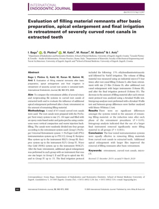
Figure 1 presents three-dimensional models of teeth
after root canal filling, after basic retreatment, after
additional root canal enlargement and after final irri-
gation protocol in each group.
There were no significant differences in the initial
filling material volume amongst the four groups
(P 0.05). Table 1 shows the initial volume of the
filling material (Volume I) in mm3
and the remaining
volume of the material after the basic retreatment
procedure (Volume II), after additional instrumenta-
tion with instrument of greater tip size (Volume III)
and after final irrigation protocol (Volume IV), for
each of the instrumentation system tested. Table 2
shows the rate of decrease (%) of the filling material
after each phase of the retreatment procedure (com-
pared to the Volume I and compared to the previous
retreatment step) for each of the instrumentation sys-
tems tested.
There were no significant differences amongst the
groups in the amount of the remaining filling mate-
rial, or the reduction rates, after each phase of the
retreatment procedures: after basic retreatment, after
additional instrumentation and after final irrigation
(P 0.05; Figures 2, 3 and 4).
Intragroup analysis indicated that the addition of a
larger instrument removed a significantly larger
amount of filling material in all groups (P 0.001).
The final irrigation protocol removed additionally sig-
nificant amounts of the filling material in all groups
(P 0.001).
There were few samples completely free from the
filling material in the apical 5 mm of the canal after
each phase of the retreatment. After the basic retreat-
ment, in Group I and IV, there was one sample, and
in Group II and III two samples were completely free
from the filling material in the apical 5 mm. One
additional sample each in Group II, III and IV was
found free of material remnants after additional
instrumentation with a larger instrument. After the
final irrigation, an additional sample in Group I and
IV was recorded to be without apical filling material.
Discussion
Debridement and disinfection of the apical third of root
canals are considered one of the more difficult chal-
lenges during root canal treatment. Due to the complex-
ity of the apical area of the root canal (intracanal
isthmuses, constriction and apical delta) and apical fora-
men, which in most cases is more oval than round (Mar-
tos et al. 2009), it is difficult to prepare and clean all
canal walls using round files (Grande et al. 2007).
Recent histological studies report significant amounts of
pulp tissue remnants and bacteria in the apical area
after single-file reciprocating instrumentation and con-
ventional sodium hypochlorite irrigation (Siqueira et al.
2018, Varela et al. 2019). According to some authors,
preparation of root canals to larger sizes allows better
removal of infected dentine (Card et al. 2002), enhances
deeper penetration of irrigants (Boutsioukis et al. 2010),
significantly reduces the amount of bacteria in the canal
system (McGurkin-Smith et al. 2005) and cleans more
debris from the canal (Plotino et al. 2019). There have
been numerous published studies that investigated the
influence of the root canal preparation size on different
outcomes: intracanal debridement (Boutsioukis et al.
2010), microbial reduction (Aminoshariae Kulild
2015), smear layer removal (Plotino et al. 2019) and
healing of periapical periodontitis (Saini et al. 2012).
The aim of this study was to evaluate the validity of per-
forming an additional larger apical preparation after a
basic retreatment, using different rotary or reciprocat-
ing files, during retreatment of filling materials in curved
root canals.
In the present study, all four tested instrumentation
systems had similar retreatment ability in curved root
canals filled with thermoplasticized gutta-percha and
Retreatment in curved root canals Bago et al.
© 2020 International Endodontic Journal. Published by John Wiley Sons Ltd
International Endodontic Journal, 53, 962–973, 2020
966
- 6. Bago et al. Retreatment in curved root canals
International Endodontic Journal, 53, 962–973, 2020
© 2020 International Endodontic Journal. Published by John Wiley Sons Ltd 967
- 7. sealer (median reduction rate 87.8–96.9%). Thus, the
null hypothesis (i) cannot be rejected. Many studies
have been published comparing the retreatment abil-
ity of different rotary or reciprocating systems. How-
ever, only three studies compared the Reciproc and
RB systems. Two of these indicated similar efficacy of
Reciproc and RB (De-Deus et al. 2019a, b). The third
study reported superior retreatment ability of the
Reciproc file (Bago et al. 2019). These conflicting
results are likely due to numerous variables that
potentially influence definitive conclusions, that is the
canal anatomy, the type of root canal filling and fill-
ing technique, the size of the file used for the retreat-
ment, the experience of the operator, the number of
samples in each group and the evaluation protocol.
For example, the differences between the present
study and a previous study by the same group (Bago
et al. 2019), where superior retreatment ability of the
Reciproc system was observed, may be due to the fact
that previous study only used a size R40 instrument,
whilst the present study used size R25 followed by
R40 instrument. Thus, it could be assumed that the
sequential use of Reciproc and RB file R25 and R40
results in similar retreatment outcomes. The superior
retreatment ability of the Reciproc and RB systems is
likely due to the instrument design, which is charac-
terized by an S-shaped cross-section with sharp cut-
ting edges and a large chip space (Giansiracusa
Rubini et al. 2014). Whilst it is reasonable to expect
that greater taper at the tip of the file R25 instrument
(Reciproc and RB; 0.08 for 3 mm) might promote
improved removal of the filling material compared to
the WOG system (size 25, .07 taper for 3 mm at the
tip), this was not observed in the present study. Thus,
in the present study, no differences were observed
between the reciprocating systems and the rotary sys-
tem. Similar results have been observed previously,
also in curved root canals (Rios et al. 2014, R€
odig
et al. 2014, Monquilhott Crozeta et al. 2016). Fur-
thermore, a review (Rossi-Fedale Aly Ahmed 2017)
concluded that reciprocating and rotary systems exhi-
bit similar retreatment abilities. Only one study has
been published on the retreatment ability of the Wave
One Gold system. Canali et al. (2019) reported similar
retreatment ability between Wave One Gold and
Wave One in mesial canals of mandibular molars. In
the present study, the WOG displayed similar retreat-
ment ability to the other tested systems, both after
basic retreatment and after additional apical enlarge-
ment, although its file size 25 has a lower apical
taper (7%) compared to other tested systems (taper
8%), and a smaller file tip size 35 was used for the
apical enlargement.
The present study demonstrated that additional
instrumentation with larger tip sizes (35–40) removed
significantly more of the remaining filling material
compared to the basic retreatment instrumentation
(size 25). Thus, the null hypothesis (ii) can be
rejected. The additional apical enlargement resulted
in a reduction rate ranging from 93.7% to 98.6%. A
Table 1 Volume (in mm3
) of the filling material initially
(Volume I) and after each phase of root canal retreatment:
after basic retreatment (Volume II), after additional larger tip
size instrumentation (Volume III) and after final irrigation
protocol (Volume IV)
Mean SD Minimum Maximum Median
Volume I
Group
PTU + PTG
9.85 4.61 1.57 17.74 9.30
Group RB 8.08 3.66 3.54 14.14 6.77
Group R 8.71 3.04 3.90 12.34 9.45
Group WOG 5.69 1.85 2.08 7.45 6.58
Volume II
Group
PTU + PTG
1.12 1.47 0.00 4.20 0.42
Group RB 0.71 1.06 0.08 3.36 0.26
Group R 2.09 2.38 0.00 6.76 1.25
Group WOG 1.15 1.17 0.00 3.11 0.68
Volume III
Group
PTU + PTG
0.45 0.68 0.00 2.22 0.16
Group RB 0.47 0.75 0.02 2.06 0.13
Group R 1.05 1.53 0.00 4.61 0.16
Group WOG 0.81 0.97 0.00 2.49 0.36
Volume IV
Group
PTU + PTG
0.16 0.27 0.00 0.85 0.05
Group RB 0.30 0.48 0.00 1.28 0.05
Group R 0.70 1.23 0.00 3.83 0.07
Group WOG 0.70 0.89 0.00 2.39 0.26
PTU + PTG, ProTaper Universal + ProTaper Gold; R, Reciproc;
RB, Reciproc Blue; WOG, Wave One Gold.
Figure 1 Three-dimensional model of a tooth (coloured according to the volume of material under investigation) after root
canal filling (a), after basic retreatment (b), after additional root canal enlargement (c) and after final irrigation protocol (d) in
four experimental groups according to the instrumentation system used: ProTaper Universal files and ProTaper Gold; Reciproc
Blue; Reciproc; Wave One Gold.
Retreatment in curved root canals Bago et al.
© 2020 International Endodontic Journal. Published by John Wiley Sons Ltd
International Endodontic Journal, 53, 962–973, 2020
968
- 8. Table 2 Reduction rate (in %) of the volume of the filling material after each phase of root canal retreatment respect to the ini-
tial filling volume (Volume I) and respect to the filling volume in the previous stages (Volume II and Volume III)
Mean (%) SD (%) Minimum (%) Maximum (%) Median (%)
Volumen II versus I reduction (%)
Group PTU + PTG 91.10 10.10 73.70 100.00 95.30
Group RB 90.10 14.20 53.40 99.40 96.90
Group R 78.10 26.30 26.40 100.00 87.80
Group WOG 82.60 16.80 57.50 100.00 89.90
Volumen III versus II reduction (%)
Group PTU + PTG 46.50 26.20 0.00 77.40 51.70
Group RB 42.30 30.50 2.90 94.50 43.40
Group R 35.10 30.40 0.00 99.20 25.10
Group WOG 37.80 27.40 0.00 79.40 23.30
Volumen III versus I reduction (%)
Group PTU + PTG 96.40 4.30 86.00 100.00 98.30
Group RB 94.10 9.00 71.00 100.00 98.60
Group R 89.70 13.90 61.00 100.00 98.30
Group WOG 87.80 14.00 66.00 100.00 93.70
Volumen IV versus III reduction (%)
Group PTU + PTG 41.20 41.50 0.00 100.00 20.10
Group RB 33.90 36.00 0.20 95.90 19.30
Group R 24.10 32.30 0.00 97.20 8.60
Group WOG 21.60 21.90 0.00 65.50 14.30
Volumen IV versus II reduction (%)
Group PTU + PTG 62.70 34.00 0.00 100.00 70.90
Group RB 62.50 30.20 22.60 98.80 64.20
Group R 48.60 35.00 0.00 99.60 52.50
Group WOG 46.70 31.30 0.00 91.50 33.70
Volumen IV versus I reduction (%)
Group PTU + PTG 98.60 1.90 94.70 100.00 99.20
Group RB 96.50 5.20 85.10 100.00 99.10
Group R 92.60 11.90 67.70 100.00 99.30
Group WOG 89.40 12.70 68.00 100.00 95.10
PTU + PTG, ProTaper Universal + ProTaper Gold; R, Reciproc; RB, Reciproc Blue; WOG, Wave One Gold.
Figure 2 Reduction rate of root canal filling material after basic retreatment (Volume II) in four experimental groups: ProTa-
per Universal + ProTaper Gold, Reciproc Blue, Reciproc and Wave One Gold.
Bago et al. Retreatment in curved root canals
International Endodontic Journal, 53, 962–973, 2020
© 2020 International Endodontic Journal. Published by John Wiley Sons Ltd 969
- 9. few studies have been published regarding the influ-
ence of additional instrumentation after basic retreat-
ment on the removal of the filling material. De-Deus
et al. (2019a) compared Reciproc, RB and XP-endo
Shaper systems for the removal of thermoplasticized
root canal fillings from straight and oval root canals
and observed improved removal after additional apical
enlargement for all three systems (reduction rate
ranging from 82.18% to 84.80%). The same group of
authors (De-Deus et al., 2019b) also published similar
results for Reciproc and RB systems in oval canals
with only 5.4–7.4% of the filling material remaining.
Although these studies also supported the benefit of
additional larger instrumentation in the retreatment
procedure, this is the first study demonstrating its
effectiveness in curved root canals.
Despite this evidence, additional instrumentation
with larger files after the basic retreatment yielded few
samples completely free from the filling material in the
apical 5 mm of the canal. One sample each in Group I
and IV and two samples each in Group II and III were
completely free from the filling material in the apical
5 mm of the canal after basic retreatment. One addi-
tional sample each in Group II, III and IV was found
free of material remnants after additional instrumenta-
tion with a larger instrument, and an additional sam-
ple in group I and IV was recorded to be without
apical filling material after the final irrigation. These
Figure 3 Reduction rate of root canal filling material after additional root canal enlargement (Volume III), compared to the
Volume II, in four experimental groups: ProTaper Universal + ProTaper Gold, Reciproc Blue, Reciproc and Wave One Gold.
Figure 4 Reduction rate of root canal filling material after final irrigation protocol (Volume IV), compared to the Volume III,
in four experimental groups: ProTaper Universal + ProTaper Gold, Reciproc Blue, Reciproc and Wave One Gold.
Retreatment in curved root canals Bago et al.
© 2020 International Endodontic Journal. Published by John Wiley Sons Ltd
International Endodontic Journal, 53, 962–973, 2020
970
- 10. findings indicate the possibility of the tested systems to
completely remove the filling material from the apical
section. However, future studies using larger sample
sizes are necessary to confirm these conclusions.
Future studies should also attempt to elucidate the
superior clinical benefits of improved intracanal filling
material removal as well as the influence on the canal
anatomy (canal transportation) in curved root canals.
In this study, the conventional syringe final irrigation
protocol reduced significantly the amount of the
remaining filling material indicating its importance in
the retreatment protocol. Recent studies pointed out
that supplementary irrigation with agitation tech-
niques enhanced the removal of filling material (Suk
et al. 2017, Silveira et al. 2018, Pedull
a et al. 2019).
However, additional studies are necessary to evaluate
whether there is an activated irrigation technique cap-
able of compensating for the limitations of mechanical
preparation and resulting in similar filling removal as
in larger instrumentation.
Conclusion
Despite the limitations of the present study, the four
tested instrumentation systems (PTU + PTG, Reciproc,
RB and WOG) were equally effective in removing root
filling materials from curved root canals in extracted
teeth. Furthermore, additional apical enlargement with
larger files improved the removal of filling remnants
above and beyond the basic retreatment. The conven-
tional final irrigation protocol proved to be important
during the retreatment by further reducing significantly
the remaining filling material.
Conflict of interest
The authors have stated explicitly that there are no
conflicts of interest in connection with this article.
References
Aksel H, K€
uc€
ukkaya E, Askerbeyli €
Ors S, Serper A, Ocak M,
Celik HH (2019) Micro-CT evaluation of the removal of
root fillings using the ProTaper Universal Retreatment sys-
tem supplemented by the XP-Endo Finisher file. Interna-
tional Endodontic Journal 52, 1070–6.
Aminoshariae A, Kulild J (2015) Master apical file size –
smaller or larger: a systematic review of microbial reduc-
tion. International Endodontic Journal 48, 1007–22.
Bago I, Suk M, Kati
c M, Gabri
c D, Ani
c I (2019) Compar-
ison of the effectiveness of various rotary and
reciprocating systems with different surface treatments to
remove gutta-percha and an epoxy resin-based sealer
from straight root canals. International Endodontic Journal
52, 105–13.
Bernardes RA, Duarte MA, Vivian RR et al. (2016) Compar-
ison of three retreatment techniques with ultrasonic acti-
vation in flattened canals using micro-computed
tomography and scanning electron microscopy. Interna-
tional Endodontic Journal 49, 890–7.
Boutsioukis C, Gogos C, Verhaagen B, Verslius M, Kastri-
nakis E, Van der Sluis LW (2010) The effect of apical
preparation size on irrigant flow in root canals evaluated
using an unsteady computational fluid dynamics model.
International Endodontic Journal 43, 874–81.
Canali LCF, Duque JA, Vivan RR, Bramante CM, S
o MVR,
Duarte MAH (2019) Comparison of efficiency of the
retreatment procedure between Wave One Gold and Wave
One systems by Micro-CT and confocal microscopy: an
in vitro study. Clinical Oral Investigation 23, 337–53.
Card SJ, Sigurdsson A, Ostravik D, Trope M (2002) The
effectiveness of increased apical enlergement in reducing
intracanal bacteria. Journal of Endodontics 28, 779–83.
Crozeta BM, de Sousa-Neto MD, Leoni GB et al. (2016)
Micro-computed tomography study of filling material
removal from oval-shaped canals by using rotary, recipro-
cating and adaptive motion systems. Clinical Oral Investiga-
tion 45, 793–7.
De-Deus G, Belladonna FG, Zuolo AS et al. (2019a) Effective-
ness of Reciproc Blue in removing canal filling material
and regaining apical patency. International Endodontic Jour-
nal 52, 250–7.
De-Deus G, Belladonna FG, Zuolo AS et al. (2019b) 3-Dimen-
sional ability assessment in removing root filling material
from pair-matched oval-shaped canals using thermal-trea-
ted instruments. Journal of Endodontics 45, 1135–43.
Delai D, Jardine AP, Mestieri LB et al. (2019) Efficacy of a
thermally treated single file compared with rotary systems
in endodontic retreatment of curved canals: a micro-CT
study. Clinical Oral Investigation 23, 1837–44.
Drukteinis S, Peciuliene V, Dummer PMH, Hupp J (2019)
Shaping ability of BioRace, ProTaper Next and Genius
nickel-titanium instruments in curved canals of mandibu-
lar molars: a Micro-CT study. International Endodontic Jour-
nal 52, 86–93.
Gambill JM, Alder M, del Rio CE (1996) Comparison of
nickel-titanium and stainless steel hand-file instrumenta-
tion using computed tomography. Journal of Endodontics
22, 369–75.
Giansiracusa Rubini A, Plotino G, Al-Sudani D et al. (2014)
A new device to test cutting efficiency of mechanical
endodontic instruments. Medical Science Monitor 20, 374–
8.
Grande NM, Plotino G, Butti A, Messina F, Pameijer CH,
Somma F (2007) Cross-sectional analysis of root canals
prepared with NiTi rotary instruments and stainless steel
Bago et al. Retreatment in curved root canals
International Endodontic Journal, 53, 962–973, 2020
© 2020 International Endodontic Journal. Published by John Wiley Sons Ltd 971
- 11. reciprocating files. Oral Surgery Oral Medicine Oral Pathol-
ogy Oral Radiology, and Endodontology 103, 120–6.
Kaloustian MK, Nehme W, El Hachem C et al. (2019) Evalu-
ation of two shaping systems and two sonic irrigation
devices in removing root canal filling material from distal
roots of mandibular molars assessed by micro-CT. Interna-
tional Endodontic Journal 52, 1635–44.
Libonati A, Montemurro E, Nardi R, Campanella V (2018)
Percentage of gutta-percha-filled areas in canals obtu-
rated by 3 different techniques with and without the
use of endodontic sealer. Journal of Endodontics 44, 506–
9.
Machado AG, Guliherme BPS, Provenzano JC et al. (2019)
Effects of preparation with the Self-Adjustible File, TRUSh-
ape and XP-endo Shaper Systems, and a supplementary
step with XP-endo Finisher R on filling material removal
during retreatment of mandibular molar canals. Interna-
tional Endodontic Journal 52, 709–15.
Marfisi K, Mercad
e M, Plotino G, Clavel T, Duran-Sindreu F,
Roig M (2015) Efficacy of Reciproc and profile instruments
in the removal of gutta-percha from straight and curved
root canals ex vivo. Journal of Oral and Maxillofacial
Research 6, e1.
Marfisi K, Mercade M, Plotino G, Duran-Sindreu F, Bueno R,
Roig M (2010) Efficacy of three different rotary files to
remove gutta-percha and Resilon from root canals. Inter-
national Endodontic Journal 43, 1022–8.
Marques da Silva B, Baratto-Filho F, Leonardi DP, Henriques
Borges H, Valpato L, Branco Barletta F (2012) Effective-
ness of ProTaper, D-RaCe, and Mtwo retreatment files with
and without supplementary instruments in the removal of
root canal filling material. International Endodontic Journal
45, 927–32.
Martins MP, Duarte MAH, Cavenago BC, Kato AS, da Sil-
veira Bueno CE (2017) Effectiveness of the ProTaper Next
and Reciproc systems in removing root canal filling mate-
rial with sonic or ultrasonic irrigation: A Micro-computed
tomographic study. Journal of Endodontics 43, 467–71.
Martos J, Ferrer-Luque C, Gonz
alez-Rodriguez MP, Castro
LAS (2009) Topographical evaluation of the major apical
foramen in permanent teeth. International Endodontic Jour-
nal 42, 329–34.
McGurkin-Smith R, Trope M, Caplan D, Sigurdsson A (2005)
Reduction of intracanal bacteria using GT rotary instru-
mentation, 5.25% NaOCl, EDTA and Ca(OH)2. Journal of
Endodontics 31, 359–63.
Monquilhott Crozeta B, Dami~
ao de Sousa-Neto M, Bianchi
Leoni G, Francisco Mazzi-Chaves J, Terezinha Corr^
ea Silva-
Souza Y, Baratto-Filho F (2016) A micro-computed
tomography assessment of the efficacy of rotary and recip-
rocating techniques for filling material removal in root
canal retreatment. Clinical Oral Investigation 20, 2235–40.
Navares G, de Albuquerque DS, Freire LG et al. (2016) Effi-
cacy of ProTaper NEXT compared with Reciproc in
removing obturation materials from severly curved root
canals: a micro-computed study. Journal of Endodontics 42,
803–8.
Ng YL, Mann V, Gulabivala K (2008) Outcome of secondary
root canal treatment: a systematic review of the literature.
International Endodontic Journal 41, 1026–46.
Pedull
a E, Abiad RS, Conte G et al. (2019) Retreatability of
two hydraulic calcium silicate-based root canal sealers
using rotary instrumentationwith supplementary irrigant
agitation protocols: a laboratory-based micro-computed
tomographic analysis. International Endodontic Journal 52,
1377–87.
Plotino G, €
Ozy€
urek T, Grande NM, G€
undogar M (2019)
Influence of size and taper of basic root canal preparation
on root canal cleanliness: a scanning electron microscopy
study. International Endodontic Journal 52, 343–51.
de Rios MA, Villela AM, Cunha RS, et al (2014) Efficacy of 2
reciprocating systems compared with a rotary retreatment
system for gutta-percharemoval. Journal of Endodontics 40,
543–6.
R€
odig T, Reicherts P, Konietschke F, Dullin C, Hahn W,
H€
ulsmann M (2014) Efficacy of reciprocating and rotary
NiTi instruments for retreatment of curved root canals
assessed by micro-CT. International Endodontic Journal 47,
942–8.
Romeiro K, de Almeida A, Cassimiro M et al. (2019) Reci-
proc and Reciproc Blue in the removal of bioceramic and
resin-based sealers in retreatment procedures. Clinical Oral
Investigation, 24, 405–16.
Rossi-Fedale G, Aly Ahmed HM (2017) Assessment of root
canal filling removal effectiveness using micro-computed
tomography: a systemic review. Journal of Endodontics 43,
20–6.
Saini HR, Tewari S, Sanwan P, Duhan J, Gupta A (2012)
Effect of different apical preparation sizes on outcome of
primary endodntic treatment: a randomized controlled
trial. Journal of Endodontics 38, 1309–15.
Schneider SW (1971) A comparison of canal preparations in
straight and curved root canals. Oral Surgery Oral Medicine
Oral Pathology 32, 271–5.
Silveira SB, Alves FRF, Marceliano-Alves MF et al. (2018)
Removal of root canal fillings in curved canals using
either mani GPR or HyFlex NT followed by passive ultra-
sonic irrigation. Journal of Endodontics 44, 299–303.
Siqueira JF Jr, Alves FR, Versiani MA et al. (2013) Correla-
tive bacteriologic and micro-computed tomographic analy-
sis of mandibular molar mesial canals prepared by self-
adjusting file, reciproc, and twisted file systems. Journal of
Endodontics 39, 1044–50.
Siqueira JF Jr, Perez AR, Marceliano-ALves MF et al. (2018)
What happens to unprepared root canal walls: a correla-
tive analysis using micro-computed tomography and his-
tology/scanning electron microscopy. International
Endodontic Journal 51, 501–8.
Retreatment in curved root canals Bago et al.
© 2020 International Endodontic Journal. Published by John Wiley Sons Ltd
International Endodontic Journal, 53, 962–973, 2020
972
- 12. Somma F, Cammarota G, Plotino G, Grande NM, Pameijer
CH (2008) The effectiveness of manual and mechanical
instrumentation for the retreatment of three different
root canal filling materials. Journal of Endodontics 34,
466–9.
Suk M, Bago I, Kati
c M,
Snjari
c D, Muniti
c
SM, Ani
c I
(2017) The efficacy of photon-initiated photoacoustic
streaming in the removal of calcium silicate-based filling
remnants from the root canal after rotary retreatment.
Lasers in Medical Science 32, 2066–62.
Varela P, Souza E, De Deus G, Duran-Sindreu F, Mercede M
(2019) Effectiveness of complementary irrigation routines
in debriding pulp tissue from root canals instrumented
with a single reciprocating file. International Endodontic
Journal 52, 475–83.
Yilmaz F, Koc
ß C, Kamburo
glu K et al. (2018) Evaluation of 3
different retreatment techniques in maxillary molar teeth
by using micro–computed tomography. Journal of Endodon-
tics 44, 480–4.
Zhao D, Shen Y, Peng B, Haapasalo M (2014) Root canal
preparation of mandibular molars with 3 nickel-titanium
rotary instruments: a micro-computed tomographic study.
Journal of Endodontics 40, 1860–4.
Bago et al. Retreatment in curved root canals
International Endodontic Journal, 53, 962–973, 2020
© 2020 International Endodontic Journal. Published by John Wiley Sons Ltd 973