1. Comparative Study of Different Novel Nickel-Titanium
Rotary Systems for Root Canal Preparation in Severely
Curved Root Canals
Ismail Davut Capar, DDS, PhD,* Huseyin Ertas, DDS, PhD,* Evren Ok, DDS, PhD,†
Hakan Arslan, DDS, PhD,* and Elif Tarim Ertas, DDS, PhD‡
Abstract
Introduction: We compared the effects of 6 different ro-
tary systems on transportation, canal curvature, centering
ratio, surface area, and volumetric changes of curved
mesial root canals of mandibular molar via cone-beam
computed tomographic (CBCT) imaging. Methods: Me-
siobuccal root canals of 120 mandibular first molars with
an angle of curvature ranging from 20
–40
were divided
into 6 groups of 20 canals. Based on CBCT images taken
before instrumentation, the groups were balanced with
respect tothe angleandradiusofcanal curvature.Root ca-
nals were shaped with the following systems with an api-
cal size of 25: OneShape (OS) (MicroMega, Besancon,
France), ProTaper Universal (PU) F2 (Dentsply Maillefer,
Ballaigues, Switzerland), ProTaper Next X2 (Dentsply
Maillefer), Reciproc (R) R25 (VDW, Munich, Germany),
Twisted File Adaptive (TFA) SM2 (SybronEndo, Orange,
CA), and WaveOne primary (Dentsply Tulsa Dental Spe-
cialties, Tulsa, OK). After root canal preparation, changes
were assessed with CBCT imaging. The significance level
was set at P = .05. Results: The R system removed a
significantly higher amount of dentin than the OS, PU,
and TFA systems (P .05). There was no significant differ-
ence among the 6 groups in transportation, canal curva-
ture, changes of surface area, and centering ratio after
instrumentation. Conclusions: The6different file systems
straightened root canal curvature similarly and produced
similar canal transportation in thepreparation of mesial ca-
nals of mandibular molars. R instrumentation exhibited su-
perior performance compared with the OS, TFA, and PU
systems with respect to volumetric change. (J Endod
2014;40:852–856)
Key Words
Cone-beam computed tomographic imaging, OneShape,
ProTaper Next, ProTaper Universal, Reciproc, root canal
transportation, root canal volume, Twisted File Adaptive,
WaveOne
Root canal instrumentation should preserve the existing apical foramen with a flared
shape from the apical to the coronal ends and not change the original canal curva-
ture (1). However, during preparation, especially when preparing curved canals,
iatrogenic errors, such as ledges, zips, perforations, and root canal transportation,
can occur (2). Technological advancements in rotary nickel-titanium (NiTi) instru-
ments have led to new design concepts and easier and faster techniques that preserve
the original canal shape with considerably less iatrogenic error (3, 4). Numerous root
canal shaping techniques with all of the NiTi systems and different kinematics have been
advanced to maintain the original canal shape and thus remain better centered (5, 6).
A new concept for NiTi files has recently been introduced with different working
motions that finish root canal shaping with only a single file. Two of these single-file
systems, Reciproc (VDW, Munich, Germany) and WaveOne (WO) (Dentsply Tulsa
Dental Specialties, Tulsa, OK), are used in a reciprocating motion and are made of a
special NiTi alloy (M-Wire) to increase flexibility and improve cyclic fatigue of the in-
strument. An instrument with a reciprocating motion turns a shorter angular distance
than a rotary instrument, providing lower stress values. Therefore, a reciprocating
instrument should have a prolonged fatigue life (7). However, for progressing to the
apex, a reciprocating file that uses an equal bidirectional movement needs more inward
pressure, will cut less effectively than a similar-sized rotary file, and is more limited in
augering debris out of the canal (8).
The Twisted File Adaptive (TFA) (SybronEndo, Orange, CA) is a novel file that uses
a combined continuous rotation and a reciprocating motion. The file uses continuous
rotation when the file is exposed to a minimal or no applied load and uses reciprocal
motion when it engages dentin and load is applied. Manufacturers claimed that this
adaptive technology and twisted file design using R-phase treatment increases debris
removaland flexibility and allowsthe file toadjust tointracanaltorsionalforces depend-
ing on the amount of pressure placed on the file.
The OneShape (OS) file (MicroMega, Besancon, France) is another single-file
system that is used in a traditional, continuous, rotational motion. The OS file has an
asymmetric cross-sectional geometry that generates traveling waves of motion along
the active part of the file.
The ProTaper Next (PN) (Dentsply Maillefer, Ballaigues, Switzerland) is another
novel NiTi file system; it has an offset design and progressive and regressive percentage
tapers on a single file and is made from M-Wire technology. Having various percentage
tapers functions to decrease the screw effect and dangerous taper lock by minimizing the
contact between a file and dentin (9). In the apical portion, PN instruments (X1, X2, and
X3) have less taper (0.04, 0.06, and 0.07, respectively) than ProTaper Universal (PU)
From the Departments of *Endodontics and ‡
Oral Diagnosis and Radiology, Faculty of Dentistry, _Izmir Katip C¸ elebi University, _Izmir, Turkey; and †
Department of
Endodontics, Faculty of Dentistry, S¸ifa University, _Izmir, Turkey.
Address requests for reprints to Dr Ismail Davut Capar, Department of Endodontics, Faculty of Dentistry, _Izmir Katip C¸ elebi University, Izmir 35620, Turkey. E-mail
address: capardt@hotmail.com
0099-2399/$ - see front matter
Copyright ª 2014 American Association of Endodontists.
http://dx.doi.org/10.1016/j.joen.2013.10.010
Basic Research—Technology
852 Capar et al. JOE — Volume 40, Number 6, June 2014
2. finishing files (F1, F2, and F3 and 0.07, 0.08, and 0.09, respectively).
The advantages of PN files include being able to cut a larger envelope
of motion compared with a similarly sized file with a symmetrical
mass and axis of rotation with the aid of having an offset asymmetric
design. Thus, smaller and more flexible PN files can cut the same size
preparation as a larger and more rigid file with a centered mass and
axis of rotation (8).
Investigations of the shaping effect of these new NiTi systems with
different design features and kinematics are important for understand-
ing how the differences affect their performance; however, the effect of
these new NiTi rotary systems on root canal geometry has not yet been
compared. Thus, we aimed to evaluate and compare the volume of
removed dentin, change of surface area, canal transportation, and canal
centering ability in extracted human teeth using CBCT scanning after
using 6 different NiTi rotary systems.
Materials and Methods
We used curved mesial roots of mandibular molars extracted for
reasons not related to this study. Radiographs of teeth in both the
buccolingual and mesiodistal directions were taken for selecting the
samples. Only teeth with 2 separated mesial canals and no significant
calcifications were included. We fixed 120 teeth in a silicone impression
material and scanned them for morphometric evaluation of the prein-
strumented root canals by using CBCT imaging (NewTom 5G; QR,
Verona, Italy). Exposure parameters were kept constant before and
after instrumentation, and an 8 Â 8 cm field of view was preferred
with a high-resolution denture scan mode using a 36-second scanning
time and a 5.4-second exposure time. Tube potential and tube current
were automatically determined from scout views by the CBCT machine.
Axial slice thickness was 0.075 mm with a pixel size of 0.075 mm.
The CBCT images of the samples were analyzed with NNT software
(New Net Technologies Ltd, Naples, FL) using a Dell Precision T5400
workstation (Dell, Round Rock, TX). Mesiobuccal canal curvature angles
of the teeth were measured according to Estrela et al (10). Briefly, 2
straight lines of the same length were used. The first line showed the con-
tinuity of the apical region, and the second line followed the middle and
coronalthirdsoftherootcanal.Themidpointofeachlinewasdetermined,
and a circle was drawn to pass over the midpoints. The center of the circle
was marked, and 2 lines representing the radii were drawn to the mid-
points. The angle between the radii was geometrically measured, and
the canal curvature was expressed in degrees. The specimens were allo-
cated to 1 of 6 groups (n = 20) based on the canal curvature angle
and radius. Teeth were accessed with a diamond bur, and the working
length determination of mesiobuccal canals was determined by inserting
a size 10 K-type file to the root canal terminus and subtracting 1 mm from
this measurement. A glide path was performed via a size 15 K-typefile. RC-
Prep (Premier Dental Products, Plymouth Meeting, PA) was used in all ca-
nal preparations, and the root canal was irrigated with 2 mL 2.5% sodium
hypochlorite solution after each instrument change. Each instrument was
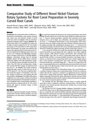
Figure 1. Measurements of root canal transportation (A) before instrumentation and (B) after instrumentation.
Figure 2. Representative images of 3-dimensional superimposed reconstructions. Red indicates preoperative area; green indicates postoperative area. (A–H)
Representative images of WaveOne group at different angles.
Basic Research—Technology
JOE — Volume 40, Number 6, June 2014 Different Novel Nickel-titanium Rotary Systems 853
3. used in 4 canals. Apical preparation was completed with a size 25 instru-
ment by using the instrument order specified by the manufacturer. Except
fortheTFAgroups,allinstrumentswereoperatedwith alow-torquemotor
(VDW Silver, VDW). The TFA groups were operated with their own motor
(Elements Motor, SybronEndo). The preparation sequences were as
follows:
1. Group 1: OS file having a taper of 0.06 and a size of 25 was used with
in-and-out movements without pressure at a rotational speed of 400
rpm and 400 gcm torque. When apical resistance was encountered,
the instrument was removed and cleaned, and the root canal was irri-
gated.
2. Group 2: For each PU file, the individual rotational speed and torque
limit programmed in the file library of the motor were used. The
sequence was as follows: SX, S1, S2, F1, and F2. The first 3 shaping
files were used with a brushing motion away from the root concav-
ities before light resistance was encountered, and the last 2 finishing
files were used with a nonbrushing action until the working length
was reached.
3. Group 3: PN files were used with the sequence PU SX, PN X1, and X2
at a rotational speed of 300 rpm and 200 gcm torque. Each file was
used with a brushing motion similar to the PU files.
4. Group 4: The Reciproc (R) R25 (VDW) file was used with the Re-
ciproc all program of the motor. The file was used in a slow in-and-
out pecking motion. The instrument was cleaned after 3 pecks, and
the root canal was irrigated.
5. Group 5: TFA (Sybron Endo) instruments were used with the TFA
program of their motor in a sequence of SM1 and SM2. The file
was advanced to the canal with a single controlled motion until
the file engaged dentin; the file was then removed and cleaned,
and the root canal was irrigated.
6. Group 6: The WO file was used with the WO all program of the mo-
tor. The file was used in a slow in-and-out pecking motion. The in-
strument was cleaned after 3 pecks, and the root canal was irrigated.
After the working length was reached inall groups, CBCTimaging of
the prepared samples was repeated using the same position and param-
eters inordertocomparepre-and post-images.Canalcurvaturesofsam-
plesweremeasuredwiththesameprotocol.Threecross-sectionalplanes
of images before and after instrumentation at 2, 5, and 8 mm from the
apical end of the root were analyzed for transportation and centering
ratio. Transportation at each level was calculated using the following for-
mula (11): (x1Àx2)À(y1Ày2). The canal centering ratio at each level
was calculated using the following formula (11): (x1Àx2)/(y1Ày2) or
(y1Ày2)/(x1Àx2); x1 and x2 represented the shortest mesial distances
from the outside of the curved root to the periphery of the uninstru-
mented and instrumented canal, respectively, and y1 and y2 represented
the shortest distal distances from the outside of the curved root to the
periphery of the uninstrumented and instrumented canal, respectively
(Fig. 1A and B). The shortest distance from the outside of the curved
root to the periphery of the instrumented canal was also recorded.
Before and after instrumentation, mesiobuccal canals of each spec-
imen were traced, and the total volume was measured. The removed
dentin volume was determined in mm3
for each root canal by subtracting
the uninstrumented canal volume from the instrumented canal volume
(Figs. 2 and 3). Volumetric measurements were obtained by using
Simplant Pro15 software (Materialise Dental NV, Leuven, Belgium).
The distribution of teeth among the 6 groups for preinstrumentation
canal curvature, radius, surface area, and volume was assessed by using
analysis of variance. Data were presented as means and standard devia-
tion. Volume, surface area, and canal curvature data (preoperatively
and postoperatively) showed a parametric distribution; thus, analysis of
variance and Tukey test were used to compare among groups. Changes
in canal transportation and centering ratio data showed a nonparametric
distribution; thus, theKruskal-Wallis testwasused to comparethe groups.
The significance level was set at P = .05. Statistical analysis wasperformed
with SPSS Statistics Version 20 for Windows (IBM, Chicago, IL).
Results
Table 1 shows the preinstrumentation curvature, radius, sur-
face area, and volume characteristics of the curved canals. No files
Figure 3. Three-dimensional reconstructions of mesiobuccal root canals of
mandibular first molars. Buccolingual views of root canals (A) before (red)
and (B) after (green) preparation.
TABLE 1. Preinstrumentation Curvature, Radius, Surface Area, and Volume Characteristics of Curved Canals and P Values among the Groups (n = 20)
Instrument
Curvature (
) Radius (mm) Volume (mm3
) Surface area (mm2)
Mean ± SD Min–Max Mean ± SD Min–Max Mean ± SD Min–Max Mean ± SD Min–Max
OneShape 28.0 Æ 5.84 20.2–40.0 6.6 Æ 1.67 3.7–10.2 2.20 Æ .64 1.0–3.25 16.8 Æ 4.04 8.0–22.7
ProTaper 28.3 Æ 5.85 20.0–40.0 6.3 Æ 1.78 3.5–9.7 2.42 Æ .51 1.78–3.16 19.5 Æ 2.94 16.2–25.0
ProTaper Next 27.9 Æ 5.70 20.0–39.2 6.5 Æ 1.61 3.8–10.8 2.63 Æ .68 2.0–3.91 20.1 Æ 3.35 15.0–26.3
Resiproc 28.0 Æ 5.71 20.4–39.3 6.7 Æ 1.72 4.3–9.8 2.12 Æ .65 1.0–3.23 18.3 Æ 4.33 13.1–25.2
Twisted Adaptive 28.2 Æ 5.81 20.7–39.8 6.6 Æ 1.85 4.1–9.7 2.17 Æ 1.37 0.82–4.78 16.5 Æ 7.67 9.3–31.0
WaveOne 28.1 Æ 5.69 20.8–39.4 6.6 Æ 2.16 3.8–10.6 2.78 Æ .97 1.3–4.4 19.7 Æ 7.34 8.0–29.9
P value 1 .989 .083 .146
SD, standard deviation.
Basic Research—Technology
854 Capar et al. JOE — Volume 40, Number 6, June 2014
4. fractured during the study. In all groups at 2, 5, and 8 mm,
maximum root canal transportation (0.3, 0.4, and 0.6 mm, respec-
tively) was less than the shortest distances from the outside of the
root to the periphery of the uninstrumented canal (0.5, 0.6, and 0.7
mm, respectively). There was no significant difference among the 6
groups in transportation, canal curvature, changes of surface area,
and centering ratio after instrumentation (P .05, Tables 2 and
3). The R group removed more dentin than the OS, TFA, and PU
groups (P .05). The other groups showed similar dentin removal
(P .05, Table 2).
Discussion
We compared the effects of five newly developed file systems that
have different designs, manufacturing methods, number of files, and
kinematics (ie, continuous rotation, reciprocating motion, and combined
reciprocating and rotation motion) on canal transportation, centering
ratio, and volume of removed dentin by using CBCT imaging. The PU
rotarysystem,which has beenusedover theyears,wasusedasareference
technique for comparison.
The distribution of the 6 groups with respect to canal curvature,
radius, and volume was well balanced (Table 1). The curvatures of
all root canals ranged between 20
and 40
, volumes of root canals
ranged between 0.82 mm3
and 4.78 mm3
, and the radii ranged between
3.5 and 10.8 mm (Table 1).
Changes in canal curvature after the use of the different NiTi file sys-
tems were not statistically significant. This is in agreement with the find-
ings of previous studies (12À15). One reason for this finding is that all
the instruments have noncutting tips that work with minimal apical
pressure and function only as a guide to allow easy penetration (16).
Single-filetechniqueshavebeensuggested for rootcanalpreparation
mostly based on opinion and simplicity rather than proven effectiveness.
Numerous studies comparing the shaping ability of single-file systems
and conventional ones using a full range of instruments have shown,
similar to our study, that both systems result in satisfactory preservation
of the original canal shape (13, 17, 18). Our results showed that both
twisted and grinded instruments have a similar effect on both canal
transportation and the centering ratio. Freire et al (19) also showed
that both twisted and grinded instruments allow the preparation of curved
canals with little transportation.
Maximum root canal transportation in all groups was less than the
shortest distances from the outside of the curved root to the periphery of
theuninstrumentedcanal.Thus,thesenewlymanufacturedrotarysystems
with size 25 and a taper of 0.06 (PN, TFA, OS) or 0.08 (WO, R, PU) could
be used in curved canals because of minimal transportation. This is in
agreement with previous studies that showed that more recent rotary
systems shaped curved root canals without significant shaping errors
(12, 15, 20, 21). However, in contrast to our findings, some previous
studies suggested that NiTi files with tapers greater than 0.04 for apical
enlargement of curved canals should not be used in older rotary
instruments, such as Profile (Dentsply Maillefer) (16) and conventional
ProTaper (22); otherwise, transportation would result. These conflicting
results might be because we used PU files, which have a ‘‘rounded safe
tip’’(23),insteadofconventionalProTaperfilesandbecauseofnewtech-
nologies (R-phase treatment, M-wire, and reciprocal motion).
We found that the OS and TFA systems with a constant 0.06 taper
removed less dentin than the R instrument (0.08 taper at the apical 3
mm followed by a regressive taper of 0.43). However, the PTN file hav-
ing an overall and apical 0.06 taper removed similar amounts of dentin
compared with other instruments having an 0.08 apical taper. This
TABLE 2. Statistical Analysis of Straightening of Canal Curvature (
), Removed Dentin Volume (mm3
), and Change of Surface Area (mm2
) for the Tested Groups
(n = 20)
Instrument
Straightening (
) Volumetric changes (mm3
) Change of surface area (mm2
)
Mean ± SD Min–Max Mean ± SD Min–Max Mean ± SD Min–Max
OneShape 4.90 Æ 3.07 0.4–10.5 2.30 Æ .90a
0.97–4.30 8.85 Æ 4.29 4.97–18.71
ProTaper 3.59 Æ 2.66 0.0–8.5 2.25 Æ 1.25a
0.42–4.17 6.24 Æ 3.40 1.51–11.92
ProTaper Next 3.76 Æ 2.16 0.3–8.5 2.78 Æ 1.35ab
0.50–4.59 8.76 Æ 5.77 0.40–18.25
Resiproc 3.48 Æ 2.60 0.0–10.1 3.51 Æ 1.73b
0.8–6.76 10.63 Æ 6.09 1.97–24.29
Twisted Adaptive 3.49 Æ 2.70 0.4–9.9 1.98 Æ 1.27a
0.57–3.88 9.18 Æ 6.63 0.92–18.96
WaveOne 3.87 Æ 2.65 0.1–8.1 2.99 Æ 1.23ab
1.09–5.30 10.34 Æ 3.70 4.78–16.16
p value .533 .004 .108
Different superscript letters indicate a significant difference between groups.
TABLE 3. Statistical Analysis of Mean Transportation (mm) and the Centering Ratio Values for Tested Groups
Instrument
Apical Middle Coronal
Mean ± SD Min–Max Mean ± SD Min–Max Mean ± SD Min–Max
OneShape Transportation 0.07 Æ 0.05 0–0.2 0.135 Æ 0.11 0–0.4 0.15 Æ 0.15 0–0.5
Centering ratio 0.44 Æ 0.45 0–1 0.36 Æ 0.43 0–1 0.53 Æ 0.45 0–1
ProTaper Transportation 0.07 Æ 0.06 0–0.2 0.120 Æ 0.09 0–0.3 0.15 Æ 0.12 0–0.3
Centering ratio 0.5 Æ 0.48 0–1 0.47 Æ 0.39 0–1 0.49 Æ 0.38 0–1
ProTaper Next Transportation 0.09 Æ 0.08 0–0.3 0.105 Æ 0.08 0–0.2 0.21 Æ 0.13 0–0.5
Centering ratio 0.41 Æ 0.48 0–1 0.39 Æ 0.45 0–1 0.32 Æ 0.35 0–1
Resiproc Transportation 0.10 Æ 0.06 0–0.2 0.130 Æ 0.12 0–0.4 0.22 Æ 0.16 0–0.6
Centering ratio 0.29 Æ 0.41 0–1 0.44 Æ 0.45 0–1 0.39 Æ 0.41 0–1
Twisted Adaptive Transportation 0.07 Æ 0.06 0–0.2 0.085 Æ 0.09 0–0.3 0.18 Æ 0.12 0–0.4
Centering ratio 0.39 Æ 0.48 0–1 0.55 Æ 0.46 0–1 0.25 Æ 0.38 0–1
WaveOne Transportation 0.06 Æ 0.06 0–0.2 0.125 Æ 0.08 0–0.3 0.19 Æ 0.13 0–0.4
Centering ratio 0.5 Æ 0.46 0–1 0.42 Æ 0.36 0–1 0.37 Æ 0.39 0–1
P value Transportation .516 .674 .453
Centering ratio .730 .780 .208
Basic Research—Technology
JOE — Volume 40, Number 6, June 2014 Different Novel Nickel-titanium Rotary Systems 855
5. might have been because of the offset asymmetric design as mentioned
previously. When we compared the similar tapered (0.08) instruments
with different kinematics for volumetric changes (R, WO, and PU), the R
instrument removed more dentin than PU, whereas WO exhibited
similar performance with R and PU.
Despite the WO and R instruments having similarities (same alloy,
reciprocation movement, and tip size), their different cross-sectional
designs may explain these results. R has a double-cutting edge S-shaped
geometry, whereas WO has a modified, convex, triangular cross-section
with radial lands at the tip and a convex triangular cross-section in the
middle and coronal portion of the instrument, similar to the PU instru-
ments (24). Stern et al (25) reported that the use of PU instruments
showed similar dentin removal with rotational or reciprocating
motions. Thus, differences might be caused by the different cross-
sectionaldesigninsteadofdifferentkinematics.Arecentstudycomparing
the shaping ability of PU and R instruments in oval canals showed that PU
removed more dentin than R instruments (24). However, in our study, R
removed more dentin than PU. This conflicting finding may be explained
by the different sizes of the instruments (25 and 40) working with
different root canal anatomy.
When we compared the 0.06 tapered instruments (OS, PN, and
TFA), there were no significant differences with respect to dentin
removal. Both twisted and grinded instruments having the same taper
had no statistically significant difference in root canal volume, which
is consistent with the results of Fayyad et al (26). Changes in surface
area were similar to volumetric changes; however, volumetric changes
were statistically significant, whereas surface area changes were not.
Conclusion
This study showed that the 6 different file systems straighten root
canal curvature similarly and produce similar canal transportation in
the preparation of curved mesial canals of mandibular molars. R
exhibited superior performance compared with OS, TFA, and PU in volu-
metric change.
Acknowledgments
The authors thank Micro Mega for providing the OneShape
instruments.
The authors deny any conflicts of interest related to this study.
References
1. Schilder H. Cleaning and shaping the root canal. Dent Clin North Am 1974;18:
269–96.
2. Weine FS, Kelly RF, Lio PJ. The effect of preparation procedures on original canal
shape and on apical foramen shape. J Endod 1975;1:255–62.
3. Gambarini G. The K3 rotary nickel titanium instrument system. Endod Top 2005;10:
179–82.
4. Peters OA. Current challenges and concepts in the preparation of root canal systems:
a review. J Endod 2004;30:559–67.
5. Schafer E, Florek H. Efficiency of rotary nickel-titanium K3 instruments compared
with stainless steel hand K-Flexofile. Part 1. Shaping ability in simulated curved
canals. Int Endod J 2003;36:199–207.
6. Weine FS. The use of non-ISO-tapered instruments for canal flaring. Compend Con-
tin Educ Dent 1996;17:651–6. 8–60, 62–3; quiz 64.
7. Wan J, Rasimick BJ, Musikant BL, et al. A comparison of cyclic fatigue resistance in
reciprocating and rotary nickel-titanium instruments. Aust Endod J 2011;37:122–7.
8. Ruddle CJ, Machtou P, West JD. The shaping movement: fifth-generation technology.
Dent Today 2013;32:94. 96–99.
9. Ruddle CJ. The ProTaper endodontic system: geometries, features, and guidelines
for use. Dent Today 2001;20:60–7.
10. Estrela C,Bueno MR, Sousa-NetoMD, etal.Methodfor determinationofrootcurvature
radius using cone-beam computed tomography images. Braz Dent J 2008;19:114–8.
11. Gambill JM, Alder M, del Rio CE. Comparison of nickel-titanium and stainless steel
hand-file instrumentation using computed tomography. J Endod 1996;22:369–75.
12. Celik D, Tasdemir T, Er K. Comparative study of 6 rotary nickel-titanium systems and
hand instrumentation for root canal preparation in severely curved root canals of
extracted teeth. J Endod 2013;39:278–82.
13. Marzouk AM, Ghoneim AG. Computed tomographic evaluation of canal shape in-
strumented by different kinematics rotary nickel-titanium systems. J Endod 2013;
39:906–9.
14. You SY, Kim HC, Bae KS, et al. Shaping ability of reciprocating motion in curved root
canals: a comparative study with micro-computed tomography. J Endod 2011;37:
1296–300.
15. Burklein S, Benten S, Schafer E. Shaping ability of different single-file systems in
severely curved root canals of extracted teeth. Int Endod J 2013;46:590–7.
16. Kum KY, Spangberg L, Cha BY, et al. Shaping ability of three ProFile rotary instru-
mentation techniques in simulated resin root canals. J Endod 2000;26:719–23.
17. Burklein S, Hinschitza K, Dammaschke T, et al. Shaping ability and cleaning
effectiveness of two single-file systems in severely curved root canals of extracted
teeth: Reciproc and WaveOne versus Mtwo and ProTaper. Int Endod J 2012;45:
449–61.
18. Berutti E, Chiandussi G, Paolino DS, et al. Canal shaping with WaveOne Primary
reciprocating files and ProTaper system: a comparative study. J Endod 2012;38:
505–9.
19. Freire LG, Gavini G, Branco-Barletta F, et al. Microscopic computerized tomographic
evaluation of root canal transportation prepared with twisted or ground nickel-
titanium rotary instruments. Oral Surg Oral Med Oral Pathol Oral Radiol Endod
2011;112:e143–8.
20. Zhao D, Shen Y, Peng B, et al. Micro-computed tomography evaluation of the prep-
aration of mesiobuccal root canals in maxillary first molars with Hyflex CM, Twisted
Files, and K3 instruments. J Endod 2013;39:385–8.
21. Guelzow A, Stamm O, Martus P, et al. Comparative study of six rotary nickel-titanium
systems and hand instrumentation for root canal preparation. Int Endod J 2005;38:
743–52.
22. Schafer E, Vlassis M. Comparative investigation of two rotary nickel-titanium instru-
ments: ProTaper versus RaCe. Part 1. Shaping ability in simulated curved canals. Int
Endod J 2004;37:229–38.
23. Camara AS, de Castro Martins R, Viana AC, et al. Flexibility and torsional strength of
ProTaper and ProTaper Universal rotary instruments assessed by mechanical tests.
J Endod 2009;35:113–6.
24. Versiani MA, Leoni GB, Steier L, et al. Micro-computed tomography study of oval-
shaped canals prepared with the Self-adjusting File, Reciproc, WaveOne, and Pro-
Taper Universal Systems. J Endod 2013;39:1060–6.
25. Stern S, Patel S, Foschi F, et al. Changes in centring and shaping ability using three
nickel-titanium instrumentation techniques analysed by micro-computed tomogra-
phy (muCT). Int Endod J 2012;45:514–23.
26. Fayyad DM, Elhakim Elgendy AA. Cutting efficiency of twisted versus machined
nickel-titanium endodontic files. J Endod 2011;37:1143–6.
Basic Research—Technology
856 Capar et al. JOE — Volume 40, Number 6, June 2014